

 PDF
PDF ePub
ePub Citation
 Print
Print


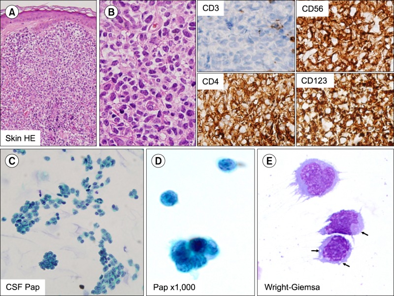
A 73-year-old man presented with skin nodules, which were composed of monomorphic, medium-sized blastic cells with irregular nuclei, fine chromatin, and scanty cytoplasm (A, B). Immunohistochemical staining revealed a typical blastic plasmacytoid dendritic cell neoplasm (BPDCN) phenotype; the nodules stained positively for CD4, CD56, and CD123, and negatively for CD3, CD8, MPO, TdT, and Epstein-Barr virus. The patient was treated with six cycles of CHOP, and achieved complete remission. Five months later, he presented with a headache and left ptosis. Examination of the cerebrospinal fluid (CSF) showed numerous medium-sized monomorphic cells, either in loose clusters or dispersed singly. The nuclei were round or irregular, showing fine chromatin and prominent nucleoli, and the cytoplasm was scant and grey-blue, with intracytoplasmic microvacuoles and pseudopodia (C, D, and E: arrows indicate intracytoplasmic vacuoles). BPDCN is an aggressive tumor with a high frequency of cutaneous, bone marrow, and peripheral blood involvement. Although most patients initially respond to combination chemotherapy, BPDCN regularly recurs in the skin, soft tissue, and central nervous system (CNS). BPDCN commonly recurs in the CNS, and this can be easily diagnosed by CSF examination, which showed characteristic monotonous tumor cells with cytoplasmic microvacuoles.
 XML Download