Journal List > Blood Res > v.51(1) > 1092308

Kang, Ko, Choi, Choi, Seo, Lee, and Lee: Humanizing NOD/SCID/IL-2Rγnull (NSG) mice using busulfan and retro-orbital injection of umbilical cord blood-derived CD34+ cells

Abstract

Background

Humanized mouse models are still under development, and various protocols exist to improve human cell engraftment and function.

Methods

Fourteen NOD/SCID/IL-2Rγnull (NSG) mice (4‒5 wk old) were conditioned with busulfan and injected with human umbilical cord blood (hUCB)-derived CD34+ hematopoietic stem cells (HSC) via retro-orbital sinuses. The bone marrow (BM), spleen, and peripheral blood (PB) were analyzed 8 and 12 weeks after HSC transplantation.

Results

Most of the NSG mice tolerated the regimen well. The percentage of hCD45+ and CD19+ cells rose significantly in a time-dependent manner. The median percentage of hCD45+cells in the BM was 55.5% at week 8, and 67.2% at week 12. The median percentage of hCD45+ cells in the spleen at weeks 8 and 12 was 42% and 51%, respectively. The median percentage of hCD19+ cells in BM at weeks 8 and 12 was 21.5% and 39%, respectively (P=0.04). Similarly, the median percentage of hCD19+ cells in the spleen at weeks 8 and 12 was 10% and 24%, respectively (P=0.04). The percentage of hCD19+ B cells in PB was 23% at week 12. At week 8, hCD3+ T cells were barely detectable, while hCD7+ was detected in the BM and spleen. The percentage of hCD3+ T cells was 2‒3% at week 12 in the BM, spleen, and PB of humanized NSG mice.

Conclusion

We adopted a simplified protocol for establishing humanized NSG mice. We observed a higher engraftment rate of human CD45+ cells than earlier studies without any significant toxicity. And human CD45+ cell engraftment at week 8 was comparable to that of week 12.

INTRODUCTION

Several human diseases do not have appropriate animal models [1], or the available animal models have significant differences from the human counterpart [1]. Non-human primates have been regarded as ideal models for translating basic research findings into clinical applications due to their similarities to humans [12]. Recently, the European governing bodies and the United States National Institutes of Health have significantly reduced the use of chimpanzees in research due to cost and ethical concerns [3]. Humanized mice carrying human hematopoietic and immune systems are considered as ideal tools for studying hematopoiesis, infectious disease, and immunology [3]. For example, dengue and human immunodeficiency virus infect humanized mice, while they do not replicate in rodents [4]. Still, humanized mouse models need further development in order to more closely recapitulate human biological systems.
Humanized mouse models are still under development, and various protocols exist to improve human cell engraftment and function. The source of stem cells can vary depending on the research objectives. Usually, CD34+ cells yield long-term engraftment and are chosen to study hematopoiesis [3]. Peripheral blood mononuclear cells are used to study transplantation immunology [56]. Total body irradiation (TBI) has been a standard conditioning regimen to achieve high levels of human cell engraftment in xenograft animal models [37] because it triggers the secretion of stem cell factor (SCF), which is critical for hematopoietic stem cell engraftment, proliferation, and survival [7]. However, TBI is a tedious procedure to implement, because it requires strict regulation for the use of irradiators, remote location from animal housing, and special animal care [8]. Recently, researchers have used chemotherapeutic agents such as busulfan to induce similar hematopoietic effects as TBI [89101112]. The route of donor cell administration is another factor to consider for successful transplantation [13]. Donor cells can be administered via vein, liver, or bone [3] The tail vein is the most frequently used intravascular access [1314], but it is technically challenging and often requires the heating of mice to enhance peripheral vasodilation [13]. The retro-orbital sinus is an alternative administration route that causes less pain and distress for animals [1516]. However, uncertainty exists about the time and duration of engraftment after human cell transplantation into the NOD/SCID/IL-2Rγnull (NSG) mice. Human CD45+ cells can be detected in the peripheral blood as early as 3 weeks after human HSC injection [8]. In other studies, high engraftment level was observed 22-24 weeks after human HSC transplantation [1718]. It was reported that NSG mice survived up to 300 days after human HSC transplantation [8].
In this study, young NSG mice (4-5 wk old) were conditioned with busulfan and injected with HSCs via their retro-orbital sinuses. They were maintained in pathogen-free sterile conditions, but without individualized ventilating cages (IVC). Most of the NSG mice did well after busulfan and HSC injection. We analyzed the bone marrow, spleen, and peripheral blood of NSG mice at weeks 8 and 12 after human HSC transplantation and examined human cell engraftment.

MATERIALS AND METHODS

Mice

NOD.Cg-PrkdcscidIl2rγtm1Wjl/SzJ (NOD-scid IL2rγnull, NSG) mice were obtained from the Jackson Laboratory (Bar Harbor, ME, USA). For all experiments, mice were bred as a homozygous line and maintained under specific pathogen-free conditions at Laboratory of Animal Research Center in Korea Institute of Radiological Medical Sciences (Seoul, Korea). Experiments were conducted according to the guidelines for ethical use of animals of our institution under an approved protocol (KIRAMS 2015-0018).

Isolation of human CD34+ cells from human umbilical cord blood

Human umbilical cord blood (hUCB) was provided by the Seoul Metropolitan Government Public Cord Blood Bank (Allcord, Korea). Mononuclear cells (MNCs) were enriched using a RosetteSep Human Progenitor Enrichment kit (StemCell Technologies, Canada) and isolated from hUCB using Ficoll-Paque PREMIUM (GE Healthcare Bio-Sciences AB, Sweden) density gradient centrifugation. MNCs were enriched for hCD34+ cells using a human CD34 MicroBead Kit UltraPure (Miltenyi Biotec, Spain) according to the manufacturer's instructions. The purity of hCD34+ cells was 87.6% as determined using a FACSCanto II flow cytometer (BD Biosciences, USA), and these hCD34+ cells were preserved in STEM-CELLBANKER medium (Zenoaq, Japan) at -80℃ until use.

Transplantation of hCD34+ cells

NSG mice were conditioned with busulfan (Korea Otsuka Pharmaceutical, Korea). Busulfan was dissolved in dimethyl sulfoxide (Sigma Chemical Co., St. Louis, MO, USA) and diluted with 0.9% saline. The liquid busulfan solution was intraperitoneally (i.p.) injected into NSG mice (25 mg/kg body weight, 500-625 µg per dose) 48 and 24 hours prior to transplantation. The next day, 1×10⁵ hCD34+ cells in 100 µL phosphate-buffered saline (PBS) were transplanted into the NSG mice via retro-orbital sinus injection. To prevent urinary tract infections, we used enrofloxacin (0.27 mg/mL) as a prophylactic antibiotic in drinking water of mice.

Analysis of engraftment

Eight and 12 weeks after transplantation, mice were sacrificed and mononuclear cells were isolated from bone marrow, spleen, and peripheral blood. Single-cell suspensions were prepared by standard procedures and were stained with the following antibodies: hCD34-fluorescein isothiocyanate (FITC), hCD45-allophycocyanin (APC) (Miltenyi Biotec, Spain), hCD3-fluorescein isothiocyanate (FITC), and hCD19-phycoerythrin (PE) (BD Biosciences, USA). Flow cytometry was performed using a FACSCanto II (BD Biosciences, USA). Ten thousand to one million events were acquired per sample and analyzed with FACSDiva software (BD Biosciences, USA). Cell lysates were prepared from bone marrow and spleen, and western blotting was performed by standard procedures using hCD45 and hCD7 antibodies (BD Biosciences, USA).

Statistical analysis

Statistical analysis was performed using GraphPad Prism software (GraphPad Software Inc., CA) and SPSS (IBM Corp., USA) and P <0.05 was considered statistically significant.

RESULTS

Reconstitution of human cells in NSG mice transplanted with hUCB-derived CD34+ cells

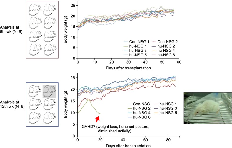
Fourteen NSG mice received i.p. busulfan and retro-orbital hCD34+ cell injection, and most of them tolerated the regimen well (Fig. 1). However, 1 mouse showed decreased activity and hunched posture and died 20 days after hCD34+ cell injection (Fig. 2). We analyzed the bone marrow, spleen, and peripheral blood of the NSG mice at 8 and 12 weeks after transplantation and found that the percentage of human CD45+ cells rose significantly in a time-dependent manner (Fig. 3). The median percentages of human CD45+ cells in the bone marrow were 55.5% at week 8 and 67.2% at week 12. Similarly, the median percentages of human CD45+ cells in the spleen at weeks 8 and 12 were 42% and 51%, respectively.

Reconstitution of human B- and T-cells in humanized NSG mice

The percentage of hCD19+ cells rose significantly in a time-dependent manner (Fig. 4A). The median percentages of human CD19+ cells in bone marrow at weeks 8 and 12 were 21.5% and 39%, respectively (P=0.04). Similarly, the median percentages of human CD19+ cells in spleen at weeks 8 and 12 were 10% and 24%, respectively (P=0.04). The percentage of hCD19+ B-cells in peripheral blood was 23% at week 12.
The percentage of human CD3+ T cells in the bone marrow, spleen, and peripheral blood of humanized NSG mice was 2-3% at week 12 (Fig. 4B). However, human CD3+ cells were barely detectable at week 8 (Fig. 4B). Therefore, we performed western blotting using anti-hCD7, a marker of early T cell lineage. Because of the limited sample volume that remained after hCD45, hCD19, and hCD3 analysis, samples from only 4 mice could be analyzed. We observed that hCD7+ cells were detectable in the bone marrow and spleen of all 4 mice at 8 weeks after hUCB-derived CD34+ cell injection (Fig. 5).

DISCUSSION

Humanized mice are gaining attention as animal models for translating basic research findings into clinical applications [3]. Currently, the NOD/SCID/IL-2Rγnull (NSG) mice, which lack T-, B-, and NK cell activity, are considered as ideal candidates to establish humanized mice [19]. Many preliminary reports have demonstrated the utility of humanized mice in infectious disease and immunology [34]. Humanized mouse model protocols are still under development to improve human cell engraftment and function [3].
With a simplified protocol, we observed a higher engraftment rate of human CD45+ cells than earlier studies. The NSG mice were conditioned with busulfan, injected with hUCB-CD34+ cells via retro-orbital sinus, and housed without IVCs. Traditionally, total body irradiation (TBI) has been used as conditioning regimen [7]. However, the administration of TBI requires strict facility regulations and results in substantial mortality [8]. It is suggested that animals receiving TBI need to be maintained in strictly controlled pathogen-free conditions [8]. Busulfan (1,4-butanediol dimethanesulfonate) is an alkylating agent that has long been used in human HSC transplantation [20]. Because it has predominantly myelosuppressive and minimally immunosuppressive properties, recent studies suggested that busulfan induces similar hematopoietic effects to TBI while being easier and less expensive for animal transplantation models [9101112]. Some studies used the same busulfan dose and cell dose as our study [810121718]. The engraftment rate varied depending on the dose of busulfan, CD34+ cells and the time point of analysis [810121718]. Choi et al. conditioned NSG mice with busulfan and infused 1×10⁵ hUCB-derived CD34+ cells and reported that human CD45+ cells comprised 76% of bone marrow CD45+ cells at the 24th week [10]. At the 12th week, the percentage of human CD45+ cells in the NSG mice injected with 1×10⁵ hUCB-derived CD34+ cells after a single dose of busulfan conditioning (20 mg/kg) was 25.33% [17]. Our median percentages of human CD45+ cells in the bone marrow of NSG mice at 8 and 12 weeks after transplantation were 59.3% and 72.3%, respectively. We adopted split dose busulfan conditioning (2 doses of 20-30 mg/kg) and assumed that this might have contributed to a higher engraftment rate. Moreover, there were no infectious complications after HSC transplantation. Our NSG mice were maintained in less strict conditions and all mice did well after HSC transplantation.
In the current study, the percentage of human CD3+ cells was in the range of 2-3% at week 12 after transplantation. Hayakawa et al. reported that their percentage of human CD3+ cells in the peripheral blood of NSG mice was 0.6% at 8 weeks after transplantation [8]. Meanwhile, the percentage of T cells was 21.15% in the mesenteric lymph node at week 12 after HSC transplantation [17]. T cell engraftment in NSG mice gradually increases after HSC transplantation [17]. The percentages of human CD3+ cells in mouse bone marrow at weeks 12 and 22 after transplantation were 3.33% and 40.6%, respectively [17]. Still, it is uncertain to what extent the transplanted human cells could reconstitute a hematopoietic system in NSG mice. Singh et al. observed a prolonged human cell chimerism over 300 days, with increasing CD3+ T cell levels [17]. On the other hand, limited engraftment of myeloid lineage cells, especially red blood cells, was reported [821]. Humans and mice differ in the growth factors and cytokines required for the development of the hematopoietic and immune systems [22]. NSG mice lack the HLA molecule for human T cell education, and have poorly organized lymphoid architecture and deficiencies in lymph node development [23]. Various studies have attempted to increase reconstitution of human hematopoietic cells. Transgenic expression of IL-3, GM-CSF, and SCF increased the percentages of human myeloid cells in the bone marrow of NSG mice engrafted with human HSC [24]. The bone marrow, liver, thymus (BLT) model showed robust and consistent engraftment of multiple human hematopoietic lineages [232526]. Cotransplantation of fetal bone tissue facilitated the development and reconstitution of human B cells in humanized NSG mice [11]. A recent study reported that intrahepatic injection of human UCB-derived CD34+ cells can facilitate human T cell development in livers of humanized NSG mice [27]. Administration of recombinant human IL-7 also improved T cell development in humanized mice [2228]. To improve human hematopoietic engraftment and myeloid differentiation, new generations of immunodeficient mouse stains that express human hematopoietic growth factors are under development [2429].
In conclusion, we observed a high rate of hUBC-derived CD34+ cell engraftment in NSG mice using a simplified protocol. The recipient NSG mice were conditioned with busulfan, injected with CD34+ cells via retro-orbital sinus, and maintained without IVCs. Most of them tolerated the regimen, and human cell engraftment after 8 weeks was comparable to the 12th week. Further studies are necessary to increase engraftment rate and function of human hematopoietic cells.

Notes

This study was supported by a grant from the Korea Institute of Radiological and Medical Sciences (KIRAMS), funded by the Ministry of Science, ICT and Future Planning, Korea (1711021931).

Authors' Disclosures of Potential Conflicts of Interest: No potential conflicts of interest relevant to this article were reported.

References

1. McClure HM. Nonhuman primate models for human disease. Adv Vet Sci Comp Med. 1984; 28:267–304. PMID: 6395673.
crossref
2. Hu SL. Non-human primate models for AIDS vaccine research. Curr Drug Targets Infect Disord. 2005; 5:193–201. PMID: 15975024.
crossref
3. Shultz LD, Brehm MA, Garcia-Martinez JV, Greiner DL. Humanized mice for immune system investigation: progress, promise and challenges. Nat Rev Immunol. 2012; 12:786–798. PMID: 23059428.
crossref
4. Brehm MA, Jouvet N, Greiner DL, Shultz LD. Humanized mice for the study of infectious diseases. Curr Opin Immunol. 2013; 25:428–435. PMID: 23751490.
crossref
5. King M, Pearson T, Shultz LD, et al. A new Hu-PBL model for the study of human islet alloreactivity based on NOD-scid mice bearing a targeted mutation in the IL-2 receptor gamma chain gene. Clin Immunol. 2008; 126:303–314. PMID: 18096436.
crossref
6. Harui A, Kiertscher SM, Roth MD. Reconstitution of huPBL-NSG mice with donor-matched dendritic cells enables antigen-specific T-cell activation. J Neuroimmune Pharmacol. 2011; 6:148–157. PMID: 20532647.
crossref
7. Sugimoto K, Adachi Y, Moriyama K, et al. Induction of the expression of SCF in mouse by lethal irradiation. Growth Factors. 2001; 19:219–231. PMID: 11811778.
crossref
8. Hayakawa J, Hsieh MM, Uchida N, Phang O, Tisdale JF. Busulfan produces efficient human cell engraftment in NOD/LtSz-Scid IL2Rgamma(null) mice. Stem Cells. 2009; 27:175–182. PMID: 18927475.
9. Robert-Richard E, Ged C, Ortet J, et al. Human cell engraftment after busulfan or irradiation conditioning of NOD/SCID mice. Haematologica. 2006; 91:1384. PMID: 17018389.
10. Choi B, Chun E, Kim M, et al. Human B cell development and antibody production in humanized NOD/SCID/IL-2Rγ(null) (NSG) mice conditioned by busulfan. J Clin Immunol. 2011; 31:253–264. PMID: 20981478.
crossref
11. Kim M, Choi B, Kim SY, et al. Co-transplantation of fetal bone tissue facilitates the development and reconstitution in human B cells in humanized NOD/SCID/IL-2Rγnull (NSG) mice. J Clin Immunol. 2011; 31:699–709. PMID: 21544592.
crossref
12. Chevaleyre J, Duchez P, Rodriguez L, et al. Busulfan administration flexibility increases the applicability of scid repopulating cell assay in NSG mouse model. PLoS One. 2013; 8:e74361. PMID: 24069300.
crossref
13. Yardeni T, Eckhaus M, Morris HD, Huizing M, Hoogstraten-Miller S. Retro-orbital injections in mice. Lab Anim (NY). 2011; 40:155–160. PMID: 21508954.
crossref
14. Suckow MA, Danneman P, Brayton C, editors. The laboratory mouse. Boca Raton, FL: CRC Press;2001.
15. Price JE, Barth RF, Johnson CW, Staubus AE. Injection of cells and monoclonal antibodies into mice: comparison of tail vein and retroorbital routes. Proc Soc Exp Biol Med. 1984; 177:347–353. PMID: 6091149.
crossref
16. Steel CD, Stephens AL, Hahto SM, Singletary SJ, Ciavarra RP. Comparison of the lateral tail vein and the retro-orbital venous sinus as routes of intravenous drug delivery in a transgenic mouse model. Lab Anim (NY). 2008; 37:26–32. PMID: 18094699.
crossref
17. Singh M, Singh P, Gaudray G, et al. An improved protocol for efficient engraftment in NOD/LTSZ-SCIDIL-2Rγnull mice allows HIV replication and development of anti-HIV immune responses. PLoS One. 2012; 7:e38491. PMID: 22675567.
crossref
18. Lee M, Jeong SY, Ha J, et al. Low immunogenicity of allogeneic human umbilical cord blood-derived mesenchymal stem cells in vitro and in vivo. Biochem Biophys Res Commun. 2014; 446:983–989. PMID: 24657442.
crossref
19. Ishikawa F. Modeling normal and malignant human hematopoiesis in vivo through newborn NSG xenotransplantation. Int J Hematol. 2013; 98:634–640. PMID: 24258713.
crossref
20. Ciurea SO, Andersson BS. Busulfan in hematopoietic stem cell transplantation. Biol Blood Marrow Transplant. 2009; 15:523–536. PMID: 19361744.
crossref
21. Li Y, Chen Q, Zheng D, et al. Induction of functional human macrophages from bone marrow promonocytes by M-CSF in humanized mice. J Immunol. 2013; 191:3192–3199. PMID: 23935193.
crossref
22. O'Connell RM, Balazs AB, Rao DS, Kivork C, Yang L, Baltimore D. Lentiviral vector delivery of human interleukin-7 (hIL-7) to human immune system (HIS) mice expands T lymphocyte populations. PLoS One. 2010; 5:e12009. PMID: 20700454.
23. Brehm MA, Shultz LD, Luban J, Greiner DL. Overcoming current limitations in humanized mouse research. J Infect Dis. 2013; 208(Suppl 2):S125–S130. PMID: 24151318.
crossref
24. Drake AC, Chen Q, Chen J. Engineering humanized mice for improved hematopoietic reconstitution. Cell Mol Immunol. 2012; 9:215–224. PMID: 22425741.
crossref
25. Covassin L, Jangalwe S, Jouvet N, et al. Human immune system development and survival of non-obese diabetic (NOD)-scid IL2rγ(null) (NSG) mice engrafted with human thymus and autologous haematopoietic stem cells. Clin Exp Immunol. 2013; 174:372–388. PMID: 23869841.
crossref
26. Lavender KJ, Messer RJ, Race B, Hasenkrug KJ. Production of bone marrow, liver, thymus (BLT) humanized mice on the C57BL/6 Rag2(-/-)γc(-/-)CD47(-/-) background. J Immunol Methods. 2014; 407:127–134. PMID: 24769067.
crossref
27. Choi B, Chun E, Kim M, et al. Human T cell development in the liver of humanized NOD/SCID/IL-2Rγ(null)(NSG) mice generated by intrahepatic injection of CD34(+) human (h) cord blood (CB) cells. Clin Immunol. 2011; 139:321–335. PMID: 21429805.
crossref
28. van Lent AU, Dontje W, Nagasawa M, et al. IL-7 enhances thymichuman T cell development in "human immune system" Rag2-/- IL-2Rgammac-/- mice without affecting peripheral T cell homeostasis. J Immunol. 2009; 183:7645–7655. PMID: 19923447.
29. Chen Q, Khoury M, Chen J. Expression of human cytokines dramatically improves reconstitution of specific human-blood lineage cells in humanized mice. Proc Natl Acad Sci U S A. 2009; 106:21783–21788. PMID: 19966223.
crossref
Fig. 1

Scheme for generating the humanized NSG mice. MNCs isolated from hUCB were enriched using a RosetteSep kit and human CD34 MicroBead Kit. NSG mice were conditioned by busulfan and CD34+ cells were injected via retro-orbital sinus.

br-51-31-g001
Fig. 2

Survival and weight changes of the NSG mice after hCD34+ cell injection. Humanized NSG mice were monitored daily after transplantation. Most of the NSG mice did well, but one mice (depicted with an arrow) showed features suggesting GVHD (weight loss, hunched posture and diminished activity as shown in the photo) 10 days after transplantation.

br-51-31-g002
Fig. 3

Human cell reconstitution of NSG mice transplanted with hUCB-derived CD34+ cells. Levels of human CD45+ cells in mouse tissues at different times after transplantation are shown. Bone marrow, spleen, and blood were isolated from the humanized NSG mice and MNCs isolated from each organ were stained and analyzed. The percentages are represented as mean±SEM in humanized mice.

br-51-31-g003
Fig. 4

Human CD19+ and CD3+ cell reconstitution from NSG mice injected with hUCB-derived CD34+ cells. The percentages are represented by mean±SEM in humanized mice.

br-51-31-g004
Fig. 5

hCD45 and hCD7 expression levels in spleen (SPL) and bone marrow (BM) of NSG mice 8 weeks after hUCB-derived CD34+ cell injection.

br-51-31-g005
TOOLS
Similar articles