TO THE EDITOR: Thrombotic thrombocytopenic purpura (TTP) was first described by Eli Moschowitz about 90 years ago [1]. TTP is caused by a severe deficiency in the protein A disintegrin and metalloprotease with thrombospondin 1 motifs 13 (ADAMTS13), a metalloprotease that cleaves ultra-large von Willebrand factor (vWF) multimers [2]. Deficiency in this enzyme causes the accumulation of large vWF multimers, which increase platelet adhesiveness and impair fibrinolytic activity with subsequent thrombotic occlusion of the microvasculature. TTP is a rare and potentially life-threatening disorder. The incidence rate of suspected TTP-hemolytic uremic syndrome (TTP-HUS) in the general population is 11 cases per 1,000,000 people [3], compared to the estimated incidence of one in 25,000 deliveries [4]. The clinical features of TTP may resemble those of the more common pregnancy complications, such as preeclampsia or of hemolytic anemia, elevated liver enzymes, and low platelet count (HELLP) syndrome. However, the management of TTP is completely different from that of preeclampsia/HELLP. Here, we present the case of a young woman who developed TTP in late gestation.
A 22-year-old, apparently healthy, Caribbean woman at 39 weeks of gestation was admitted from the outpatient obstetrics clinic for proteinuria and decreased fetal heart rate. She did not have any complications during previous pregnancies. She denied having fever, cough, abdominal pain, headache, blurry vision, recent infection, contacts with sick persons, or experiencing bleeding. Upon physical examination, the patient's blood pressure was 130/70 mmHg, and her heart rate was 103 beats per minute. She was alert, awake, and oriented. Abdominal examination revealed a non-tender gravid uterus of about 36-week size, and a vaginal examination showed a closed cervical os. A few petechiae were noted on the lower extremities. The rest of the physical examination was not significant.
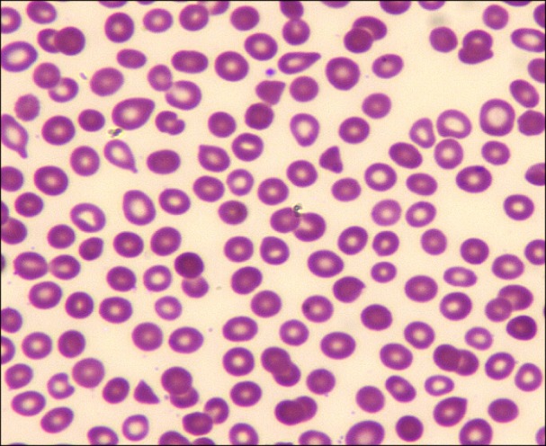
Laboratory tests performed on admission revealed a hemoglobin concentration of 10 g/dL, white blood cell count of 12.3×10³/µL, platelet count of 12×103/µL, and creatinine concentration of 0.6 mg/dL. Her coagulation profile, fibrinogen level, and liver function tests were normal, thus ruling out HELLP syndrome and disseminated intravascular coagulation (DIC). She had an elevated lactate dehydrogenase level of 560 mg/dL, reticulocyte count of 3.2%, and a very low haptoglobin level (<8 mg/dL). Autoimmune hemolysis was excluded by a normal direct antiglobulin test. A HIV test and her viral hepatitis profiles (hepatitis A, B, and C) were negative. A peripheral blood smear examination revealed the presence of occasional schistocytes (2–3 schistocytes per high power field) and markedly decreased platelet count (Fig. 1). A presumed TTP diagnosis was made based on these clinical and laboratory findings. Tests for ADAMTS13 activity and ADAMTS13 inhibitor level were sent, and arrangements were made to initiate plasma exchange (PEX).
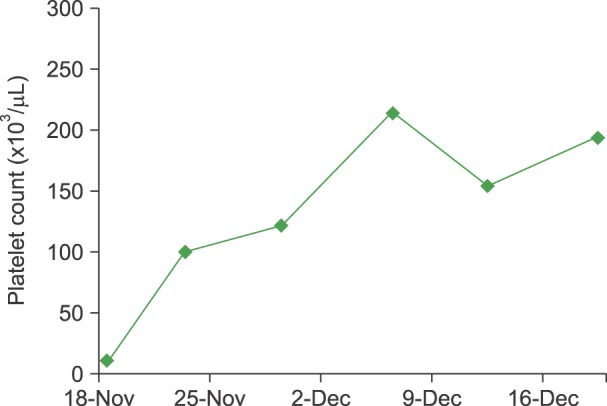
The patient underwent a cesarean section and was transfused with 2 units of single-donor platelets and 2 units of fresh frozen plasma during both the pre- and intra-operative periods. She delivered a healthy full-term baby boy without any complications. Daily one-plasma-volume PEX was initiated immediately after delivery. Because of the concern for delayed wound healing, prednisone was not initially started. She was also given folic acid. Her platelet count began to increase on day 3 of PEX. Her hospital course was complicated by healthcare-associated pneumonia, and she was also treated with broad-spectrum antibiotics. The results of the ADAMTS13 activity and ADAMTS13 inhibitor tests were returned on the fifth day of PEX, and were reported as <3% and 1.7 Bethesda units/mL, respectively, confirming the diagnosis of acquired TTP (normal <0.4 Bethesda units/mL). Prednisone (1 mg/kg) was initiated on day 7 of PEX. Platelet count was normalized after two weeks (Fig. 2); the delayed platelet recovery could be due to concurrent infection and medications. The patient was discharged on day 18 of hospitalization and followed-up with as an outpatient. Due to a slight drop in platelet count, she continued to receive five more PEX sessions in the two weeks following discharge. Five weeks after her delivery, she was maintaining a stable platelet count and the prednisone was tapered off.
TTP occurring in association with pregnancy was first reported in 1955 by Miner et al. [5]. In a review of 45 cases of pregnancy-associated thrombotic microangiopathies between 1966 and 1988, a maternal mortality of 44% and an associated fetal loss rate of 80% were reported [6]. Historically, TTP had a mortality rate as high as 90% when left untreated [7]. Prompt recognition and initiation of early therapy have drastically reduced the mortality rate to 10–20% [8].
Pregnancy is a commonly recognized risk factor for triggering an acute episode of TTP. The plausible contributing factors include an increase in concentration of procoagulant factors, decrease in fibrinolytic activity, loss of endothelial cell thrombomodulin, and progressive ADAMTS13 deficiency over the course of pregnancy [9].
Causes of TTP-HUS during pregnancy include familial TTP (Upshaw-Schulman syndrome, caused by congenital ADAMTS13 deficiency), congenital complement-mediated HUS, and acquired TTP-HUS. In a prospective series of women from the United Kingdom TTP registry, 23 (66%) of 35 pregnant women who presented with TTP were classified as having congenital TTP, and 12 cases (34%) were acquired [10]. In contrast, in an earlier series of 42 patients with a first episode of TTP during pregnancy, only 10 cases (24%) were attributable to congenital TTP [11]. In a larger series of 166 pregnancies, the median time of TTP onset was 23–24 weeks with 12%, 55%, and 33% of TTP occurring in the first, second, and third trimesters, respectively [12].
The classic "pentad" of fever, hemolytic anemia, thrombocytopenia, renal impairment, and neurologic manifestations is not always present. It is of utmost importance to have a high clinical vigilance in TTP recognition and treatment. It is also important to differentiate TTP from other serious pregnancy complications such as DIC and preeclampsia/eclampsia/HELLP. A complete blood count and review of the peripheral blood smear for microangiopathic hemolytic anemia and thrombocytopenia play a crucial role in the diagnosis of TTP. Coagulation testing should also be done to exclude the possibility of DIC. Additional testing includes a chemistry profile to determine renal function, serum lactate dehydrogenase and bilirubin to assess hemolysis, and measuring ADAMTS13 activity, with testing for an inhibitor level (anti-ADAMTS13 autoantibody) if activity is <10%.
PEX is the mainstay for TTP treatment. Once TTP is suspected, PEX should be initiated immediately. PEX removes the ADAMTS13 autoantibody and replenishes ADAMTS13 levels. Fresh frozen plasma could be infused to increase ADAMTS13 levels if PEX is not available immediately. A meta-analysis of six randomized controlled trials found that PEX is more effective than plasma infusion in improving overall survival rates [13]. The use of corticosteroids in TTP is controversial. Further prospective studies are required to define the benefit of the addition of steroids to PEX compared to PEX alone.
Patients with severe ADAMTS13 deficiency exhibit variable clinical courses. Twice daily PEX may be performed in some of the patients who fail to respond to the initial daily PEX or have exacerbation of symptoms. If response to PEX is insufficient, administration of weekly rituximab should be considered in addition to daily PEX. In an analysis of 100 patients with acute refractory or chronic relapsing idiopathic TTP treated with rituximab, complete remission was seen in 98% of patients, while 2% were non-responsive to treatment, and 9% relapsed after complete remission. Testing positive for ADAMTS13 inhibitor and severe ADAMTS13 deficiency were both found to be highly predictive of response to rituximab [14]. Once a normal platelet count is achieved, PEX may be discontinued abruptly or tapered gradually, and corticosteroids should be quickly tapered.
Many women may subsequently have successful pregnancy outcomes after recovery from TTP. Women with congenital TTP have a far higher chance of developing recurrent TTP episodes in subsequent pregnancies unless appropriate prophylaxis with plasma infusion is provided. On the other hand, the risk of recurrence in women with acquired TTP in subsequent pregnancies is low [8]. In an analysis of 16 pregnancies in 10 women who had recovered from acquired TTP associated with severe acquired ADAMTS13 deficiency (activity <10%), successful full-term deliveries were reported in 13 pregnancies (81%) [15]. Studies suggest that severe ADAMTS13 deficiency or the presence of an inhibitor may be useful in relapse prediction. The risk of recurrence may be monitored by performing the serial measurements of ADAMTS13 activity during pregnancy and elective PEX should be initiated and immunosuppressive treatments could be considered in women with ADAMTS13 deficiency.
In conclusion, although the occurrence of TTP is rare in pregnancy, clinicians should be highly vigilant of its potential presentation in pregnant women with thrombocytopenia. If TTP is clinically suspected, PEX should be promptly instituted while waiting for the results of ADAMTS13 activity level tests. Because of the potential risk of recurrent TTP, pregnant women with a history of TTP should be monitored carefully by a multidisciplinary team that includes an obstetrician, a hematologist, and a neonatologist.


 PDF
PDF ePub
ePub Citation
 Print
Print


 XML Download