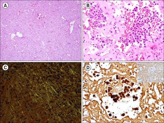
A 53-year-old man presented with a history of weight loss of 10 kg over 3 months. A peripheral blood count indicated a leukocyte count of 49.0×10³/L and 80% segmented neutrophils. The neutrophils showed toxic changes and appeared in hypersegmented forms. Bone marrow aspiration revealed marked granulocytic hyperplasia and nodular and interstitial infiltration of plasma cells. Serum and urine immunofixation electrophoresis revealed an abnormal zone of restriction in the lambda light chain. While the patient was being evaluated for multiple myeloma with concurrent chronic neutrophilic leukemia, he was rushed to the emergency room because of loss of consciousness and hypotension. Computed tomography (CT) showed spontaneous splenic rupture, but the liver contour was normal. The spleen weighed 280 g, and microscopically showed diffuse deposition of pinkish, amorphous materials in the red pulp, with effacement of the normal structures (A, ×200 and B, ×400, hematoxylin-eosin staining). On Congo red staining, the deposits showed typical apple-green birefringence under polarized light (C, ×400). The sinusoids contained numerous neutrophils and plasma cells, which showed lambda light chain restriction (D, Lambda stain and Inset, Kappa stain, ×400). Although abdominal CT revealed diffuse swelling of both kidneys, this was not confirmed histologically.


 PDF
PDF ePub
ePub Citation
 Print
Print


 XML Download