Journal List > Blood Res > v.50(4) > 1092223

Park, Yoo, and Kim: Hematopoietic stem cell expansion and generation: the ways to make a breakthrough

Abstract

Hematopoietic stem cell transplantation (HSCT) is the first field where human stem cell therapy was successful. Flooding interest on human stem cell therapy to cure previously incurable diseases is largely indebted to HSCT success. Allogeneic HSCT has been an important modality to cure various diseases including hematologic malignancies, various non-malignant hematologic diseases, primary immunodeficiency diseases, and inborn errors of metabolism, while autologous HSCT is generally performed to rescue bone marrow aplasia following high-dose chemotherapy for solid tumors or multiple myeloma. Recently, HSCs are also spotlighted in the field of regenerative medicine for the amelioration of symptoms caused by neurodegenerative diseases, heart diseases, and others. Although the demand for HSCs has been growing, their supply often fails to meet the demand of the patients needing transplant due to a lack of histocompatible donors or a limited cell number. This review focuses on the generation and large-scale expansion of HSCs, which might overcome current limitations in the application of HSCs for clinical use. Furthermore, current proof of concept to replenish hematological homeostasis from non-hematological origin will be covered.

INTRODUCTION

Hematopoietic stem cells (HSCs) reside in bone marrow and can be differentiated into multiple lineages of mature hematologic cells. The first successful hematopoietic stem cell transplantation (HSCT) using human bone marrow form a relative donor was accomplished in 1968 in a boy with X-linked severe combined immunodeficiency disease (SCID) [1]. Since the first successful achievement in human, numerous 'trial and error's had been repeated until the early 1980s when allogeneic HSCT began to be actively performed under a better recognition of transplant immunology. Dr. Donnall Thomas, who received Nobel Prize for his pioneering work in bone marrow transplantation to cure leukemia and other hematologic malignancies, must be recognized and apprised as human endeavor to cure previously incurable diseases. The outcome of allogeneic HSCT has improved over the last 30 years owing to better supportive care, better disease control prior to transplant, better human leukocyte antigen (HLA) matching, and better control of post-transplant complications. However, 30 years of successful HSCT cannot ignore the current problem of donor shortage. Insufficiency of HLA-matched donors in northern America discourages more than 40% of patients who desperately need HSCT, and it is estimated that only 20-45% of African American and other minority patients will be able to find a suitably matched unrelated donor [23]. Umbilical cord blood (CB), a currently available HSC source along with bone marrow and mobilized peripheral blood, has an advantage over other HSC sources in that it requires less stringent HLA matching with a transplant recipient. However, limited cell dose in a CB unit may lead to significant problems such as primary engraftment failure, delayed engraftment, and severe infectious complications, which often makes it difficult to be used in adult patients although multiple unit CB transplantation may partially overcome those problems [245]. The 3-year overall survival rate of allogeneic HSCT shows less than 50% in the intermediate-risk and advanced hematological malignancies according to a recent Center for International Blood and Marrow Transplant Research (CIBMTR) report [6]. Thus, providing a solution to the limited resources of HSCs may contribute to an increase in the survival rate following HSCT.
Stem cell therapy with various sources has been largely bolstered by the success story of HSCT [78910]. With recent advances in the stem cell harvest techniques, stem cells, mostly adult bone marrow stromal stem cells, are now produced and sold as a drug to treat various diseases [1112]. Stem cell therapy could be the next generation medicine harboring a potential to treat incurable diseases including cancers and degenerative diseases. The Nobel Prize winning induced pluripotent stem cell (iPSC) technology opened further application of stem cell therapy. Personalized, HLA-matched organ regeneration from patients' own tissue could broaden the application of stem cells. Recently, iPSC-derived retinal pigment epithelial cells were implanted in a person to reverse age-related macular degeneration [13].
In this review, authors will discuss current success of HSCT and ongoing trials with ex vivo HSC expansion for stem cell therapy. Proof of concept trials with preclinical data with regenerated HSCs from non-hematological origin stem cells and humanized animal-based HSC studies will also be discussed.

FINDING A SUITABLE DONOR IN UNRELATED DONOR HSCT

Despite the early success of HSCT in 1968, it had not been a very attractive therapeutic option until researchers solved HLA compatibility issues. Since only 25-30% of patients can find an HLA matched familial donor, the majority of allogeneic transplants are performed using grafts from an unrelated donor. Unrelated donor HSCT requires allele level HLA matching between the donor and recipient, which is profiled by complex molecular genetic techniques. Considering that the current standard of 'suitable' HLA matching is more stringent than that in the past, finding an HLA matched donor under current standard has become more difficult in unrelated donor transplant settings. Public awareness, clinical/social network, and large registry databases may help reduce the burden to find an HLA matched donor [1415]. In the absence of an HLA matched unrelated donor, CB can serve as an alternative HSC source. CB is preferentially used when a transplant is urgent owing to its rapid availability or when an HLA matched unrelated donor is not available due to its less stringent HLA restriction. However, as aforementioned, the major limitation of CB is low cell number. Thus, many investigators have tested ways to overcome the cell dose limitation in CB transplantation through ex vivo expansion [7891016]. It is a very tempting idea to replenish HLA-typed HSCs and to have a banking system to provide HSCs upon patient's request [17]. Ex vivo expansion of HSCs for an application in stem cell therapy would be further discussed.

EFFORTS FOR EX VIVO EXPANSION OF HSCS

In an effort to overcome the inadequate cell dose, double unit CB transplantation has been performed in the clinical field [7891016]. However, the chance of finding two HLA-matched CB donors for a patient is extremely low. Furthermore, a recent randomized clinical trial comparing one-unit versus two-unit CB transplantation for hematologic cancers revealed that double unit CB transplantation did not show any survival benefit, but instead was associated with higher incidence of severe graft-versus-host disease (GVHD), which implicates the limitation of double unit approach [18].
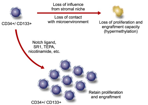
On the other hand, It has been constantly debated that expanded HSCs have less potency for engraftment and hematogenesis compared to unexpanded HSCs [3910], while preclinical and clinical studies have continuously been focusing on increasing the potency of expanded HSCs [27]. Current preclinical and clinical data suggest that long-term engrafted HSCs are only from unexpanded HSCs [7891016]. Recently, mechanistic studies have been conducted to understand the potency of expanded HSCs. Using CB derived CD34+ cell populations, researchers divided cells into two groups; the ex vivo expanded versus the unexpanded. Unexpectedly, they observed DNA hypermethylation during culture expansion of HSCs [19]. Loss of HSC forming niche during in vitro expansion could be a reason for the epigenetic changes of HSCs. Furthermore, loss of proliferation and engraftment capacity was well correlated with the expansion duration and the degree of hypermethylation [19]. Among various genes that were methylated during ex vivo expansion, genes that play a central role in the hematopoietic development and immune response were mostly affected, which indicates that those are relevant for the rapid loss of stemness during in vitro manipulation [19]. The detailed mechanistic pathway to reveal the exact genes or modifiers is remained to be elucidated, however, pan-inhibitors of DNA methylation are considered for preclinical and clinical application. The DNA methyltransferase inhibitors (DNMTI) including histone deacetylase (HDAC) inhibitors (valproic acid) [202122], 5-aza-2'-deoxycytidine (AZA) [2023], and nicotinamide (NAM, class3 HDAC-SIRT1 inhibitor) [82024] are currently being tested for their efficacy in reducing hypermethylation of the regulatory or marker genes in hematopoiesis during ex vivo expansion. Also a large scale chemical library screening has been applied to increase long-term survival of cultured HSCs [20].
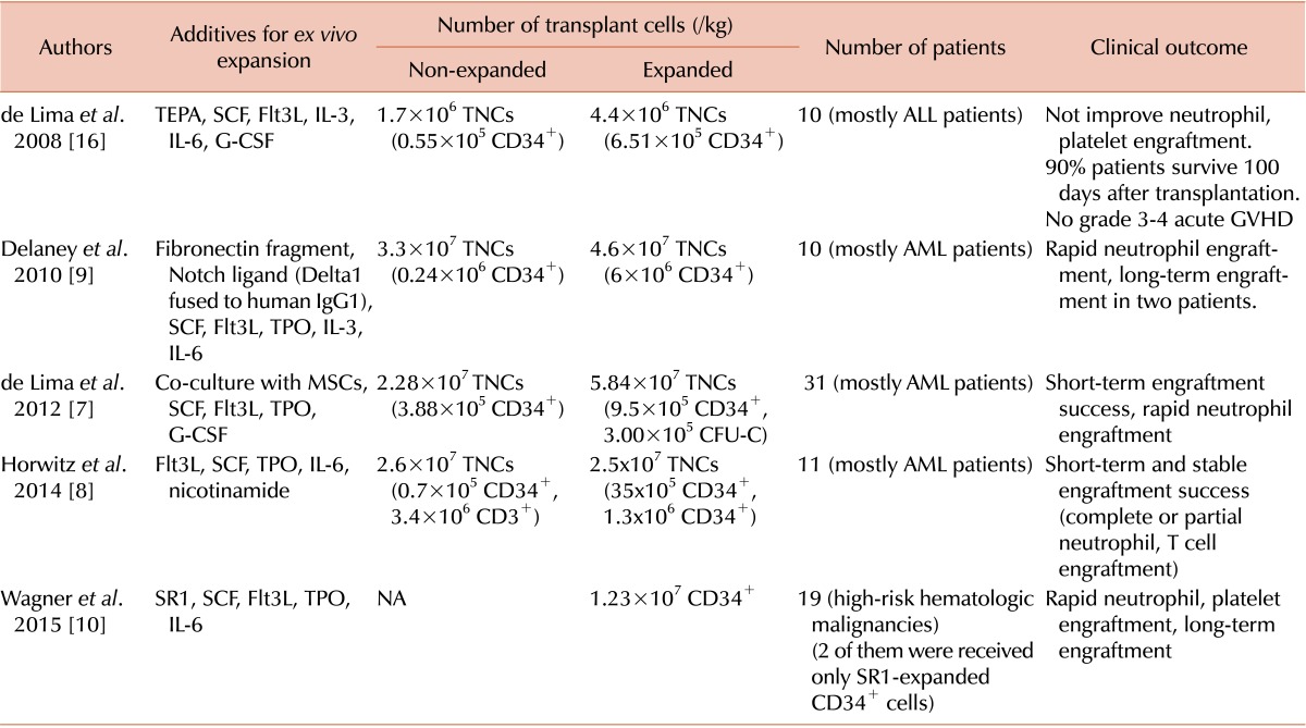
Among various ex vivo expansion methods, notch ligand [9] and stemregenin 1 (SR1) [10] are newly recognized HSC expansion reagents (Table 1). Compared to two allogeneic HSC grafts, expanded HSCs together with unexpanded HSCs from the same donor might have less chance of developing GVHD. On the other hand, transplantation of non-manipulated CB unit along with CD34+ cell-expanded HSCs from another CB demonstrated an engraftment facilitating effect, which suggested that culture-expanded CB-HSCs contribute to the early myeloid reconstitution and later are replaced by non-expanded CB cells [9]. The CB-HSCs expanded with notch ligand led to rapid myeloid recovery within 16 days after transplantation. Although long-term engraftment was derived from co-transplanted non-manipulated CB, it was the first demonstration of rapid hematopoietic engraftment supported by ex vivo expanded CB-HSCs (Table 1) [9]. Moreover, SR1 expanded CD34+ CB-HSCs have shown a neutrophil engraftment within 11 days [10].
Recent progress in CB-HSC expansion methods is summarized in Table 1 and Fig. 1. De Lima et al. [716] reported that tetraethylenepentamine (TEPA)-treated CD133+ CB culture exhibited enhanced self-renewal and expansion without loss of HSC potency. Although co-transplantation of TEPA-expanded HSCs with unexpanded HSCs in patients did not improve neutrophil and platelet engraftments, 9 out of 10 patients showed no liver or gut acute GVHD [16]. Horwitz et al. [8] transplanted a NAM-expanded unit in addition to an unexpanded unit and non-cultured T cells. As a result, neutrophil and T cell engraftments were moderately successful with rapid short-term engraftment from the NAM-expanded unit and long-term hematopoiesis from the unexpanded unit [8]. De Lima et al. [7] also reported that mesenchymal stromal cell (MSC) co-cultured CB-HSCs brought about short-term engraftment success and rapid recovery of neutrophils and platelets [7].

HUMANIZED ANIMAL-BASED HSC ENGRAFTMENT AND PRECLINICAL APPLICATIONS

Newly developed therapy must be tested for its efficacy and toxicity using preclinical animal models. However, clinical expectations are often suggested by cell-based and biochemical- based assays before being introduced into animal models. For HSCT, the long-term and short-term safety and efficacy can only be evaluated using humanized/immunocompromised animal models [2526].
Colony forming unit (CFU) and long-term culture initiating cells (LTC-IC) are often used to test the long-term survival and hematopoietic potency of HSCs. However, these in vitro measurements do not always correlate with the clinical outcome of the HSCT, and in vitro culture assays only provide a limited prediction. Also, immunophenotypic profiles such as CD34+ and CD133+ may not fully recapitulate in vivo engraftment ability either [27].
Understanding the human immunology and predicting the clinical outcome from stem cell therapy are greatly indebted to a humanized animal modeling [28]. In order to study the long-term engraftment and differentiation of HSCs, various immunodeficient animal models have been developed. Among them, genetically modified murine model has been widely used for engraftment and quantitation of HSCs [2829]. The mutation of PRKDC (protein kinase, DNA activated, catalytic polypeptide), a DNA repair enzyme, blocks V(D)J recombination. The B and T cell lineage blockage using recombinase-mutated SCID mice was commonly used for studying allogeneic or xenogeneic HSCT. Non-obese diabetes (NOD) mice showed severe reduction in the number of natural killer (NK) cells. By combining NOD and SCID background, NOD/SCID mice have been used for the transplantation of human HSCs. However, long-term survival of injected cells often cannot be identified in NOD/SCID animals due to the presence of little but active NK cells and high incidence of thymus lymphoma arising in the animals [2830].
The mutation in the interleukin-2R gamma (IL2Rγ) chain gene also causes severe immunodeficiency in mice. The IL2Rγ mutation in NOD/SCID mice (called NSG mice) greatly improved engraftment efficacy of HSCs enough to allow long-term tracking of transplanted HSCs and for studying their differentiation potential [28]. Since a slight difference in genetic background of a mouse can cause a severe disturbance in B and T cell development, the data from studies using animals of different genetic backgrounds should vary significantly, which requires a careful interpretation [28].
The number of SCID repopulating cells (SRCs) is commonly used to describe the potency of transplanted human HSCs [3132]. The details of hematopoietic lineage cells originated from the HSCs engrafted into the immunodeficient mice now can be monitored by a system called genetic barcoding. The genetic barcoding system using the lentiviral mapping technology have provided us with strong quantitative tools to profile HSC engraftment in vivo [33]. In brief, single HSC can be infected with lentivirus which has the 33bp DNA barcode composed of a 6bp sequence at the 5' end (called 'library ID') followed by a random 27bp sequence. Once permanently transduced with the lentiviral gene, the genetically barcoded (a lentiviral gene inserted into the chromosome) single HSC engrafted into an experimental mouse will replenish its bone marrow. Consequently, the mouse is able to produce the progenies of the clonal HSCs which can be counted by the next generation sequencing technology. In this way, HSCs transplanted into experimental animals can be characterized and quantified with great fidelity using the genetic barcoding system.
Earlier failure of human HSCT in 1950s was due to a lack of understanding about major histocompatibility complex (MHC) molecules. After considerable trials and failures in dogs, researchers developed molecular techniques to type human MHC (HLA) in order to find matched human donors. Because the MHC complexity of the small rodents is not comparable to humans or other larger animals, large animal models would be more desirable for studies using human HSCs. However, the high cost of generating and maintaining genetically modified animals forced most studies to favor small murine models. Nevertheless, results of immunocompromised large animals started to be accumulated owing to spontaneously mutated immunodeficient animals. Recently, genetically modified pigs with SCID phenotype were used in human stem cell transplant assays. Despite limited comparison data with representative transgenic animals, the study reported that a recombination-activating gene 2 (RAG2) mutated pig and RAG2 knockout mice shared similar phenotype with a statistical significance. These animals with such genetic background showed reduced B and T cell activity, however, RAG2 pig showed even less or undetectable CD21+CD3+ cells [30]. Furthermore, RAG2 pig showed complete loss of thymus (3 out of 4 animals, one had severely underdeveloped thymus) indicating that large animals, such as pigs, may serve as better models than the murine models for studying human HSCs [30]. The zinc finger nucleases (ZFNs), transcription activator-like effector nucleases (TALENs), and the CRISPR/Cas system are currently utilized in transgenic animal generation [34353637383940]. Considering the failures in preclinical murine models, we may seek for more human-like large animal models like the humanized mini-pig model [30].

STEM CELL-BASED HUMAN THERAPY AND HSC DIFFERENTIATION

The first human trial using patient's own iPSC-derived macular tissues has been applied in Japan in 2014. Furthermore, treatment of macular degeneration with embryonic stem cell (ESC)-derived macular tissues was carried out in 2012 [41] and 2014, and currently a phase II trial is being performed in Korea showing a promising result without any side effects so far [42]. Although Japanese human trials with iPSCs were halted recently due to an unexpected chromosomal instability in transplanted tissues, human iPSC-based cell therapy has a potential to be the next generation therapeutic regimen.
Stem cells have unique characteristics. One is self-renewal and the other is the capacity to differentiate into various cell lineages. Stem cells can be categorized into totipotent, pluripotent, and multipotent stem cells based on their differentiation capacity [34]. Totipotent stem cells, such as fertilized eggs, have the ability to develop into a new organism. Pluripotent stem cells can be named when the cells are able to differentiate into three germ layers, namely, mesoderm, ectoderm, and endoderm. The ESCs and iPSCs [43] are considered pluripotent stem cells, which can give rise to all three germ layers and germ cell lineages. In ideal conditions, HSCs are able to differentiate into a fully differentiated tissue quite equivalent to ESCs [37]. Since the usage of undifferentiated ESCs or iPSCs is limited due to a potential risk of developing teratoma [44], fully differentiated or partially differentiated progenitor cells from ESCs/iPSCs are considered for cell therapy. Details of iPSC generation and applications have been documented in many reviews [4345]. These reviews focus on the current progress in HSC differentiation from iPSCs/ESCs as well as a direct conversion from the fibroblast.
Aforementioned age-related macular disease (AMD) therapy with autologous iPSCs was a clever approach to introduce stem cell-derived tissues [13]. The lack of blood vessels in macular area prevents one's immune response from rejecting foreign tissues. Thus, unlike tissues such as bone marrow, blood, neuron, and heart muscle, the macular retinal tissue from allogeneic ESCs can be transplanted with little or no immune suppression [42]. Among other stem cell based therapeutic trials, oligodendrocytes derived from human ESCs were suggested for the treatment of spinal cord injury [46]. Continuous optimizations have been ongoing to reduce the risk of unwanted side effects, such as cancer formation from ESC/iPSC-derived tissues. We are facing an increasing demand on cell therapy using stem cells of non-hematological origin to treat a variety of incurable diseases.
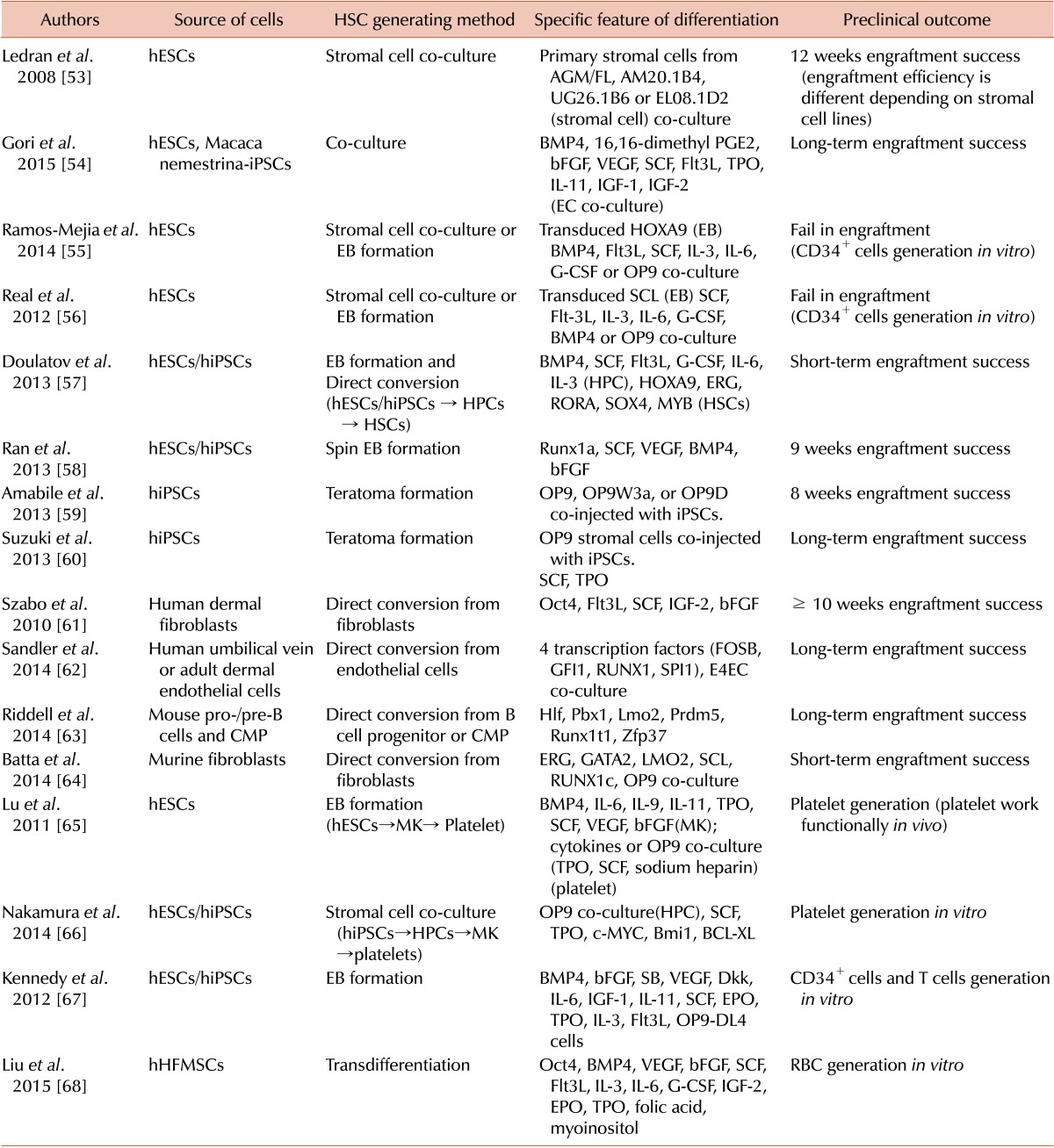
The first successful treatment of sickle cell anemia using autologous iPSCs in experimental animals in 2007 [47] was a promising start of non-hematological HSCs for HSCT. From early 2000s, B cells, T cells, NK cells and various lineage specific blood cells have been generated from human ESCs or iPSCs [4849505152]. Hematopoietic differentiations from ESCs/iPSCs with preclinical data have been well documented in reviews listed in Table 2 [53545556575859606162636465666768].
There are three ways for HSCs to be differentiated and generated from stem cells (Fig. 2). First, HSC differentiation can be stimulated by co-culturing with stromal cells [53]. Bone marrow stromal cells have been used for the expansion, maturation, and differentiation of HSCs from ESCs [55566970]. Second, cytokines can be added for HSC differentiation from stem cells [5556586771]. Lastly, direct conversion or transdifferentiation from non-hematological fibroblasts to HSCs is possible [6162636468]. HSCs could be converted from any fibroblasts or epithelial cells. However, there have been critics about a lack of workable clinical grade HSC differentiation methods. Considering that the potency of HSCs in both long-term and short-term homeostasis can only be tested in animal models [535455565758596061626364], studies using large humanized animal HSCT models would be beneficial for the progression of HSC differentiation methods [72]. Representative methods to generate stem cell-derived HSCs are summarized in Table 2. The main reason for using stromal cells in HSC generation from stem cells is to provide a microenvriomental niche for a proper HSC differentiation. Various mechanical and cell biological niches are the key components for HSC maintenance. Ledran et al. [53] used three stromal cell lines for HSC differentiation, which is known to be important in embryonic HSC development including stromal cells of aorta-gonad-mesonephros (AGM) origin. Those co-culture based HSCs from human ESCs were successfully engrafted in NSG mice although stromal cells themselves could not differentiate into HSCs [53]. Gori et al. [73] advanced a microenvironmental cue with vascular niche components jagged-1 (JAG-1) and delta-like ligand4 (DLL4). The ESCs and iPSCs co-cultured with JAG-1 and DLL4 overexpressing endothelial cells were differentiated into CD34+ CD45+ cells which showed an increased engraftment in NSG mice [54].
Suzuki et al. [60] reported a very interesting concept of differentiation with both exogenous OP9 and cytokines. The iPSCs were subcutaneously injected into a SCID mouse and induced a formation of teratoma. HSCs from teratoma migrated into the bone marrow of the SCID mouse. Those teratoma-originated HSCs were isolated and transplanted into a different mouse to replenish hematopoiesis with multilineage differentiation and long-term engraftment. Teratomabased in vivo HSC differentiation method indicated the possibility of xenogeneic in vivo based HSC generation, which utilizes a live animal as an HSC niche [60].
The direct conversion into HSCs from cells of non-hematological and non-stem cell origin are also summarized in Table 2. Doulatov et al. [57] combined two methods to differentiate human ESCs/iPSCs into HSCs. Myeloid precursors defined as CD34+CD45+ cells were generated from human ESCs/iPSCs using a cytokine-based differentiation method. Lineage specified CD34+CD45+ cells were directly converted to CD34+CD38+ HSCs by lentiviral infection with HOXA9, ERG, RORA, SOX4, and MYB. As a result, directly converted HSCs were successfully engrafted in NSG mice [57]. Szabo et al. [61] also reported the direct conversion of human dermal fibroblasts into CD45+ cells. Human dermal fibroblasts were transduced with Oct4 lentivirus and cultured with SCF and FLT3L. Converted CD45+ cells have shown a multilineage differentiation capacity. Liu et al. [68] generated red blood cells (RBCs) from human hair follicle mesenchymal stem cells (hHFMSCs) through transdifferentiation. The hHFMSCs were transduced with Oct4 lentivirus and cultured with cytokines. The generated RBCs expressed β-globin chain and their morphology was similar to that of adult RBCs [68].
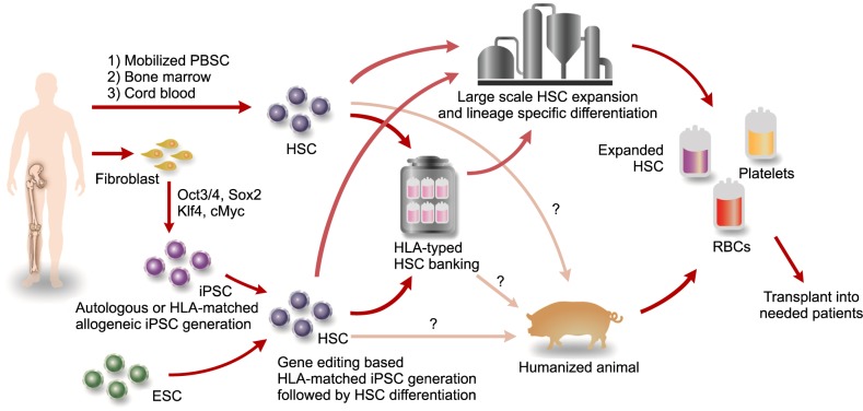
The HLA-matched human ESC/iPSC banking (Frozen bank) has been envisioned by many researchers. In theory, HLA-homozygote HSCs can cover up to 95% of the population in Japan or Great Britain with only 20 or little more iPSC lines [17]. It comes with an assumption that we have reasonably well established clinical grade HSC differentiation protocols which are currently being used in many laboratories.

CONCLUSIONS

Human bone marrow transplantation was initiated from an absolute necessity to cure patients who were previously incurable, and a lack of treatment options for incurable diseases has forced human beings to develop novel therapies. Current limitations in HSCT such as HLA-matched donor shortage and limited HSC number drive us to advance and create novel therapies. A recent report on megakaryocyte generation from stem cells to generate medical grade platelet production strongly suggests the possibility of obtaining large scale clinical grade human stem cells [66]. It is very feasible to generate HLA-matched HSCs using non-hematological origin cells such as iPSCs and ESCs, which have been used in clinical and preclinical trials. Continued studies to generate safe HSCs from stem cells will bring about improved outcome of HSCT. It would also be interesting to be able to produce clinical grade blood products generated from stem cells of humanized animals under large scale GMP. The HSC ex vivo expansion using stem cell niche modulants, such as SR1, notch ligand, HDAC inhibitors, and TEPA would compensate the use of co-cultured stromal cells which interfere with the large scale HSC expansion for therapeutic application. Continuous efforts to replenish the microenvironment niche with small molecules and media supplement would bring a breakthrough in HSCT.

Notes

Authors' Disclosures of Potential Conflicts of Interest: No potential conflicts of interest relevant to this article were reported.

References

1. Meuwissen HJ, Gatti RA, Terasaki PI, Hong R, Good RA. Treatment of lymphopenic hypogammaglobulinemia and bone-marrow aplasia by transplantation of allogeneic marrow. Crucial role of histocompatiility matching. N Engl J Med. 1969; 281:691–697. PMID: 4186068.
2. Oran B, Shpall E. Umbilical cord blood transplantation: a maturing technology. Hematology Am Soc Hematol Educ Program. 2012; 2012:215–222. PMID: 23233584.
crossref
3. Pineault N, Abu-Khader A. Advances in umbilical cord blood stem cell expansion and clinical translation. Exp Hematol. 2015; 43:498–513. PMID: 25970610.
crossref
4. Scaradavou A, Brunstein CG, Eapen M, et al. Double unit grafts successfully extend the application of umbilical cord blood transplantation in adults with acute leukemia. Blood. 2013; 121:752–758. PMID: 23223509.
crossref
5. Barker JN, Weisdorf DJ, DeFor TE, et al. Transplantation of 2 partially HLA-matched umbilical cord blood units to enhance engraftment in adults with hematologic malignancy. Blood. 2005; 105:1343–1347. PMID: 15466923.
crossref
6. Pasquini MC, Zhu X. Current uses and outcomes of hematopoietic stem cell transplantation: 2014 CIBMTR summary slides. Minneapolis, MN: Center for International Blood and Marrow Transplantation Research (CIBMTR);2015. Accessed October 11, 2015. https://www.cibmtr.org/ReferenceCenter/SlidesReports/SummarySlides/pages/index.aspx.
7. de Lima M, McNiece I, Robinson SN, et al. Cord-blood engraftment with ex vivo mesenchymal-cell coculture. N Engl J Med. 2012; 367:2305–2315. PMID: 23234514.
8. Horwitz ME, Chao NJ, Rizzieri DA, et al. Umbilical cord blood expansion with nicotinamide provides long-term multilineage engraftment. J Clin Invest. 2014; 124:3121–3128. PMID: 24911148.
crossref
9. Delaney C, Heimfeld S, Brashem-Stein C, Voorhies H, Manger RL, Bernstein ID. Notch-mediated expansion of human cord blood progenitor cells capable of rapid myeloid reconstitution. Nat Med. 2010; 16:232–236. PMID: 20081862.
crossref
10. Wagner JE, Brunstein C, McKenna D, et al. Acceleration of umbilical cord blood (UCB) stem engraftment: Results of a phase I clinical trial with stemregenin-1 (SR1) expansion culture. Biol Blood Marrow Transplant. 2015; 21:S48–S49.
crossref
11. Silva LC, Ortigosa LC, Benard G. Anti-TNF-α agents in the treatment of immune-mediated inflammatory diseases: mechanisms of action and pitfalls. Immunotherapy. 2010; 2:817–833. PMID: 21091114.
crossref
12. Elman JS, Li M, Wang F, Gimble JM, Parekkadan B. A comparison of adipose and bone marrow-derived mesenchymal stromal cell secreted factors in the treatment of systemic inflammation. J Inflamm (Lond). 2014; 11:1. PMID: 24397734.
crossref
13. Reardon S, Cyranoski D. Japan stem-cell trial stirs envy. Nature. 2014; 513:287–288. PMID: 25230622.
crossref
14. Petersdorf EW, Gooley TA, Anasetti C, et al. Optimizing outcome after unrelated marrow transplantation by comprehensive matching of HLA class I and II alleles in the donor and recipient. Blood. 1998; 92:3515–3520. PMID: 9808542.
crossref
15. Hansen JA, Gooley TA, Martin PJ, et al. Bone marrow transplants from unrelated donors for patients with chronic myeloid leukemia. N Engl J Med. 1998; 338:962–968. PMID: 9521984.
crossref
16. de Lima M, McMannis J, Gee A, et al. Transplantation of ex vivo expanded cord blood cells using the copper chelator tetraethylenepentamine: a phase I/II clinical trial. Bone Marrow Transplant. 2008; 41:771–778. PMID: 18209724.
crossref
17. Taylor CJ, Peacock S, Chaudhry AN, Bradley JA, Bolton EM. Generating an iPSC bank for HLA-matched tissue transplantation based on known donor and recipient HLA types. Cell Stem Cell. 2012; 11:147–152. PMID: 22862941.
crossref
18. Wagner JE Jr, Eapen M, Carter S, Wang Y, et al. One-unit versus two-unit cord-blood transplantation for hematologic cancers. N Engl J Med. 2014; 371:1685–1694. PMID: 25354103.
crossref
19. Weidner CI, Walenda T, Lin Q, et al. Hematopoietic stem and progenitor cells acquire distinct DNA-hypermethylation during in vitro culture. Sci Rep. 2013; 3:3372. PMID: 24284763.
crossref
20. Xie J, Zhang C. Ex vivo expansion of hematopoietic stem cells. Sci China Life Sci. 2015; 58:839–853. PMID: 26246379.
crossref
21. Chaurasia P, Gajzer DC, Schaniel C, D'Souza S, Hoffman R. Epigenetic reprogramming induces the expansion of cord blood stem cells. J Clin Invest. 2014; 124:2378–2395. PMID: 24762436.
crossref
22. Young JC, Wu S, Hansteen G, et al. Inhibitors of histone deacetylases promote hematopoietic stem cell self-renewal. Cytotherapy. 2004; 6:328–336. PMID: 16146885.
crossref
23. Araki H, Yoshinaga K, Boccuni P, Zhao Y, Hoffman R, Mahmud N. Chromatin-modifying agents permit human hematopoietic stem cells to undergo multiple cell divisions while retaining their repopulating potential. Blood. 2007; 109:3570–3578. PMID: 17185465.
crossref
24. Peled T, Shoham H, Aschengrau D, et al. Nicotinamide, a SIRT1 inhibitor, inhibits differentiation and facilitates expansion of hematopoietic progenitor cells with enhanced bone marrow homing and engraftment. Exp Hematol. 2012; 40:342–355.e1. PMID: 22198152.
25. Lang J, Weiss N, Freed BM, Torres RM, Pelanda R. Generation of hematopoietic humanized mice in the newborn BALB/c-Rag2null Il2rγnull mouse model: a multivariable optimization approach. Clin Immunol. 2011; 140:102–116. PMID: 21536497.
26. Berges BK, Wheat WH, Palmer BE, Connick E, Akkina R. HIV-1 infection and CD4 T cell depletion in the humanized Rag2-/-gamma c-/- (RAG-hu) mouse model. Retrovirology. 2006; 3:76. PMID: 17078891.
crossref
27. Matsuoka Y, Sumide K, Kawamura H, et al. Human cord blood-derived primitive CD34-negative hematopoietic stem cells (HSCs) are myeloid-biased long-term repopulating HSCs. Blood Cancer J. 2015; 5:e290. PMID: 25768404.
crossref
28. Tanner A, Taylor SE, Decottignies W, Berges BK. Humanized mice as a model to study human hematopoietic stem cell transplantation. Stem Cells Dev. 2014; 23:76–82. PMID: 23962058.
crossref
29. Brehm MA, Shultz LD, Luban J, Greiner DL. Overcoming current limitations in humanized mouse research. J Infect Dis. 2013; 208(Suppl 2):S125–S130. PMID: 24151318.
crossref
30. Lee K, Kwon DN, Ezashi T, et al. Engraftment of human iPS cells and allogeneic porcine cells into pigs with inactivated RAG2 and accompanying severe combined immunodeficiency. Proc Natl Acad Sci U S A. 2014; 111:7260–7265. PMID: 24799706.
crossref
31. Larochelle A, Vormoor J, Hanenberg H, et al. Identification of primitive human hematopoietic cells capable of repopulating NOD/SCID mouse bone marrow: implications for gene therapy. Nat Med. 1996; 2:1329–1337. PMID: 8946831.
crossref
32. Ishii M, Matsuoka Y, Sasaki Y, et al. Development of a high-resolution purification method for precise functional characterization of primitive human cord blood-derived CD34-negative SCID-repopulating cells. Exp Hematol. 2011; 39:203–213.e1. PMID: 21112372.
crossref
33. Lu R, Neff NF, Quake SR, Weissman IL. Tracking single hematopoietic stem cells in vivo using high-throughput sequencing in conjunction with viral genetic barcoding. Nat Biotechnol. 2011; 29:928–933. PMID: 21964413.
crossref
34. Kim C. iPSC technology-Powerful hand for disease modeling and therapeutic screen. BMB Rep. 2015; 48:256–265. PMID: 25104399.
crossref
35. Kim H, Kim JS. A guide to genome engineering with programmable nucleases. Nat Rev Genet. 2014; 15:321–334. PMID: 24690881.
crossref
36. Maeder ML, Linder SJ, Cascio VM, Fu Y, Ho QH, Joung JK. CRISPR RNA-guided activation of endogenous human genes. Nat Methods. 2013; 10:977–979. PMID: 23892898.
crossref
37. Kim C. Disease modeling and cell based therapy with iPSC: future therapeutic option with fast and safe application. Blood Res. 2014; 49:7–14. PMID: 24724061.
crossref
38. Ding Q, Lee YK, Schaefer EA, et al. A TALEN genome-editing system for generating human stem cell-based disease models. Cell Stem Cell. 2013; 12:238–251. PMID: 23246482.
crossref
39. Gaj T, Gersbach CA, Barbas CF 3rd. ZFN, TALEN, and CRISPR/Cas-based methods for genome engineering. Trends Biotechnol. 2013; 31:397–405. PMID: 23664777.
crossref
40. Joung JK, Sander JD. TALENs: a widely applicable technology for targeted genome editing. Nat Rev Mol Cell Biol. 2013; 14:49–55. PMID: 23169466.
crossref
41. Schwartz SD, Hubschman JP, Heilwell G, et al. Embryonic stem cell trials for macular degeneration: a preliminary report. Lancet. 2012; 379:713–720. PMID: 22281388.
crossref
42. Song WK, Park KM, Kim HJ, et al. Treatment of macular degeneration using embryonic stem cell-derived retinal pigment epithelium: preliminary results in Asian patients. Stem Cell Reports. 2015; 4:860–872. PMID: 25937371.
crossref
43. Takahashi K, Yamanaka S. Induction of pluripotent stem cells from mouse embryonic and adult fibroblast cultures by defined factors. Cell. 2006; 126:663–676. PMID: 16904174.
crossref
44. Kanemura H, Go MJ, Shikamura M, et al. Tumorigenicity studies of induced pluripotent stem cell (iPSC)-derived retinal pigment epithelium (RPE) for the treatment of age-related macular degeneration. PLoS One. 2014; 9:e85336. PMID: 24454843.
crossref
45. Schlaeger TM, Daheron L, Brickler TR, et al. A comparison of non-integrating reprogramming methods. Nat Biotechnol. 2015; 33:58–63. PMID: 25437882.
crossref
46. Geron Corporation. World's first clinical trial of human embryonic stem cell therapy cleared. Regen Med. 2009; 4:161. PMID: 19322956.
47. Hanna J, Wernig M, Markoulaki S, et al. Treatment of sickle cell anemia mouse model with iPS cells generated from autologous skin. Science. 2007; 318:1920–1923. PMID: 18063756.
crossref
48. Wang L, Li L, Menendez P, Cerdan C, Bhatia M. Human embryonic stem cells maintained in the absence of mouse embryonic fibroblasts or conditioned media are capable of hematopoietic development. Blood. 2005; 105:4598–4603. PMID: 15718421.
crossref
49. Woll PS, Grzywacz B, Tian X, et al. Human embryonic stem cells differentiate into a homogeneous population of natural killer cells with potent in vivo antitumor activity. Blood. 2009; 113:6094–6101. PMID: 19365083.
crossref
50. Galic Z, Kitchen SG, Kacena A, et al. T lineage differentiation from human embryonic stem cells. Proc Natl Acad Sci U S A. 2006; 103:11742–11747. PMID: 16844782.
crossref
51. Carpenter L, Malladi R, Yang CT, et al. Human induced pluripotent stem cells are capable of B-cell lymphopoiesis. Blood. 2011; 117:4008–4011. PMID: 21343609.
crossref
52. Knorr DA, Ni Z, Hermanson D, et al. Clinical-scale derivation of natural killer cells from human pluripotent stem cells for cancer therapy. Stem Cells Transl Med. 2013; 2:274–283. PMID: 23515118.
crossref
53. Ledran MH, Krassowska A, Armstrong L, et al. Efficient hematopoietic differentiation of human embryonic stem cells on stromal cells derived from hematopoietic niches. Cell Stem Cell. 2008; 3:85–98. PMID: 18593561.
crossref
54. Gori JL, Butler JM, Chan YY, et al. Vascular niche promotes hematopoietic multipotent progenitor formation from pluripotent stem cells. J Clin Invest. 2015; 125:1243–1254. PMID: 25664855.
crossref
55. Ramos-Mejía V, Navarro-Montero O, Ayllón V, et al. HOXA9 promotes hematopoietic commitment of human embryonic stem cells. Blood. 2014; 124:3065–3075. PMID: 25185710.
crossref
56. Real PJ, Ligero G, Ayllon V, et al. SCL/TAL1 regulates hematopoietic specification from human embryonic stem cells. Mol Ther. 2012; 20:1443–1453. PMID: 22491213.
crossref
57. Doulatov S, Vo LT, Chou SS, et al. Induction of multipotential hematopoietic progenitors from human pluripotent stem cells via respecification of lineage-restricted precursors. Cell Stem Cell. 2013; 13:459–470. PMID: 24094326.
crossref
58. Ran D, Shia WJ, Lo MC, et al. RUNX1a enhances hematopoietic lineage commitment from human embryonic stem cells and inducible pluripotent stem cells. Blood. 2013; 121:2882–2890. PMID: 23372166.
crossref
59. Amabile G, Welner RS, Nombela-Arrieta C, et al. In vivo generation of transplantable human hematopoietic cells from induced pluripotent stem cells. Blood. 2013; 121:1255–1264. PMID: 23212524.
crossref
60. Suzuki N, Yamazaki S, Yamaguchi T, et al. Generation of engraftable hematopoietic stem cells from induced pluripotent stem cells by way of teratoma formation. Mol Ther. 2013; 21:1424–1431. PMID: 23670574.
crossref
61. Szabo E, Rampalli S, Risueño RM, et al. Direct conversion of human fibroblasts to multilineage blood progenitors. Nature. 2010; 468:521–526. PMID: 21057492.
crossref
62. Sandler VM, Lis R, Liu Y, et al. Reprogramming human endothelial cells to haematopoietic cells requires vascular induction. Nature. 2014; 511:312–318. PMID: 25030167.
crossref
63. Riddell J, Gazit R, Garrison BS, et al. Reprogramming committed murine blood cells to induced hematopoietic stem cells with defined factors. Cell. 2014; 157:549–564. PMID: 24766805.
crossref
64. Batta K, Florkowska M, Kouskoff V, Lacaud G. Direct reprogramming of murine fibroblasts to hematopoietic progenitor cells. Cell Rep. 2014; 9:1871–1884. PMID: 25466247.
crossref
65. Lu SJ, Li F, Yin H, et al. Platelets generated from human embryonic stem cells are functional in vitro and in the microcirculation of living mice. Cell Res. 2011; 21:530–545. PMID: 21221130.
crossref
66. Nakamura S, Takayama N, Hirata S, et al. Expandable megakaryocyte cell lines enable clinically applicable generation of platelets from human induced pluripotent stem cells. Cell Stem Cell. 2014; 14:535–548. PMID: 24529595.
crossref
67. Kennedy M, Awong G, Sturgeon CM, et al. T lymphocyte potential marks the emergence of definitive hematopoietic progenitors in human pluripotent stem cell differentiation cultures. Cell Rep. 2012; 2:1722–1735. PMID: 23219550.
crossref
68. Liu Z, Lu SJ, Lu Y, et al. Transdifferentiation of Human Hair Follicle Mesenchymal Stem Cells into Red Blood Cells by OCT4. Stem Cells Int. 2015; 2015:389628. PMID: 25755671.
crossref
69. Vodyanik MA, Bork JA, Thomson JA, Slukvin II. Human embryonic stem cell-derived CD34+ cells: efficient production in the coculture with OP9 stromal cells and analysis of lymphohematopoietic potential. Blood. 2005; 105:617–626. PMID: 15374881.
crossref
70. Wada H, Kojo S, Kusama C, et al. Successful differentiation to T cells, but unsuccessful B-cell generation, from B-cell-derived induced pluripotent stem cells. Int Immunol. 2011; 23:65–74. PMID: 21135032.
crossref
71. Sturgeon CM, Ditadi A, Awong G, Kennedy M, Keller G. Wnt signaling controls the specification of definitive and primitive hematopoiesis from human pluripotent stem cells. Nat Biotechnol. 2014; 32:554–561. PMID: 24837661.
crossref
72. Chen U. Do we have a workable clinical protocol for differentiating lympho-hematopoietic stem cells from the source of embryonic stem cells and induced pluripotent stem cells in culture. Scand J Immunol. 2014; 80:247–249. PMID: 25041639.
crossref
73. Gori JL, Chandrasekaran D, Kowalski JP, et al. Efficient generation, purification, and expansion of CD34(+) hematopoietic progenitor cells from nonhuman primate-induced pluripotent stem cells. Blood. 2012; 120:e35–e44. PMID: 22898598.
crossref
Fig. 1

HSC proliferation and engraftment ability enhanced by stem cell niche replacing components. Current usage of SR1 and Notch ligand supplemented HSC ex vivo expansion could benefit the HSC engraftment ability and epigenetic regulation.

br-50-194-g001
Fig. 2

Schematic view of HSC expansion, non-hematological origin HSC generation, and application. Considering the current shortage of HLA-matched HSCs for patients who need allogeneic HSCT, ex vivo expanded and HLA-matched iPSC-derived HSCs could be the next generation solution. Humanized large animals could be used for the blood product generation applicable to human and for the long-term engraftment studies, because they mimic human immune complexity and might replace the artificial large scale HSC expansion.

br-50-194-g002
Table 1

Representative clinical trials with ex vivo expanded CB-HSCs.

br-50-194-i001

Abbreviations: TEPA, tertraethylenepentamine; TNC, Total nucleated cells; ALL, Acute lymphoblastic leukemia; AML, acute myeloid leukemia; SCF, stem cell factor; Flt-3L, Fms-related tyrosine kinase 3 ligand; TPO, thrombopoietin; MSC, mesenchymal stromal cell; G-CSF, granulocyte-colony stimulating factor; SR1, stemregenin-1; NA, Not applicable.

Table 2

HSC and blood generation from non-hematological cells.

br-50-194-i002

Abbreviations: hESC, human embryonic stem cell; iPSC, induced pluripotent stem cell; hiPSC, human iPSC; BMP-4, bone morphogenetic protein 4; PGE2, prostaglandin E2; bFGF, Basic fibroblast growth factor; VEGF, Vascular endothelial growth factor; SCF, stem cell factor; Flt3L, Fms-related tyrosine kinase 3 ligand; TPO, thrombopoietin; IGF-1, insulin-like growth factor; EC, endothelial cell; EB, Embryoid body; HOXA9, Homeobox protein-A9; ERG, erythroblast transformation-specific (ETS)-related gene; RORA, retinoic acid receptor (RAR)-related orphan receptor A; SOX4, sex-determining region Y (SRY)-related high mobility group box 4; MYB, myeloblastosis proto-oncogene protein; RUNX1, Runt-related transcription factor 1; FOSB, Finkel-Biskis-Jinkins murine osteosarcoma viral oncogene homolog B; GFI1, Growth factor independent 1 transcription repressor; HLF, Hepatic leukemia factor; PBX1, Pre-B-cell leukemia homeobox 1; LMO2, LIM domain only 2; PRDM5, PR Domain Containing zinc finger protein 5; ZFP37, Zinc Finger Protein 37; GATA2, GATA binding protein 2; SCL, stem cell leukemia gene product; MK, megakaryocyte; BMI1, B lymphoma Mo-MLV insertion region 1 homolog; EPO, erythropoietin; hHFMSC, human hair follicle mesenchymal stem cell; Oct-4, octamer-binding transcription factor 4.

TOOLS
Similar articles