TO THE EDITOR: Histoplasmosis is caused by Histoplasma capsulatum variant capsulatum and H. capsulatum variant duboisiis. It is a systemic fungal infection with a disease spectrum ranging from asymptomatic primary infection to disseminated disease. Disseminated histoplasmosis is usually seen in immunocompromised patients such as those with AIDS or hematological malignancies, or patients who have undergone a transplant or are receiving steroids [1]. Life threatening infections may be seen in infants [2]. We present a case of disseminated histoplasmosis, diagnosed incidentally on bone marrow aspiration, in a young, immunocompetent woman.
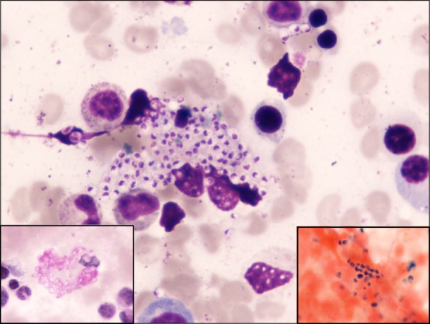
A 35-year-old woman presented with pallor, intermittent fever associated with chills, and body ache that had started 6 months earlier. There was no history of joint pain, vomiting, loose stools, cough, or dysuria. Respiratory and cardiovascular system examinations were normal. In the abdomen, non-tender hepatosplenomegaly was present, 5 cm and 1 cm below the costal margin. The patient had no history of steroid intake or diabetes, the results of the recombinant K39 and rapid malaria antigen tests were negative, and human immunodeficiency virus serology was non-reactive. However, a complete blood count revealed pancytopenia, and bone marrow aspirate smears showed many intracellular and extracellular yeast forms of H. capsulatum. These organisms were periodic acid Schiff-positive and gave a positive Prussian blue reaction with Perl's staining (Fig. 1). A diagnosis of disseminated histoplasmosis was made, and the patient started receiving amphotericin B, but was then lost in follow up.
Symptomatic infection is uncommon in immunocompetent patients exposed to H. capsulatum. The disease spectrum in these patients includes acute pulmonary histoplasmosis, chronic cavitary histoplasmosis, granulomatous mediastinitis, mediastinal fibrosis and, uncommonly, pericarditis, pleural disease, and broncholithiasis [2]. A high index of clinical suspicion is required to diagnose disseminated histoplasmosis in an immunocompetent patient, although timely diagnosis is important because it is associated with a high mortality.
Yeast forms of H. capsulatum are observed intracellularly and, rarely, in the extracellular space. On routine Romanowsky staining, the organisms are often overlooked because of the dense staining of hematopoietic cells [3]. The Grocott methenamine silver technique provides good staining of the yeast forms with minimal background staining. Perl's staining, which is routinely performed in all bone marrow aspirates, can aid in the diagnosis of histoplasmosis, avoiding the requirement for additional cytochemistry. Yeast cells give a positive Prussian blue reaction and are then highlighted against a red background [3]. The significance of this finding is unknown.
Most fungi proliferate in the presence of increased iron, and the monocyte phagocyte system of the bone marrow that is rich in iron stores may increase the growth rate of yeast cells. It is also possible that large stores of iron function as part of the host defense mechanism. Yeast cells might be inhibited by high iron concentrations within the histiocytes [3]. Further studies are required to determine the changes and deviations in iron metabolism in disseminated histoplasmosis.
References
1. Kauffman CA. Histoplasmosis: a clinical and laboratory update. Clin Microbiol Rev. 2007; 20:115–132. PMID: 17223625.

2. Pamnani R, Rajab JA, Githang'a J, Kasmani R. Disseminated histoplasmosis diagnosed on bone marrow aspirate cytology: report of four cases. East Afr Med J. 2009; 86(Suppl 12):S102–S105. PMID: 21591519.

3. Caldwell CW, Taylor H. Visualization of histoplasma capsulatum in bone marrow with Prussian blue iron stain. J Clin Microbiol. 1982; 15:156–158. PMID: 7186903.


 PDF
PDF ePub
ePub Citation
 Print
Print


 XML Download