

 PDF
PDF ePub
ePub Citation
 Print
Print


TO THE EDITOR: Extramedullary or extra-osseous plasmacytomas (EMP/EOP) are localized plasma cell neoplasms that arise in tissues other than bone. EMP comprises 3-5% of all plasma cell neoplasms. The upper respiratory tract and the oral cavity are the most common sites for EMP [1], and the thyroid gland is one of the rare sites for this neoplasm. Here, we report a case of EMP of the thyroid in a 53-year-old male who presented to the surgery outpatient department (OPD) with a left-sided thyroid swelling of six months duration.
CASE
A 53-year-old male patient presented to the surgery OPD with history of swelling of the left side of his neck for the last six months. The swelling had gradually increased to its present size over this time period and was not associated with pain, changes in his voice, or difficulty in breathing. A well-healed scar was present on the front of the patient's neck, as he had undergone surgery four years earlier for which no further details were available. The patient had not received any post-operative chemotherapy or radiotherapy. On examination, a soft, non-tender swelling measuring 8×10 cm was noted on the front of the neck toward the left side that moved with deglutition but not with protrusion of the tongue. The cervical lymph nodes were not enlarged. There were no signs of pressure effects on the trachea, larynx, esophagus, or major veins of the thorax, and no sign of hypothyroidism or hyperthyroidism.
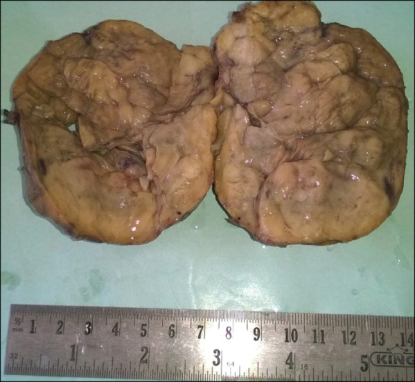
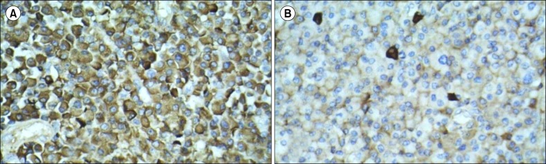
All routine investigations, including complete blood count, liver function test and renal function test, were within normal limits. A thyroid profile showed slightly increased thyroid-stimulating hormone levels (TSH, 5.92 mIU/mL; normal range, 0.5-5.0 mIU/mL) and slightly decreased T4 levels (3.65 µ/dL; normal range, 4.5-12.6 µ/dL). Contrast-enhanced computed tomography of the neck and chest showed a well-defined mass lesion of 6×6×10 cm in the region of the left lobe of the thyroid gland extending up to the hyoid bone. Inferiorly, the mass extended 2 cm above the level of the aortic arch. Ultrasonography of the abdomen showed fatty liver with an enlarged prostate, and no enlarged lymph nodes or hepato-splenomegaly were observed. Fine-needle aspiration cytology of the swollen area was performed, revealing a dispersed population of atypical plasma cells along with a few benign follicular epithelial cells that were infiltrated by lymphocytes. The possibility of plasmacytoma of the thyroid in a background of thyroiditis was suggested. Following a cytology report, an extensive workup for multiple myeloma (MM) was performed, which showed normal bone marrow, and radiographs of the skull, chest, spine, and pelvis did not show any abnormalities. Serum protein electrophoresis showed normal levels of serum proteins with a very low serum M protein level of 0.25 g/dL. Other values were as follows: serum total protein 6.90 g/dL, albumin 4.41 g/dL, alpha 1 globulin 0.35 g/dL, alpha 2 globulin 0.77 g/dL, and gamma globulin 0.82 g/dL with an A:G ratio of 1.77. The serum kappa-free light chain level was increased (24.07 mg/L; normal range, 3.30-19.40 mg/L), while the level of lambda free light chain and the kappa/lambda ratio were within normal limits (19.06 mg/L and 1.26, respectively). Serum β2 microglobulin and calcium levels were within the normal ranges. The patient was prepared for surgery, and a total thyroidectomy was performed. Intra-operatively, there was a 10×8 cm swelling on the left lobe of the thyroid with a shift of the trachea to the right side without any infiltration of the adjacent structures or capsule, and there was no lymphadenopathy. Gross examination of the specimen showed a globular soft piece of tissue measuring 8.5×6×5 cm. The external surface was nodular with an adherent capsule and showed prominent blood vessels. The cut section of the specimen was gray, firm, and fleshy in appearance (Fig. 1). Microscopically, multiple sections taken from the tumor showed sheets of plasma cells infiltrating the thyroid parenchyma with few entrapped atrophic thyroid follicles (Fig. 2A). These cells have eccentric, round nuclei with abundant eosinophilic cytoplasm. Sporadic binucleated and multinucleated plasma cells were also observed (Fig. 2B). Many lymphoid follicles with germinal centers were also present (Fig. 2C). Thus, a diagnosis of EMP of the thyroid in a background of Hashimoto's thyroiditis was made. Immunohistochemistry (IHC) showed CD138-positive tumor cells while CD20 was negative, thus clinching the diagnosis of EMP (Fig. 2D). IHC for kappa light chain showed strong cytoplasmic positivity in the tumor cells while that of the lambda light chain was negative (Fig. 3). The patient is undergoing regular follow-up and has been asymptomatic for the past eight months.
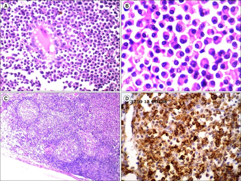 | Fig. 2Microscopy of extramedullary plasmacytoma of the thyroid. (A) Sheets of plasma cells infiltrating the thyroid parenchyma with few entrapped atrophic thyroid follicles (H&E, ×100). (B) Sheets of plasma cells with some binucleated forms (H&E, ×400). (C) Many lymphoid follicles with germinal centers (H&E, ×100). (D) CD138-positive tumor cells on immunohistochemistry.
|
Go to : 
DISCUSSION
Plasma cell neoplasms consist of monoclonal gammopathy of undetermined significance (MGUS), plasma cell myeloma, plasmacytoma, immunoglobulin deposition disorders, and osteosclerotic myeloma (POEMS syndrome) [1]. Plasmacytomas are further divided into solitary plasmacytoma of the bone (SPB) and EMP that involve the soft tissue without any signs of systemic spread [1]. In contrast to SPB, which frequently converts into MM, EMP remains localized [2]. EMP is extremely rare and comprises 3-5% of all plasma cell neoplasms. It occurs more commonly in men (M:F=2-3:1) at 40-70 years of age, with the median age at diagnosis being 55 years [1, 3]. The head and neck regions are the most common site for EMP (80-90%), and about 0.4% of all head and neck cancers is the result of EMP [4]. EMP has also been reported in other locations, such as the breast, pancreas, ovary, kidney, spermatic cord, pleura, mediastinum, etc. [3]. The thyroid gland is a very unusual site for EMP. Wiltshaw [5] reported 7 cases of EMP of the thyroid out of 272 cases of EMP, while Hazard and Schildecker [6] reported EMP in only two cases out of 14,000 thyroid operations. Macpherson et al. [7] found only one case of EMP out of 870 thyroid tumors, while Aozasa et al. [8] reported EMP of the thyroid in 6 out of 62 cases. Galieni et al. [9] reported only one case out of 46 cases of EMP. As the current case, EMP of the thyroid in a background of lymphocytic thyroiditis has also been reported by other authors [3, 8, 10].
While rendering a diagnosis of EMP of the thyroid, it is essential to exclude the possibility of MM. A diagnosis of EMP of the thyroid is confirmed by a normal bone marrow examination, absence of osteolytic lesions on a bone survey and normal levels of serum proteins on electrophoresis. Specific diagnostic criteria for EMP have been established by Galieni et al. [9]: (i) monoclonal plasma cell histology on tissue biopsy, (ii) plasma cells in the bone marrow representing <5% of all nucleated cells, (iii) absence of lytic skeletal lesions or other tissue involvement, (iv) a lack of hypercalcemia or renal failure, and (v) a low level of serum M protein, if present. Our case met all of these diagnostic criteria for EMP.
The treatment of EMP remains controversial; all three modalities, including radiotherapy alone, surgery alone, or a combined approach, are advocated by various authors [11]. Although EMP is very sensitive to radiotherapy and complete remission can be achieved by irradiation alone in most cases, surgery remains the treatment of choice if the tumor is resectable. A combined approach is advocated if surgical removal is not possible or is incomplete and if lymph node involvement is present. In the present case, since the tumor was limited to the left lobe of the thyroid gland with no extracapsular extension, surgical removal with close follow-up for recurrence was advocated. The patient has been followed up regularly in the surgical OPD for the last eight months and has remained asymptomatic to date. Long-term follow-up of EMP is advised, as around 20% cases of EMP may progress to MM [11].
EMP of the thyroid is a very rare entity, and its diagnosis is made only after ruling out the diagnosis of MM through appropriate investigations. The prognosis of localized plasmacytoma is favorable, but regular follow-up is essential as EMP may progress to MM in approximately 20% of cases.
Go to :
 XML Download