Journal List > Blood Res > v.49(2) > 1092131

Lee, Choi, Kim, Kim, Kim, Kim, Lee, Kim, Kim, Park, Park, Shim, Eom, Lee, Park, Cheong, and Park: R-CHOP chemoimmunotherapy followed by autologous transplantation for the treatment of diffuse large B-cell lymphoma

Abstract

Background

We investigated factors that influence outcomes in diffuse large B-cell lymphoma (DLBCL) patients treated with rituximab combined with the CHOP regimen (R-CHOP) followed by upfront autologous stem cell transplantation (Auto-SCT).

Methods

We retrospectively evaluated survival differences between subgroups based on the age-adjusted International Prognostic Index (aaIPI) and revised-IPI (R-IPI) at diagnosis, disease status, and positron emission tomographic/computerized tomographic (PET/CT) status at transplantation in 51 CD20-positive DLBCL patients treated with R-CHOP followed by upfront Auto-SCT.

Results

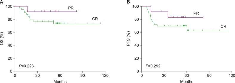
Patients had either stage I/II bulky disease (5.9%) or stage III/IV disease (94.1%). The median patient age at diagnosis was 47 years (range, 22-66 years); 53.3% and 26.7% had high-intermediate and high risks according to aaIPI, respectively. At the time of Auto-SCT, 72.5% and 27.5% experienced complete (CR) and partial remission (PR) after R-CHOP, respectively. The median time from diagnosis to Auto-SCT was 7.27 months (range, 3.4-13.4 months). The 5-year overall (OS) and progression-free survival (PFS) were 77.3% and 72.4%, respectively. The 5-year OS and PFS rates according to aaIPI, R-IPI, and PET/CT status did not differ between the subgroups. More importantly, the 5-year OS and PFS rates of the patients who achieved PR at the time of Auto-SCT were not inferior to those of the patients who achieved CR (P=0.223 and 0.292, respectively).

Conclusion

Survival was not influenced by the aaIPI and R-IPI at diagnosis, disease status, or PET/CT status at transplantation, suggesting that upfront Auto-SCT might overcome unfavorable outcomes attributed to PR after induction chemoimmunotherapy.

INTRODUCTION

Rituximab-containing chemotherapy regimens provide superior long-term progression-free survival (PFS) and overall survival (OS) relative to regimens without rituximab in patients with diffuse large B-cell lymphoma (DLBCL), regardless of age [1, 2]. However, even in the rituximab era, the survival rates of patients with high-intermediate- and high-risk International Prognostic Index (IPI) scores remained unsatisfactory [3]. Therefore, several randomized trials have prospectively evaluated the role of upfront autologous stem cell transplantation (Auto-SCT) following therapy with rituximab plus CHOP regimen (cyclophosphamide+hydroxydaunorubicin+oncovin+prednisone/prednisolone;R-CHOP) for aggressive DLBCL [4, 5, 6]. The interim evaluation in a study conducted by Vitolo et al. [4] found no significant differences between the arms treated with R-CHOP alone and R-CHOP followed by Auto-SCT regarding the 2-year OS; however, the 2-year PFS was significantly higher in the Auto-SCT arm. In contrast to the outcomes of high-intermediate-risk patients, Stiff et al. [5] reported that in the subset of high-risk patients alone, induction chemoimmunotherapy followed by early Auto-SCT significantly improved the 2-year PFS and OS relative to chemoimmunotherapy alone. However, Le Gouill et al. [6] failed to demonstrate any survival benefit of upfront Auto-SCT. The role of upfront high-dose therapy with stem cell rescue remains to be established during long-term follow-up in this high-risk group of patients. Meanwhile, we face questions regarding the factors that influence outcomes in patients with DLBCL treated with R-CHOP followed by Auto-SCT.
To investigate whether the age-adjusted IPI (aaIPI) and revised IPI (R-IPI) at diagnosis, the disease response to induction therapy, and the 18F-fluoro-2-deoxy-D-glucose positron emission tomographic/computerized tomographic (FDG PET/CT) status at transplantation could predict outcomes in patients with CD20-positive DLBCL, we retrospectively evaluated the treatment and survival outcomes of patients treated with R-CHOP followed by high-dose therapy with autologous stem cell rescue (HDT/ASCR).

MATERIALS AND METHODS

Data sources

The data were collected from the Korean Blood and Marrow Transplant Registry (KBMTR). The KBMTR is a voluntary organization comprising 43 transplantation centers located in South Korea. The Transplant Registration Committee requires participating centers to submit detailed data from consecutive patients to the KBMTR. Informed consent is obtained locally according to the KBMTR regulations. The KBMTR database was used to identify adult patients with DLBCL who underwent an initial Auto-SCT while in complete remission (CR) or partial remission (PR) after R-CHOP chemoimmunotherapy between January 2005 and April 2013. Additional data were obtained from each center to complete this study.

Patients

We analyzed 51 CD20+ DLBCL patients who underwent R-CHOP therapy followed by a consolidative HDT/ASCR between January 2005 and May 2013 as reported to the KBMTR by 14 centers. Adult patients aged ≥20 years were included. In Korea, the majority of medical expenses are covered and tightly regulated by the National Health Insurance System. All types of hematopoietic stem cell transplantation, including Auto-SCT, are reviewed in advance by the Health Care Review and Evaluation Committee. The regulations allow Auto-SCT for patients ≤65 years of age. Therefore, the majority of patients enrolled in this study were <65 years old. Three stage I or II patients with bulky disease underwent an upfront Auto-SCT because their diseases were considered advanced. This study was approved by the institutional review board of Konkuk University Medical Center.

Assessment of responses

The 2007 revised guidelines of the International Harmonization Project were adopted to describe the response criteria for DLBCL [7]. CR was defined as the complete disappearance of all detectable clinical evidence of disease and disease-related symptoms if these were present before therapy. PR was defined as a ≥50% decrease in the sum of the product of the diameters (SPD) of up to 6 of the largest dominant nodes or nodal masses. Stable disease (SD) was defined as a case that failed to attain the criteria needed for CR or PR but did not fulfill those for progressive disease (PD). We exactly followed the guidelines provided by the Consensus of the Imaging Subcommittee of the International Harmonization Project in Lymphoma with respect to performing and interpreting the PET imaging data for treatment assessments [8]. OS was defined as the time interval from the date of diagnosis to the date of death as a result of any cause or the last follow-up. PFS was defined as the time interval from the date of diagnosis to the date of lymphoma progression, relapse from CR, or death as a result of any cause.

Statistical analysis

The Kaplan-Meier method was used to estimate the probabilities of OS and PFS. To analyze the relapse probability after Auto-SCT, patients who remained alive without relapse and those who died without relapse were censored. The probability of relapse after Auto-SCT was calculated according to the 1 minus Kaplan-Meier method. The log-rank test was used in a univariate analysis to identify the prognostic factors for OS, PFS, and relapse after Auto-SCT. The statistical analyses were performed with the SPSS version 17.0 software (SPSS, Inc., Chicago, IL, USA). For all analyses, the P values were 2-sided; a P<0.05 was considered statistically significant.

RESULTS

Patient and disease characteristics

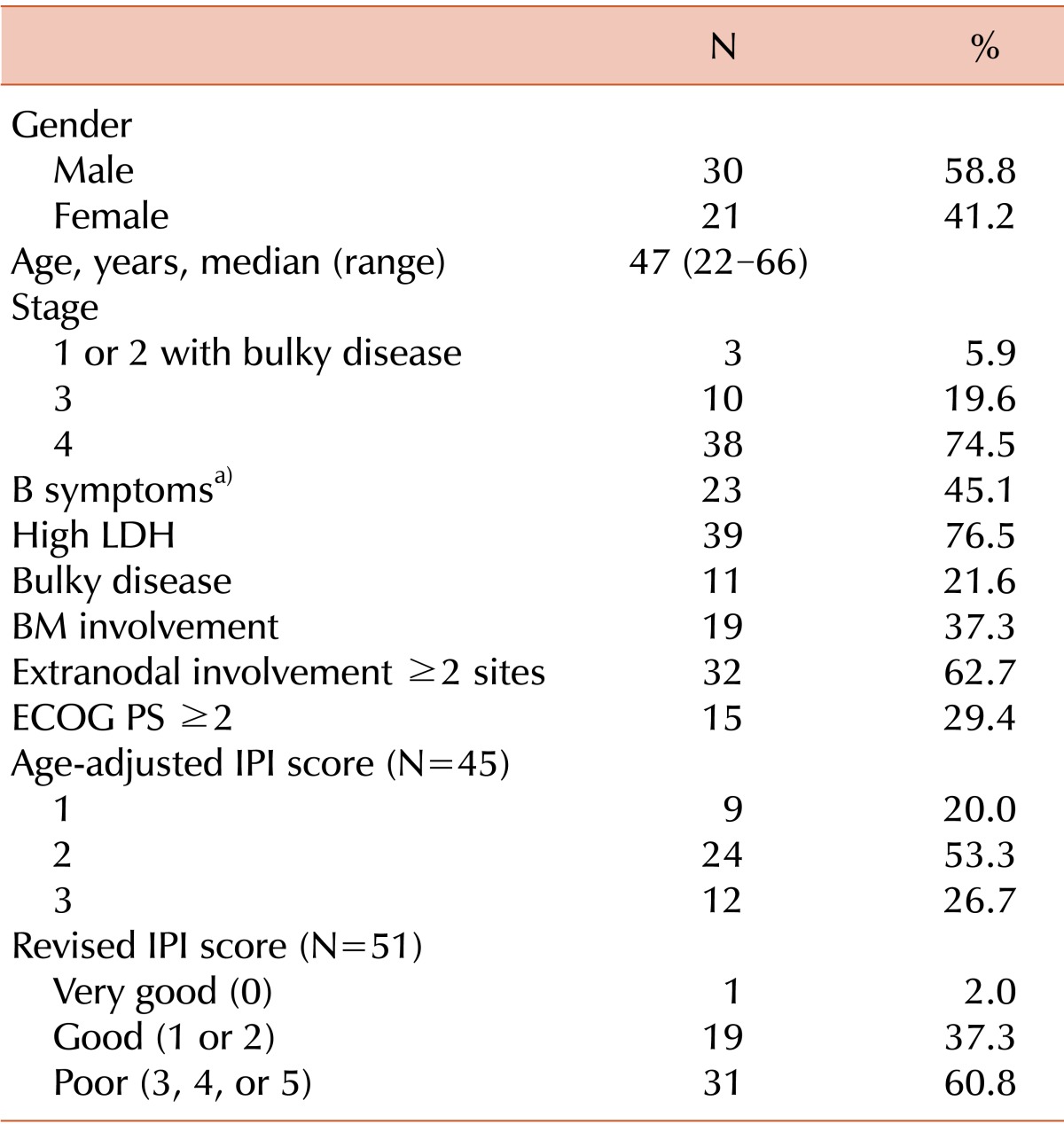
The patient and disease characteristics at diagnosis are summarized in Table 1. A total of 51 patients were evaluable in this study. The patients were classified as either stage I/II with bulky disease (5.9%) or stage III/IV (94.1%). Bulky disease was defined by the presence of 1 of the following 2 findings: (1) an abdominal node or nodal mass with a largest dimension of ≥10 cm as determined by an imaging study and (2) a mediastinal mass with a maximum width equal to or greater than one-third of the internal transverse diameter of the thorax at the T5/6 level as determined on a imaging study. At diagnosis, the aaIPI was available for 45 patients; the scores were 1, 2, and 3 in 20.0%, 53.3%, and 26.7% of the patients, respectively. The R-IPI identified 3 risk groups as follows: very good (2.0%), good (37.3%), and poor (60.8%).

Treatment before Auto-SCT and transplantation characteristics

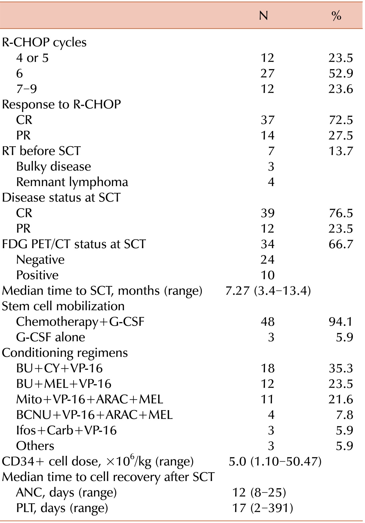
The treatments before Auto-SCT and transplantation characteristics are shown in Table 2. The CR and PR rates following R-CHOP therapy were 72.5% and 27.5%, respectively. Seven patients received involved field radiotherapy for bulky disease or remnant lymphoma prior to Auto-SCT. Consequently, CR and PR were achieved in 76.5% and 23.5% of patients, respectively, at the time of ASCT. PET/CT was available to evaluate the responses to R-CHOP before Auto-SCT in 34 of the 51 patients (66.7%). Of these patients, a negative PET/CT conversion was reported in 70.6% (24/34). The median time to Auto-SCT after diagnosis was 7.27 months (range, 3.4-13.4 months).

Response to treatment and outcomes

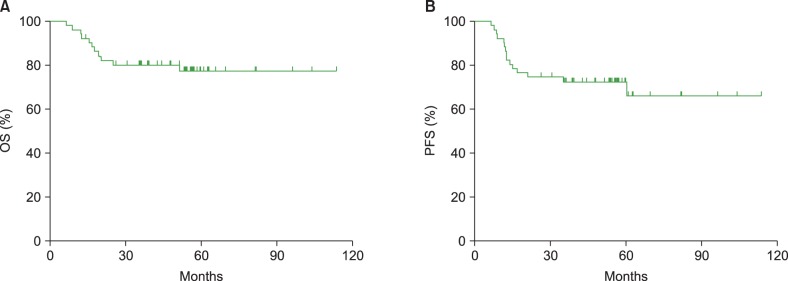
The median follow-up durations after diagnosis and Auto-SCT for the surviving patients were 56.7 months (range, 14.2-113.7 months) and 49.0 months (range, 8.2-109.2 months), respectively. All the patients achieved CR after Auto-SCT. During the follow-up period, 2 patients died of pneumonia, and 1 patient died of lactic acidosis, whereas 12 patients eventually relapsed after Auto-SCT. Eight of the 12 relapsed patients died at the time of data analysis. The 3-year OS and PFS were 80.1% and 72.4%, respectively (Fig. 1A and 1B). The 5-year OS and PFS were 77.3% and 72.4%, respectively, and the 5-year relapse probability was 23.5%. The median time to relapse after Auto-SCT was 5.8 months (range, 0.9-48.4 months).

Survival rates according to the aaIPI at diagnosis

The 5-year OS rates of the patients with aaIPI scores of 1 (N=9), 2 (N=24), and 3 (N=12) were 100%, 75.0%, and 56.2%, respectively (P=0.209; Fig. 2A). The 5-year PFS rates of the patients in the same categories were 77.8%, 70.8%, and 66.7%, respectively (P=0.840; Fig. 2B). Finally, the 5-year relapse probabilities of patients in these categories were 22.2%, 25.0%, and 25.0%, respectively (P=0.986).

Survival rates according to the R-IPI at diagnosis

The 5-year OS rates of the patients with R-IPI scores of 0 (N=1), 1 or 2 (N=19), and 3, 4, or 5 (N=31) were 100%, 78.0%, and 75.3%, respectively (P=0.867; Fig. 3A). The 5-year PFS rates of the patients in the same categories were 100%, 63.2%, and 77.4%, respectively (P=0.336; Fig. 3B). Finally, the 5-year relapse probabilities of the patients in these categories were 0.0%, 42.0%, and 12.9%, respectively (P=0.064).

Survival rates according to disease status at transplantation

The 5-year OS rates of the patients who achieved CR (N=39) and PR (N=12) were 73.1% and 91.7%, respectively (P=0.223; Fig. 4A). The 5-year PFS rates for those who achieved CR (N=39) and PR (N=12) were 69.2% and 82.5%, respectively (P=0.292; Fig. 4B). Furthermore, the 5-year relapse probabilities for those who achieved CR (N=39) and PR (N=12) were 25.6% and 16.7%, respectively (P=0.519).
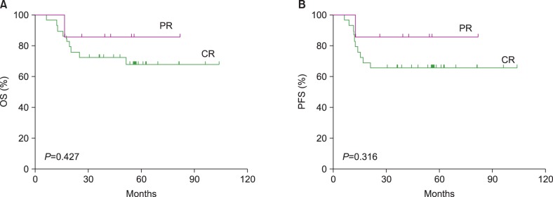
In addition, patients with aaIPI scores of 2 and 3 (N=36) were subjected to a survival analysis that excluded those with an aaIPI score of 1 (N=9). The 5-year OS rates of the patients in this subgroup who achieved CR (N=29) and PR (N=7) were 67.9% and 85.7%, respectively (P=0.427; Fig. 5A). The 5-year PFS rates for the patients in the same subgroup who achieved CR (N=29) and PR (N=7) were 65.5% and 85.7%, respectively (P=0.316; Fig. 5B). The 5-year relapse probabilities for the patients in this subgroup who achieved CR (N=29) and PR (N=7) were 27.6% and 14.3%, respectively (P=0.425).

Survival rates according to the FDG PET/CT status at transplantation

The 5-year OS rates of the patients who were PET negative (N=24) and PET positive (N=10) at the time of Auto-SCT were 73.9% and 90.0%, respectively (P=0.353; Fig. 6A). The 5-year PFS rates of the patients who were PET negative (N=24) and PET positive (N=10) at the time of Auto-SCT were 62.5% and 90.0%, respectively (P=0.337; Fig. 5B). The 5-year relapse probabilities of the patients who were PET negative (N=24) and PET positive (N=10) at the time of Auto-SCT were 37.5% and 10.0%, respectively (P=0.149).

DISCUSSION

Despite the recent prospective randomized trials involving aggressive DLBCL, the role of upfront Auto-SCT remains to be established in patients with high-risk DLBCL [4, 5, 6]. Therefore, we should keep in mind an important lesson from previous experience. Haioun et al. [9] published a paper in 1997, in which upfront Auto-SCT failed to improve the 5-year OS relative to sequential chemotherapy in high-risk aggressive non-Hodgkin's lymphoma patients who had achieved CR after induction treatment, although upfront Auto-SCT was superior to sequential chemotherapy with respect to 5-year disease-free survival (DFS). After a median follow-up duration of 8 years, the authors published the results of their final analysis in 2000 [10]. Upfront Auto-SCT was superior to sequential chemotherapy with respect to either 8-year DFS or OS. Based on this study, we think a longer follow-up is warranted to clarify the role of upfront Auto-SCT in high-risk DLBCL patients. In other words, it is too early to dismiss upfront Auto-SCT for patients with high-intermediate and high-risk non-Hodgkin's lymphoma.
According to a prospective randomized study that compared R-CHOP-21 with R-CHOP-14 for the treatment of DLBCL, CR and PR were documented in 63% and 25% DLBCL patients, respectively, in the R-CHOP-21 treatment group [11]. When patients with chemotherapy-sensitive relapses of non-Hodgkin's lymphoma were randomly assigned to receive either salvage therapy followed by HDT/ASCR or salvage therapy alone, the event-free survival (EFS) and OS rates significantly increased in the Auto-SCT group (P=0.001 and 0.038, respectively) [12]. As of 2014, the National Comprehensive Cancer Network Guidelines recommend salvage therapy followed by HDT/ASCR, when feasible, for patients who have achieved PR at best after R-CHOP-like therapy (NCCN Guidelines Version 1.2014) (www.nccn.org).
We retrospectively investigated whether the aaIPI and R-IPI at diagnosis, disease status, and PET/CT status at transplantation could predict the outcomes of patients with CD20-positive DLBCL.
In our study, the Kaplan-Meier curves of the 3 risk groups stratified according to aaIPI scores did not differ significantly with respect to the 5-year OS and PFS rates. A similar finding was described by Dilhuydy et al. [13], who performed a prospective trial to evaluate the efficacy of the addition of rituximab to front-line high-dose therapy followed by Auto-SCT in intermediate-high and high-risk patients with CD20-positive DLBCL. In that study, the 5-year OS and EFS rates were 74%±4% and 55%±5%, respectively. This difference was not statistically significant when the OS and EFS were analyzed according to the aaIPI. The IPI and its variants are the primary prognostic tools used in patients with DLBCL. In the pre-rituximab era, when the aaIPI was applied to patients with chemosensitive recurrent or refractory aggressive non-Hodgkin's lymphomas who had been treated with high-dose therapy followed by Auto-SCT, the scores were predictive of OS (P=0.034) [14]. A recent prospective study reported a 2-year PFS and OS of 63% and 73%, respectively, in aaIPI high-intermediate- and high-risk patients with aggressive B-cell lymphoma who had been treated with R-CHOP alone [5]. In our study, the respective 3-year PFS and OS were 69.4% and 75% for the high-intermediate- and high-risk groups. The 5-year PFS and OS for these groups were 69.4% and 71.1%, respectively.
In addition, we applied the R-IPI to the survival predictions in our study because Sehn et al. [15] had proposed that the revised IPI was a better outcome predictor than the standard IPI in the rituximab era in their retrospective analysis of patients with DLBCL who had been treated with R-CHOP alone. The authors reported that the 4-year PFS in the very good-risk, good-risk, and poor-risk groups were 94%, 80%, and 53%, respectively (P<0.001). Furthermore, the 4-year OS in the same prognostic risk groups were 94%, 79%, and 55%, respectively (P<0.001). As with the aaIPI, the R-IPI failed to predict survival differences between the 3 R-IPI risk groups in our study. The 5-year PFS in the 3 risk groups were 100%, 63.2%, and 77.4%, respectively (P=0.336), and the 5-year OS were 100%, 78.0%, and 75.3%, respectively (P=0.867).
Our study showed no significant differences in the 5-year OS and PFS rates between the PET-negative and PET-positive groups or between the CR and PR groups at the time of transplantation. In addition, the long-term OS and PFS did not differ significantly between the CR and PR groups, when the survival analysis was applied to patients with aaIPI scores of 2 and 3 (N=36) after excluding those with an aaIPI score of 1 (N=9). In our study, no tissue confirmations were made in patients from the PET-positive group. We presupposed that either the majority of the PET-positive true lesions converted to a lymphoma-free status after Auto-SCT or some of the PET-positive lesions might already have represented negative disease at the time of transplantation. Evaluations to check the disease response to induction chemoimmunotherapy might not play a role in prognosis prediction in the context of upfront Auto-SCT for DLBCL.
The role of interim PET/CT for predicting the outcomes in DLBCL patients has been debatable. Dupuis et al. [16] evaluated the prognostic impact of PET after 2 and 4 cycles of CHOP or CHOP-like chemotherapy with or without rituximab in 103 patients with DLBCL. The 5-year EFS rates were significantly higher in the PET-negative group than in the PET-positive group after 4 cycles of chemotherapy (80% vs. 36%, P<0.0001). In contrast, Moskowitz et al. [17] conducted a prospective study to clarify the significance of interim PET by obtaining biopsies from 38 patients with an interim positive PET after 4 cycles of accelerated R-CHOP from among 97 patients with advanced-stage DLBCL. Thirty-three of the 38 PET positive patients had negative biopsy results, and the PFS in the 33 with PET-positive but biopsy negative disease did not significantly differ from that of the 59 PET-negative patients. The authors concluded that the interim FDG-PET evaluation did not predict the outcomes in response to an R-CHOP-like regimen in the DLBCL patients.
The role of pre-Auto-SCT PET/CT has also been debatable even in cases treated with upfront Auto-SCT following induction chemoimmunotherapy. Dickinson et al. [18] retrospectively analyzed 39 patients with refractory or relapsed DLBCL to explore the predictive value of pre-Auto-SCT FDG-PET scans. Compared with those who had positive PET scans, patients with negative PET scans prior to Auto-SCT had a superior 3-year PFS (81% vs. 35%, P=0.003) and 3-year OS (81% vs. 39%, P=0.01). In contrast, after performing a retrospective analysis of 42 patients with DLBCL who were treated with rituximab-containing chemotherapy regimens followed by upfront Auto-SCT, Roland et al. [19] reported that patients with pre-Auto-SCT-positive PET scans who achieved PET negativity after Auto-SCT had EFS and OS rates equivalent to those with pre-Auto-SCT-negative PET scans. The authors suggested that chemosensitive DLBCL patients with positive pre-Auto-ASCT PET scans were likely eligible candidates for upfront Auto-SCT.
In conclusion, this registry-based retrospective analysis demonstrated a favorable long-term outcome of upfront consolidative HDT/ASCR for DLBCL. The OS and PFS rates according to the aaIPI, R-IPI, and PET status did not significantly differ between the subgroups. The outcome of the patients who achieved PR at the time of Auto-SCT did not significantly differ from that of the patients who achieved CR. R-CHOP induction therapy followed by Auto-SCT could bridge the survival gap between the CR and PR groups at the time of Auto-SCT.

ACKNOWLEDGMENTS

The clinical data for this study were collected from the Korean Blood and Marrow Transplant Registry (KBMTR), The Korean Society of Blood and Marrow Transplantation.

Notes

No potential conflicts of interest relevant to this article were reported.

Data were collected from the Korean Blood and Marrow Transplant Registry (KBMTR).

References

1. Coiffier B, Thieblemont C, Van Den Neste E, et al. Long-term outcome of patients in the LNH-98.5 trial, the first randomized study comparing rituximab-CHOP to standard CHOP chemotherapy in DLBCL patients: a study by the Groupe d'Etudes des Lymphomes de l'Adulte. Blood. 2010; 116:2040–2045. PMID: 20548096.
crossref
2. Pfreundschuh M, Kuhnt E, Trumper L, et al. CHOP-like chemotherapy with or without rituximab in young patients with good-prognosis diffuse large-B-cell lymphoma: 6-year results of an open-label randomised study of the MabThera International Trial (MInT) Group. Lancet Oncol. 2011; 12:1013–1022. PMID: 21940214.
crossref
3. Ziepert M, Hasenclever D, Kuhnt E, et al. Standard International prognostic index remains a valid predictor of outcome for patients with aggressive CD20+ B-cell lymphoma in the rituximab era. J Clin Oncol. 2010; 28:2373–2380. PMID: 20385988.
4. Vitolo U, Chiappella A, Brusamolino E, et al. A randomized multicenter phase III study for first line treatment of young patients with high risk (AAIPI 2-3) diffuse large B-cell lymphoma (DLBCL): Rituximab (R) plus dose-dense chemotherapy CHOP14/MEGA CHOP14 with or without intensified high-dose chemotherapy (HDT) and autologous stem cell transplantation (ASCT). Results of DLCL04 trial of Italian lymphoma foundation (FIL). Ann Oncol. 2011; 22(Suppl 4):iv106. (abst 72).
5. Stiff PJ, Unger JM, Cook JR, et al. Autologous transplantation as consolidation for aggressive non-Hodgkin's lymphoma. N Engl J Med. 2013; 369:1681–1690. PMID: 24171516.
crossref
6. Le Gouill S, Milpied NJ, Lamy T, et al. First-line rituximab (R) high-dose therapy (R-HDT) versus R-CHOP14 for young adults with diffuse large B-cell lymphoma: Preliminary results of the GOELAMS 075 prospective multicenter randomized trial. J Clin Oncol. 2011; 29:abst 8003. ASCO Annual Meeting.
crossref
7. Cheson BD, Pfistner B, Juweid ME, et al. Revised response criteria for malignant lymphoma. J Clin Oncol. 2007; 25:579–586. PMID: 17242396.
8. Juweid ME, Stroobants S, Hoekstra OS, et al. Use of positron emission tomography for response assessment of lymphoma: consensus of the Imaging Subcommittee of International Harmonization Project in Lymphoma. J Clin Oncol. 2007; 25:571–578. PMID: 17242397.
crossref
9. Haioun C, Lepage E, Gisselbrecht C, et al. Groupe d'Etude des Lymphomes de l'Adulte. Benefit of autologous bone marrow transplantation over sequential chemotherapy in poor-risk aggressive non-Hodgkin's lymphoma: updated results of the prospective study LNH87-2. J Clin Oncol. 1997; 15:1131–1137. PMID: 9060555.
10. Haioun C, Lepage E, Gisselbrecht C, et al. Survival benefit of high-dose therapy in poor-risk aggressive non-Hodgkin's lymphoma: final analysis of the prospective LNH87-2 protocol-a groupe d'Etude des lymphomes de l'Adulte study. J Clin Oncol. 2000; 18:3025–3030. PMID: 10944137.
crossref
11. Cunningham D, Hawkes EA, Jack A, et al. Rituximab plus cyclophosphamide, doxorubicin, vincristine, and prednisolone in patients with newly diagnosed diffuse large B-cell non-Hodgkin lymphoma: a phase 3 comparison of dose intensification with 14-day versus 21-day cycles. Lancet. 2013; 381:1817–1826. PMID: 23615461.
12. Philip T, Guglielmi C, Hagenbeek A, et al. Autologous bone marrow transplantation as compared with salvage chemotherapy in relapses of chemotherapy-sensitive non-Hodgkin's lymphoma. N Engl J Med. 1995; 333:1540–1545. PMID: 7477169.
crossref
13. Dilhuydy MS, Lamy T, Foussard C, et al. Front-line high-dose chemotherapy with rituximab showed excellent long-term survival in adults with aggressive large b-cell lymphoma: final results of a Phase II GOELAMS Study. Biol Blood Marrow Transplant. 2010; 16:672–677. PMID: 20045738.
crossref
14. Jabbour E, Peslin N, Arnaud P, et al. Prognostic value of the age-adjusted International Prognostic Index in chemosensitive recurrent or refractory non-Hodgkin's lymphomas treated with high-dose BEAM therapy and autologous stem cell transplantation. Leuk Lymphoma. 2005; 46:861–867. PMID: 16019530.
crossref
15. Sehn LH, Berry B, Chhanabhai M, et al. The revised International Prognostic Index (R-IPI) is a better predictor of outcome than the standard IPI for patients with diffuse large B-cell lymphoma treated with R-CHOP. Blood. 2007; 109:1857–1861. PMID: 17105812.
crossref
16. Dupuis J, Itti E, Rahmouni A, et al. Response assessment after an inductive CHOP or CHOP-like regimen with or without rituximab in 103 patients with diffuse large B-cell lymphoma: integrating 18fluorodeoxyglucose positron emission tomography to the International Workshop Criteria. Ann Oncol. 2009; 20:503–507. PMID: 19074215.
crossref
17. Moskowitz CH, Schoder H, Teruya-Feldstein J, et al. Risk-adapted dose-dense immunochemotherapy determined by interim FDG-PET in Advanced-stage diffuse large B-Cell lymphoma. J Clin Oncol. 2010; 28:1896–1903. PMID: 20212248.
crossref
18. Dickinson M, Hoyt R, Roberts AW, et al. Improved survival for relapsed diffuse large B cell lymphoma is predicted by a negative pre-transplant FDG-PET scan following salvage chemotherapy. Br J Haematol. 2010; 150:39–45. PMID: 20507301.
crossref
19. Roland V, Bodet-Milin C, Moreau A, et al. Impact of high-dose chemotherapy followed by auto-SCT for positive interim [18F] FDG-PET diffuse large B-cell lymphoma patients. Bone Marrow Transplant. 2011; 46:393–399. PMID: 20577223.
crossref
Fig. 1
(A) Probability of overall survival after autologous stem cell transplantation (Auto-SCT) for 51 diffuse large B-cell lymphoma (DLBCL) patients. (B) Probability of progression-free survival after Auto-SCT for 51 DLBCL patients.
br-49-107-g001
Fig. 2
(A) Probability of overall survival after autologous stem cell transplantation (Auto-SCT) according to the age-adjusted International Prognostic Index (aaIPI) score at diagnosis. (B) Probability of progression-free survival after Auto-SCT according to the aaIPI score at diagnosis.
br-49-107-g002
Fig. 3
(A) Probability of overall survival after autologous stem cell transplantation (Auto-SCT) according to the revised International Prognostic Index (IPI) score at diagnosis. (B) Probability of progression-free survival after Auto-SCT according to the revised IPI score at diagnosis.
br-49-107-g003
Fig. 4
(A) Probability of overall survival after autologous stem cell transplantation (Auto-SCT) according to the disease status at transplantation. (B) Probability of progression-free survival after Auto-SCT according to the disease status at transplantation.
br-49-107-g004
Fig. 5
(A) Probability of overall survival after autologous stem cell transplantation (Auto-SCT) according to the disease status at transplantation for patients classified into the age-adjusted International Prognostic Index (aaIPI) high-intermediate- and high-risk groups. (B) Probability of progression-free survival after Auto-SCT according to the disease status after transplantation for patients classified into the aaIPI high-intermediate and high-risk groups.
br-49-107-g005
Fig. 6
(A) Probability of overall survival after autologous stem cell transplantation (Auto-SCT) according to the 18F-fluoro-2-deoxy-D-glucose positron emission tomographic/computed tomographic (FDG PET/CT) status at transplantation. (B) Probability of progression-free survival after Auto-SCT according to the FDG PET/CT status at transplantation.
br-49-107-g006
Table 1
Patient and disease characteristics at diagnosis.
br-49-107-i001

a)B symptoms indicate systemic symptoms such as fever, night sweats, and weight loss, which are associated with non-Hodgkin's lymphoma.

Abbreviations: LDH, lactate dehydrogenase; BM, bone marrow; ECOG, Eastern Cooperative Oncology Group; PS, performance status; IPI, International Prognostic Index.

Table 2
Treatments before ASCT and transplantation characteristics.
br-49-107-i002

Abbreviations: ASCT, autologous stem cell transplantation; RCHOP, rituximab+cyclophosphamide+adriamycin+vincristine+prednisolone; CR, complete remission; PR, partial remission; RT, radiation therapy; SCT, stem cell transplantation; FDG, 18F-fluoro-2-deoxy-D-glucose; PET, positron emission tomography; CT, computed tomography; G-CSF, granulocyte colony-stimulating factor; BU, busulfan; CY, cyclophosphamide; VP-16, etoposide; MEL, melphalan; Mito, mitoxantrone; ARAC, cytosine arabinoside; BCNU, carmustine; Ifos, ifosfamide; Carb, carboplatine; CD, cluster of differentiation; ANC, absolute neutrophil count; PLT, platelet.

TOOLS
Similar articles