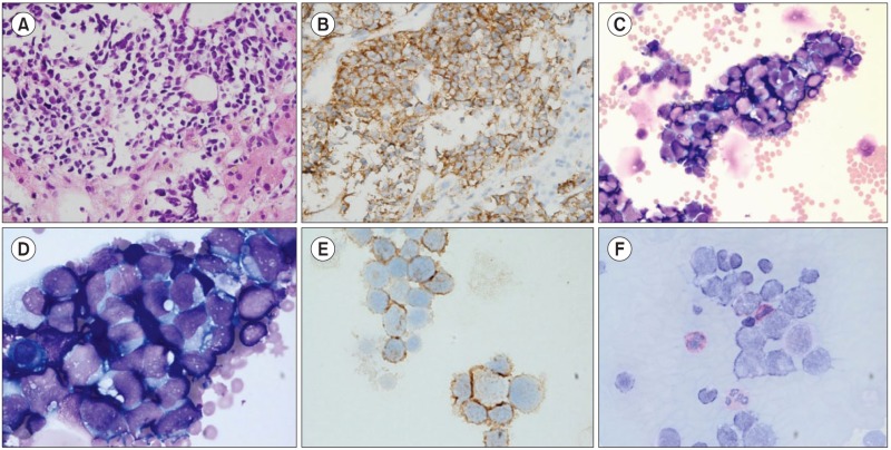
Neuroendocrine tumor (NET) of the gallbladder (GB) (GB-NET) is a very rare condition, representing 0.5% of all NET cases. Here, we report a case of GB-NET with peritoneal metastasis. A 55-year-old man was admitted to our institution for workup for an abdominal distension, which developed 5 weeks ago. Computed tomography (CT) of his abdomen showed ascites and an 18-cm heterogeneous enhancing mass surrounding the GB and invading the liver. Needle biopsy of the GB revealed poorly differentiated sheets of neoplastic cells with hyperchromatic nuclei and indistinct cytoplasm (A, hematoxylin-eosin, ×400). Neoplastic cells were positive for synaptophysin on immunohistochemical staining (B, ×400), but negative for cytokeratin and leukocyte common antigen, indicating neuroendocrine carcinoma, small cell type. Ascitic fluid was centrifuged, and numerous clusters of small to medium-sized neoplastic cells with back-to-back appearance were observed at a frequency of 74% (C, Wright stain, ×400; D, Wright stain, ×1,000). Neoplastic cells were positive for CD56 on immunohistochemical staining (E, ×1,000) but negative for periodic acid-Schiff on cytochemical staining (F, ×1,000). On the basis of these results, the patient was diagnosed as having GB-NET with peritoneal metastasis.


 PDF
PDF ePub
ePub Citation
 Print
Print


 XML Download