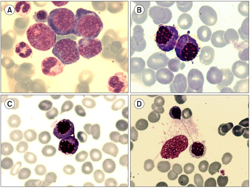
The terminal stage of cell division is represented by a binucleated cell with the 2 nuclei separated within the undivided cytoplasm (A, binucleated basophilic erythroblasts). The cytoplasm divides later to form 2 distinct cells. In myelodysplastic syndromes, cellular division of erythroblasts may occur asynchronously: cytoplasmic division and separation occur first followed by nuclear division. Microscopically, the internuclear bridge appears as a narrow strand of chromatin, binding the 2 nuclei. The nuclear border often appears distorted and stretched, resembling a teardrop shape. The 3 phases of internuclear bridging illustrated herein are from the bone marrow of a 72-year-old man affected by refractory anemia with ringed sideroblasts: in phase I, after the cytoplasm divides, nuclei of both cells remain connected with a bundle of thin fibrils (B, two erythroblasts with asynchronous maturation [pyknotic nuclei and basophilic cytoplasm] that have undergone mitosis but are still connected with thin chromatin filaments); in phase II, the bundle of thin fibrils is thickened and shorter (C, the erythroblasts with thickened, shorter chromatin filaments and intact nuclear boundary); and in phase III, the 2 cells recede and stretch the filament, which is then completely separated (D, the erythroblasts are greatly alienated, with a long and thin connecting thread [like chewing gum], and nuclear contours are modified and formed a teardrop shape [upper cell]).


 PDF
PDF ePub
ePub Citation
 Print
Print


 XML Download