Abstract
Acquired hemophilia A (AHA) is a bleeding disorder caused by the development of an auto-antibody against endogenous factor VIII (FVIII). In this study, the epitope of the autoantibody was identified in a 67-year-old female patient with AHA. A prolonged activated partial thromboplastin time (77.4 s) that failed to correct in an incubation mixing test (68.2 s), a decreased FVIII activity, and a high FVIII inhibitor (14.6 Bethesda units/mL) were observed. Enzyme-linked immunosorbent assay demonstrated that the antibody belonged to the immunoglobulin G4 subclass. An immunoblotting assay revealed the light chain (A3/C1/C2 domain) of FVIII as the binding region of the antibody. The bleeding experienced by our patient resulted from the interference of FVIII binding to both FIX by anti-A3 antibodies and phospholipids and von Willebrand factor by anti-C2 antibodies. To the best of our knowledge, this is the first study in Korea characterizing an autoantibody in the context of AHA.
Acquired hemophilia A (AHA), a bleeding disorder that occurs in patients without a family history of hemophilia, is caused by the development of autoantibodies against endogenous factor VIII (FVIII). The incidence of AHA is 1.3-1.5 cases per million people per year [1]. Up to 85% of affected individuals are more than 60 years of age [2]. The age distribution of AHA is typically biphasic, with a small peak between 20 and 30 years of age and the major peak between 68 and 80 years of age [3]. In approximately 50% of AHA cases, FVIII autoantibodies are associated with the postpartum period, autoimmune diseases, underlying hematologic or solid cancers, infections, vaccination, or the use of certain medications [4-6]. Clinical manifestations of AHA include bleeding at any location in the body; however, hemarthrosis, the hallmark of congenital hemophilia, is uncommon in AHA. Diagnosis of AHA requires identifying hemophilia with inhibitors and excluding congenital hemophilia. The two major therapeutic strategies for AHA are the control of bleeding and the eradication of autoantibodies [7].
Antibodies against FVIII may either block the function of FVIII or promote the clearance of factor VIII from circulation [8, 9]. Identification of the FVIII epitopes to which inhibitor antibodies bind is crucial for understanding the mechanisms of inhibitor activity [9]. In this study, we attempted to identify the epitope of the autoantibody against FVIII in a patient with AHA.
A 67-year-old female, experiencing 12 h of acute abdominal and right flank pain, was admitted to our hospital through the emergency department. Her medical history was unremarkable, except for hypertension controlled with amlodipine. The patient had no family history of abnormal bleeding. One week prior to hospital admittance, she developed bruises on her left lower extremity and gross hematuria. She did not receive medical care, and these conditions resolved spontaneously.
Initial examination revealed pale conjunctiva and both soft-tissue swelling and bruising on her left lower extremity. Tenderness to palpation over the lower abdomen and rebound tenderness were noted. Her vital signs were within normal ranges. Complete blood cell count indicated hypochromic anemia with normal white blood cell counts (hemoglobin, 8.2 g/dL; hematocrit, 23.9%; white blood cell counts, 9.1×10⁹/L; platelet counts, 342×109/L). The patient's prothrombin time (PT, 12.5 s; international normalized ratio [INR], 1.11) and thrombin time (TT, 14.3 s) were normal. Her activated partial thromboplastin time (APTT, 77.4 s) was prolonged and not corrected by incubation mixing test (APTT, 68.2 s).
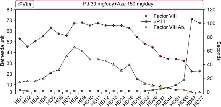
Abdominal computed tomography scan demonstrated multifocal fluid collections throughout the abdomen and pelvis, localized mainly on the right side. An enlarged psoas muscle, intramuscular hematomas in the right iliopsoas, and a paravertebral muscle with active bleeding were noted. Multiple calcifications within the pancreas and atrophy of the pancreatic parenchyma suggested chronic pancreatitis. The spleen, liver, and gall bladder appeared normal. Angiography of the abdominal arteries demonstrated suspicious extravasation from a right lumbar artery. Gelfoam embolization of the right second and third lumbar arteries was performed. The patient was immediately treated with recombinant FVIIa (90 IU/kg every 6 to 8 h; NovoSeven) for 2 days because of active bleeding. The FVIII level was 2%, and the titer of FVIII inhibitor was 14.6 Bethesda units (BU)/mL. The levels of FIX (121%), FXI (90%), and FXII (94%) were all within normal ranges. The patient was treated with oral prednisolone (30 mg/d) and azathioprine (150 mg/d) to eradicate the autoantibody. The tests for anti-nuclear antibody, lupus anticoagulant, hepatitis B virus surface antigen, hepatitis C virus antibody, and human immunodeficiency virus antigen and antibody were all negative, with the exception of anti-Ro antibody (3+). After 2 days of immunosuppressive treatment, the patient's flank pain and hematuria resolved and, 2 weeks later, the hematoma disappeared. On the 45th hospital day, the patient was discharged on oral prednisolone (20 mg/d) and azathioprine (150 mg/d), without further complications. The prednisolone treatment was discontinued approximately 6 months later. The patient's APTT normalized; her FVIII level reached the upper 50%, and the inhibitor was undetectable (Fig. 1).
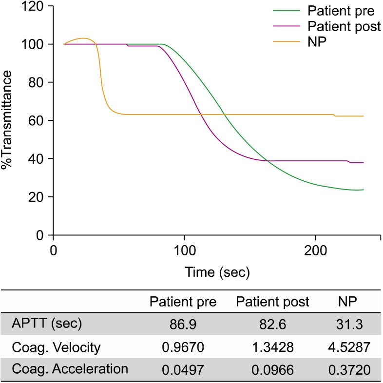
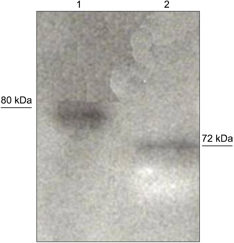
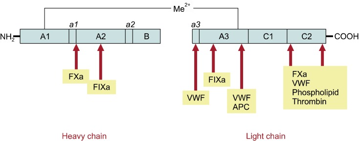
APTT waveform analysis (MDA series; Organon, Technika) of the patient's plasma showed a delayed clotting pattern compared to normal pooled plasma (Fig. 2). Thrombin generation assay (Thermo Fisher Scientific, Waltham, MA, USA) showed decreased production of thrombin in the patient's plasma compared to normal plasma. Virtually no hemostatic effect occurred after the infusion of plasma. Slight improvement in the waveform analysis, but not in the thrombin generation test, was noted (not shown). Enzyme-linked immunosorbent assay demonstrated that the patient's antibody belonged to the immunoglobulin (Ig) G4 subclass. Immunoblotting assays demonstrated that the light chain (A3/C1/C2 domain) of FVIII is the binding region of the autoantibody (Figs. 3, 4).
FVIII functions as a cofactor for FIXa in the tenase complex, and deficiency of FVIII reduces the generation of thrombin on the surface of activated platelets. Synthesized as a 330-kDa precursor protein with an A1-a1-A2-a2-B-a3-A3-C1-C2 domain structure, the single-chain FVIII protein is proteolytically processed to yield a series of amino-terminal heavy-chain fragments of approximately 92-200 kDa and a carboxy-terminal light chain of 80 kDa [8]. Most acquired FVIII inhibitors are IgG1 and IgG4 autoantibodies, directed toward epitopes within the A2, A3, and C2 domains of the FVIII molecule. The IgG subclass of inhibitor in our case was also IgG4, and the binding region was the 72-kDa A3/C1/C2 domain of the light chain (common in AHA). Depending on the location of the target epitope, different mechanisms of action for the anti-FVIII antibodies have been reported. Anti-A3 antibodies neutralize the procoagulant activity of FVIII by preventing its interaction with FIXa. Anti-C2 antibodies disrupt the binding of FVIII to phospholipid and von Willebrand factor (VWF), whereas antibodies to A2 and A3 interfere with FVIII binding to factor X and factor IXa. The role of the C1 domain has not been discovered [9-12]. The bleeding that occurred in our patient developed from the interference of FVIII binding to both FIX, by anti-A3 antibodies, and phospholipids and VWF, by anti-C2 antibodies.
Several studies have investigated FVIII-specific CD4+ T cells as a plausible source of the observed inhibitor production in AHA [13, 14]. First, Bray et al. [13] observed that the FVIII inhibitors may spontaneously disappear in conjunction with an HIV-associated decline in CD4+ T cell counts. Reding et al. [14] demonstrated that a strong positive correlation exists between inhibitor titer and the proportion of Th2-driven IgG4 anti-FVIII antibodies, and that Th1 cells may be involved in the long-term maintenance of anti-FVIII antibody synthesis. Those results could lead to the assumption that FVIII-specific CD4+ T cells are crucial for the development of AHA. Based on these results, immunosuppressive treatments targeting CD4+ T cells might effectively prevent the synthesis of anti-FVIII antibody.
Patients with AHA are at high risk of bleeding-related mortality, and this is likely due to the delay in the diagnosis of AHA [1, 2]. Thus, AHA should be suspected by the physician immediately for patients especially those who are elderly, have an autoimmune disease, are pregnant, or have a malignancy, or experiencing recent onset of bleeding with isolated prolongation of APTT that is not corrected in mixing studies. We speculate that the cause of the present case is autoimmune disease because of positive results with the anti-Ro antibody; yet, the patient had no other symptoms. Therefore, we deduce that the cause of our case is idiopathic. Most AHA patients have hemorrhaging of the skin, muscles, or soft tissues and mucous membranes (e.g., epistaxis, gastro-intestinal and urological bleeds, and retroperitoneal hematomas), whereas hemarthrosis, a typical feature of congenital FVIII deficiency, is uncommon. We summarized all reported cases of AHA in the Korean journals (Table 1). If a physician suspects AHA, laboratory tests, including specific factor assays and the Bethesda assay for the quantification of inhibitors, should be performed for confirmatory diagnosis. Recently, the Korean Society of Thrombosis and Hemostasis (KSTH) proposed guidelines for the management of acquired hemophilia [15]. In summary, this is the first demonstration of epitope localization for a FVIII inhibitor in a patient with AHA in Korea. More studies are necessary for further clarification of the pathophysiology of AHA.
References
1. Collins P, Macartney N, Davies R, Lees S, Giddings J, Majer R. A population based, unselected, consecutive cohort of patients with acquired haemophilia A. Br J Haematol. 2004; 124:86–90. PMID: 14675412.
2. Collins PW, Hirsch S, Baglin TP, et al. Acquired hemophilia A in the United Kingdom: a 2-year national surveillance study by the United Kingdom Haemophilia Centre Doctors' Organisation. Blood. 2007; 109:1870–1877. PMID: 17047148.
3. Franchini M, Montagnana M, Lippi G. Clinical, laboratory and therapeutic aspects of platelet-type von Willebrand disease. Int J Lab Hematol. 2008; 30:91–94. PMID: 18333841.
4. Meiklejohn DJ, Watson HG. Acquired haemophilia in association with organ-specific autoimmune disease. Haemophilia. 2001; 7:523–525. PMID: 11554945.
5. Sallah S, Wan JY. Inhibitors against factor VIII in patients with cancer. Analysis of 41 patients. Cancer. 2001; 91:1067–1074. PMID: 11267950.
6. Solymoss S. Postpartum acquired factor VIII inhibitors: results of a survey. Am J Hematol. 1998; 59:1–4. PMID: 9723568.
7. Defendenti C, Atzeni F, Spina MF, et al. Clinical and laboratory aspects of Ro/SSA-52 autoantibodies. Autoimmun Rev. 2011; 10:150–154. PMID: 20854935.
8. Lavigne-Lissalde G, Schved JF, Granier C, Villard S. Anti-factor VIII antibodies: a 2005 update. Thromb Haemost. 2005; 94:760–769. PMID: 16270627.
9. Lavigne-Lissalde G, Rothschild C, Pouplard C, et al. Characteristics, mechanisms of action, and epitope mapping of anti-factor VIII antibodies. Clin Rev Allergy Immunol. 2009; 37:67–79. PMID: 19172415.
10. Scandella D, Mattingly M, de Graaf S, Fulcher CA. Localization of epitopes for human factor VIII inhibitor antibodies by immunoblotting and antibody neutralization. Blood. 1989; 74:1618–1626. PMID: 2477082.
11. Fulcher CA, de Graaf Mahoney S, Roberts JR, Kasper CK, Zimmerman TS. Localization of human factor FVIII inhibitor epitopes to two polypeptide fragments. Proc Natl Acad Sci U S A. 1985; 82:7728–7732. PMID: 2415970.
12. Scandella D, Gilbert GE, Shima M, et al. Some factor VIII inhibitor antibodies recognize a common epitope corresponding to C2 domain amino acids 2248 through 2312, which overlap a phospholipid-binding site. Blood. 1995; 86:1811–1819. PMID: 7544643.
13. Bray GL, Kroner BL, Arkin S, et al. Loss of high-responder inhibitors in patients with severe hemophilia A and human immunodeficiency virus type 1 infection: a report from the Multi-Center Hemophilia Cohort Study. Am J Hematol. 1993; 42:375–379. PMID: 8493988.
14. Reding MT, Lei S, Lei H, Green D, Gill J, Conti-Fine BM. Distribution of Th1- and Th2-induced anti-factor VIII IgG subclasses in congenital and acquired hemophilia patients. Thromb Haemost. 2002; 88:568–575. PMID: 12362225.
15. The Korean Society on Thrombosis and Hemostasis. The guidelines of Hemophilia. 2011. Seoul, Korea: Han Kuk Medical Publisher;p. 115–120.
16. Song MH, Ahn SJ, Yoo SH, Kim BH, Park JH. Total knee arthroplasty-associated acquired hemophilia: a case report. J Korean Orthop Assoc. 2006; 41:167–169.
17. Chung BH, Kim YJ, Kim MS, et al. A case of acquired hemophilia detected after influenza vaccination. Korean J Med. 2004; 66:298–301.
18. Lee N, Yoon SH, Lim W, et al. Post-operative bleeding due to acquired hemophilia successfully treated with recombinant factor VIIa: case report. Korean J Hematol. 2009; 44:163–167.
19. Hwang HW, Kong JH, Yu DW, Kim WT, Kim HS, Lee CI. A patient with acquired hemophilia A induced by clopidogrel. Korean J Hematol. 2012; 47:80–82. PMID: 22479283.