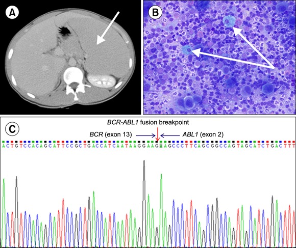
A 17-year-old boy with no specific medical history presented with abdominal distension. A complete blood analysis revealed a leukocyte count of 151.6×10⁹/L (segmented neutrophils, 46%; lymphocytes, 4%; eosinophils, 6%; basophils, 15%; metamyelocytes, 1%; myelocytes, 15%; and blasts, 13%), a hemoglobin level of 99.0 g/L, and a platelet count of 568.0×10⁹/L. A contrast-enhanced abdominal computed tomography image showed marked splenomegaly (A) and mildly enlarged left para-aortic lymph nodes. Bone marrow aspiration revealed trilineage proliferation, including prominently small megakaryocytes with dyspoietic features, as well as eosinophilia and pseudo-Gaucher cells (B) and a normal beta-glucocerebrosidase level. The estimated M : E (Myeloid : Erythroid) ratio was approximately 6.8 : 1, and the blast cell counted was as high as 4.5%. The Philadelphia chromosome was found in all 20 analyzed metaphase cells. BCR/ABL1 (b2a2 type) gene rearrangement was detected with both fluorescence in situ hybridization (FISH) and reverse transcriptase-polymerase chain reaction (RT-PCR; C). We believe that this is a unique case of adolescent chronic myeloid leukemia that is characterized by an accelerated phase onset, extreme splenomegaly, and the presence of pseudo-Gaucher cells.


 PDF
PDF ePub
ePub Citation
 Print
Print


 XML Download