

 PDF
PDF ePub
ePub Citation
 Print
Print


I. Introduction
Encephalocele is a serious congenital anomaly characterized by herniation of the brain tissue and meninges through a defect either along the midline of the cranial vault or at the base of the skull1. The anomaly is found to occur with an estimated frequency of 1 to 4 cases per 10,000 live births2. Basal encephaloceles are rare craniofacial anomalies, with an estimated frequency of 1 case per 100,000 live births3. Based on the anatomical site of the defect in the cranium, they are classified into transsphenoidal, sphenoethmoidal, sphenoorbital, and transethmoidal variants3.
Among the types of basal encephaloceles, the transsphenoidal variant is the rarest and accounts for 5% of all basal encephaloceles (1 in 70,000 live births). Associated congenital anomalies occur in about one-third of these patients and include hypertelorism, medial nasal fissure, broad nasal roots, and cleft lip and cleft palate with or without optic and cerebral malformations. The most significant clinical features of transsphenoidal basal encephaloceles are respiratory difficulties, episodes of recurrent meningitis, cerebrospinal fluid (CSF) leaks, and endocrinal abnormalities. Rarely has this anomaly been found to be asymptomatic. We hereby report an asymptomatic case of transsphenoidal basal encephalocele with cleft palate and highlight a modified version of two-flap palatoplasty for achieving closure of the cleft palate.
II. Case Report
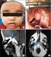
A full-term baby of 18 months born by normal vaginal delivery to parents of nonconsaguinous marriage was referred to our unit for management of cleft palate. All developmental milestones were found to be normal in the child. Hypertelorism and a broad nasal root were observed on extraoral examination.(Fig. 1. A) The child was surprisingly asymptomatic, with no history of meningitis, CSF rhinorrhoea, or nasal obstruction at presentation.
Examination of the oral cavity revealed a completely cleft palate, which allowed straightforward visualization of a round and whitish cystic mass having a smooth surface and located in the midline measuring roughly 2.5×1.5 cm.(Fig. 1. B) The hematological, biochemical, and endocrine profiles were within normal limits. A non-contrast computed tomography (CT) scan of the brain revealed a bony defect in the body of the sphenoid bone measuring 6.4 mm, with a defect in the hard palate.(Fig. 1. C) There was evidence of a mass extending from the anterior wall of the sella turcica into the left nasal cavity and up to the roof of the oral cavity, measuring 3.6×1.4×3.1 cm in the greatest dimension.(Fig. 1. D) Based on the CT findings, a diagnosis of transsphenoidal basal encephalocele was made. The opinion of a neurosurgeon was sought, and since the patient was devoid of any symptoms, they were referred back to us for palatoplasty. Informed written consent was sought from the patient's parents for the surgical procedure under general anesthesia.
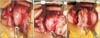
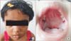
The technique we performed for closure of the cleft palate was similar to Bardach's two-flap palatoplasty. An incision was made along the cleft and alveolar margins, with the incisions along the cleft margins in the region of the encephalocele made 0.5 mm more laterally onto the sound bone to avoid inadvertent entry into the encephalocele. Palatal mucoperiosteal flaps were raised pedicled on the greater palatine vessels, per standard protocol.(Fig. 2. A) The nasal mucosa posterior to the mass was meticulously released and sutured with 5-0 Vicryl sutures in a buried fashion. The abnormal position of the levator palatine muscle was released and sutured to its counterpart to form a muscle sling, as in intravelar veloplasty. With great caution, the mucosa over the encephalocele was de-epithelialized, and the mucoperiosteal flaps were closed over the mass.(Fig. 2. B) Thus, the soft palate was closed in three layers, nasal, muscular, and oral, similar to Bardach's two-flap palatoplasty, and the hard palate was closed by mucoperiosteal flaps over the de-epithelialized surface of the encephalocele.(Fig. 2. C) The raw areas exposed along the alveolar margins were allowed to heal by secondary intention. The postoperative recovery was uneventful. The patient has been regularly followed for the last three years without any complications.(Fig. 3)
III. Discussion
Transsphenoidal encephaloceles are the rarest of the basal encephaloceles. Defective ossification of the body of the sphenoidal bone with persistence of the craniofacial canal best explains the reason for the development of basal transsphenoidal encephaloceles4.
Sphenoidal encephaloceles remain asymptomatic in most instances, unlike other types of encephaloceles, and are not detected unless they emerge through a defect in the palate, similar to in our patient5. The most significant clinical features are respiratory difficulties, feeding difficulties, episodes of recurrent meningitis, and endocrine abnormalities. Associated congenital anomalies are seen in about one-third of patients and include hypertelorism, broad nasal roots, median nasal fissures, and cleft lip and palate. Optic malformations like anophthalmia, colobomas, retinal abnormalities, and morning glory syndrome and cerebral malformations like agenesis of the corpus callosum, hydrocephalus, and pituitary hypoplasia have also been reported678. Encephaloceles herniated from the nasal cavity can be differentiated from nasal polyps by the presence of pulsations, presenting medially from the septum and widening the nasal bridge. Nasal polyps, by contrast, originate from the turbinates and do not widen the nasal bridge3.
Advanced imaging studies play a decisive role in establishing the diagnosis of encephalocele, as well as to define any neural or vascular elements in the sac7. We opted for a noncontrast CT scan of the brain that allowed for precise identification of the skull base defect. Many clinicians also perform magnetic resonance imaging of the brain to define the contents of the herniated sac. Endocrine assessment should also be carried out in every patient, as hypothalamic pituitary dysfunction and deficiency of antidiuretic and growth hormones are common findings9.
The surgical management of transsphenoidal encephaloceles is typically attempted via either transcranial, transoral, or transnasal endoscopic approaches10, with most surgeons opting for the transoral route for cases of transsphenoidal encephaloceles with cleft palate, because there is less risk of damaging the functioning tissues within the wall of the encephalocele, and the herniated sac can be separated from the nasal mucosa, enabling adequate closure.
Surgery is usually indicated in symptomatic cases or in cases in which life threatening complications, like CSF leaks, meningitis, or visual disturbance secondary to the lesion, might be result from failure to intervene10. Furthermore, the prognosis in cases treated by neurosurgical intervention is reserved, with only slightly more than half of treated cases presenting a favorable evolution, as described in a study by Macfarlane et al.11 who demonstrated that only 59% of 114 cases resulted in normal development. Also, a number of cases have been documented in the literature where the encephaloceles have remained asymptomatic and have been incidentally discovered, without causing any problems to the patient1213. Based on the asymptomatic nature of this case of encephalocele, the neurosurgeon's clearance for the palatoplasty procedure, and the strong evidence presented in the literature that asymptomatic encephaloceles do not affect normal development, a decision was made to perform only a palatoplasty procedure to protect the herniated brain tissue from any form of trauma. The procedure performed was similar to Bardach's two-flap palatoplasty, except that the nasal layer was not closed in the area of the encephalocele; instead, the palatal mucoperiosteum was placed directly over the deepithelialized mucosa of the transsphenoidal encephalocele.
In conclusion, transsphenoidal basal encephaloceles with cleft palate may sometimes manifest asymptomatically. In such cases, following neurosurgical evaluation and clearance, our technique of the modified two-flap palatoplasty can be safely employed. However, these patients require periodic long-term follow-up.
 XML Download