

 PDF
PDF ePub
ePub Citation
 Print
Print


I. Introduction
Congenital auricular deformities are relatively common and most frequently involve the upper one-third of the auricle. The lower part of the auricle is affected in 1:15,000 of live births1, and the vast majority are cleft earlobe anomalies2. Various techniques have been introduced for congenital cleft earlobe repair depending on the size and form2. The purpose of this report was to introduce a useful operative technique for repair.
II. Technical Note
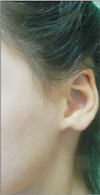
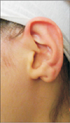
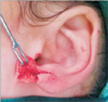
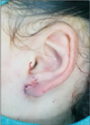
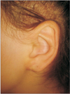
Between September 2015 and May 2017, two patients who presented with a congenital earlobe cleft underwent surgical correction at our institution. The patients were 11- and 17-year-old females. The anomalies were unilateral in both cases and occurred on the patient's left side. The first patient demonstrated a tag and a hypoplasia-type cleft earlobe malformation according to Park's classification2.(Fig. 1) The second patient had a defective type of cleft earlobe anomaly.(Fig. 2)
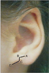
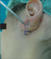
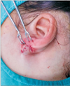
The auricle was compared to the unaffected side, and the amount of tissue loss on the affected side was determined. We designed an operative technique composed of a Z-plasty anteriorly and a Y-V advancement posteriorly. The Z-plasty incision was marked with a right angled anterior and continued on the marginal surfaces for a corrective intervention. The lateral (line AB) and vertical (line BC) lines were 5 mm, while the medial component along line CD was 6 mm in length.(Fig. 3) An incision that ran half of the total thickness was drawn beginning from the anterior surface of the earlobe to the cleft margin and ending at the inferior point of the medial component.(Fig. 4) After the incision was made, the flaps were undermined and separated from the posterior skin to produce anterior and posterior subdermal flaps of equal thickness.(Fig. 5) The first flap composed the front layer. The key suture of this layer stretched from the lateral component to the inferior point of the medial component, accomplishing the Z-plasty transposition. The anterior skin was closed with 6/0 prolene sutures (Ethicon Inc., Somerville, NJ, USA). (Fig. 6) A slit was prepared on the posterior surface, and the second subdermal flap section of the medial component was adapted as a Y-V advancement flap.(Fig. 7) The second flap created the back layer and supplied sufficient earlobe bulk to pull the earlobe medially.(Fig. 8)
This technique was applied to two patients to repair their specific deformities. The patients were followed up for a period ranging from 3 to 14 months. She has been wearing earrings since the sixth month postoperatively.(Fig. 9) Ultimately, the consecutive application of the front and back flaps created a well-shaped earlobe. The patients healed uneventfully.
III. Discussion
Malformational auricular anomalies reveal themselves by their absent structural tissue and are caused by damage during embryologic or fetal development. The auricle is formed between the fifth and ninth weeks of gestation in the embryologic period. Failure of fusion of auricular components hillock 1 and hillock 6 is probably the cause of cleft earlobe3. Lower auricular malformations most commonly comprise cleft earlobe deformities, and they can be classified into four subtypes based on the corrective method required; defective, tag and cleft, tag and cleft with hypoplasia, and simple type2.
Acquired cleft earlobe deformity is extensively seen worldwide. However, congenital cleft earlobe presents as a distinct entity. Various reconstructive methods have been described according to the severity of the anomaly45678. These approaches have used local flaps, grafts and simple suture techniques. The Z-plasty, 7-plasty or other applications can be used to correct simple clefts8. The disadvantage of these methods is that a scar line ends from the free margin of the lobule. Alternatively, a method that creates a sutureless-free lobule margin may be preferred6.
We used two distinct subdermal flaps. In this technique, the front flap was used for the Z-plasty, while the back became the Y-V advancement flap. This method allowed for control of the rotation of the earlobe as well. To avoid any ischemic condition, we did not use local anesthetics, but we know it is not necessary constantly. Park2 corrected this type of anomaly by the using buttoning procedure and a chondrocutaneous postauricular arterial flap. We selected a simple technique that is applicable to clefts with a wide transverse diameter of the lobule. The Z-plasty on the front side is a safe intervention for this condition. After the flap transposition is completed on the front side, it becomes clear which process to use for the second flap on the back side. We chose this method because we consider it to be easier than Park's validated method2. The medial component contains thick tissue, so it is suited to various reconstructive options. A common feature of many techniques is that they design a two-layer repair of the lobule2468. Cases that are more serious than a simple cleft will still require a local tissue transfer therefore contains extending incisions2910.
We advised our patients that they could begin wearing earrings during the sixth month postoperatively. This time frame was chosen to allow the influence of gravity to overcome possible earlobe contraction and prevent creating a different deformity. We report a useful method that may be worth considering for some patients with cleft earlobe.
 XML Download