

 PDF
PDF ePub
ePub Citation
 Print
Print


I. Introduction
A transconjunctival approach has been used frequently to treat fractures of the infraorbital rim and orbital floor1, as well as other types of cosmetic and pathological situations. Advantages to this approach are easy approach to the bone, rapid closure once the procedure is complete and favorable aesthetic results including minimal skin scarring2.
Some complications have been associated with this approach showing it is a complex procedure. Baumann and Ewers3 reported 2% complications and Malhotra et al.4 reported 10% complications at 2-year follow-up. This case report presents an important complication of the transconjunctival approach to treating an orbital fracture.
II. Case Report
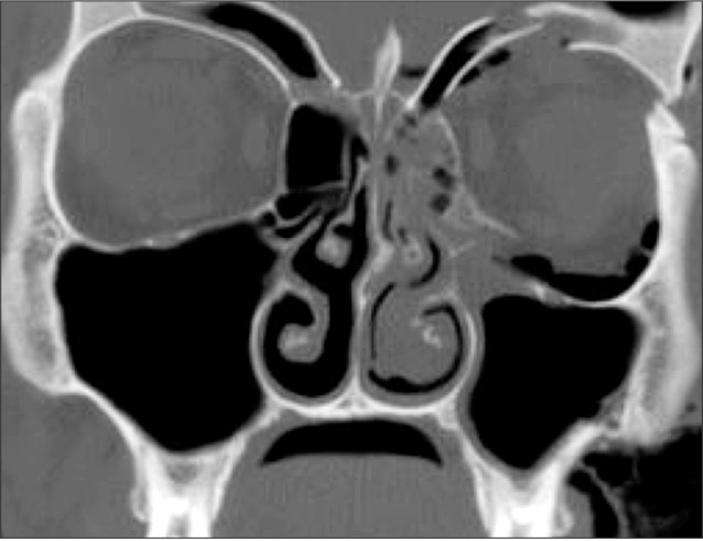
A 30-year-old male patient was admitted to the Hospital at Temuco, Chile, due to a high-energy trauma caused by a fall from a height of 4 m. During admission the patient's condition was assessed and a fracture of the left orbital complex was diagnosed, in addition to fractures of the orbital rim and floor, orbital sidewall, zygomaticomaxillary pillar and ipsilateral zygomatic arch.(Fig. 1) The patient was in intensive care for one week, at which point it was possible to provide surgical treatment. No medical history was present, including diabetes, Metabolic or facial syndromes or nutritional conditions and no smoking was reported.
The immediate preoperative stage showed severe left periorbital edema, ophthalmoplegia and diplopia in different fields. The surgical procedure was executed by a maxillofacial surgeon with 6 years experience in facial trauma; an upper approach was made for the left supraorbitaral rim, a left intraoral approach and a transconjunctival approach with lateral canthotomy. The procedure was performed conventionally and routinely, without complications. According to the proposed technique, internal rigid fixation was used with 1.5 plates, and no problem in screw insertion was present; at some stage during surgery, a forced retraction of the conjunctival flap was undertaken. The final suture to the conjunctiva was made using two stitches with a reverse knot and a 5-0 absorbable suture.
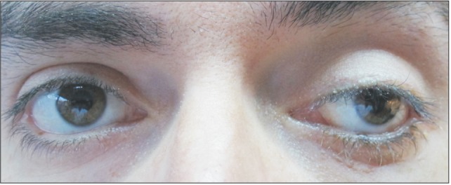
In the initial postoperative days, increased sclera exposure was observed, with no symptoms of pain or signs of enophthalmos or dystopia. On the fourth postoperative day an acute low-level conjunctivitis associated with chemosis and a 3 mm palpebral conjunctiva exposure was seen.(Fig. 2) The patient was discharged 1 week after surgery and during the hospital stay was kept on a course of endovenous corticoids (12 mg intravenous [IV] per day for 3 days, 24 mg IV per day during the last 4 days; Fresenius Kabi AG, Bad Homburg, Germany).
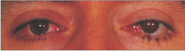
At the first out-patient check-up 10 days after the surgery, the patient presented with severe chemosis of the left eye with 5 mm of exposure (Fig. 3), slight ophthalmoplegia and ptosis of the upper eyelid, with no signs of pain or visual alterations. Ciprofloxacin ophthalmic solution with corticosteroids (drops, 2.5 mL per day; S.M.B. Farma, Santiago, Chile) was introduced as well as an eye patch to avoid major complications associated with lack of lubrication. Physiotherapy was also prescribed to improve mobility of the upper eyelid. The ophthalmologist's assessment reported positive visual acuity in all fields with functional impotence in the infraversion.
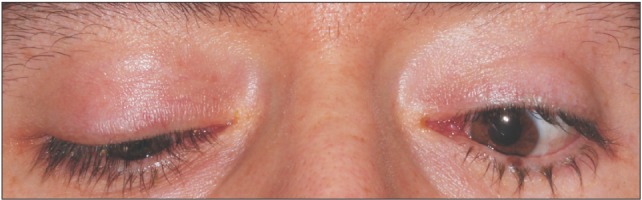
The condition evolved in a limited fashion over the following 8 days (18 days after surgery), at which point the chemosis began to subside. One month after the surgery (Fig. 4) the patient showed symptom resolution, with resolved chemosis and associated conjunctivitis, but persistence of post-traumatic strabismus, which was subsequently solved with surgical treatment to manage the extrinsic muscles of the eye involved in the anomalous position of the eyeball. After two years of follow-up, the patient had an adequate recovery from the facial trauma with a slight ptosis of the upper left eyelid, limited infraversion and no signs of alterations in visual acuity.(Fig. 5, 6)
The written informed consent for the publication of the photographs was obtained from the patient.
III. Discussion
Treatment of orbital fractures and fractures of the zygomatic orbital complex has been linked to different conditions, where variables such as the patient's age and gender as well as the presence of other facial fractures are combined5. In a series by Manganello-Souza and Rodrigues de Freitas6 reporting on 40 patients treated with the transconjunctival approach, only five presented with related complications such ectropion, corneal ulceration and ptosis of the lateral canthus. Another series with 56 patients reported only 4 patients with complications associated with entropion and trichiasis7. Baumann and Ewers' study3 of 99 patients reported 2% complications, describing a case with entropion and another case with tarsal laceration, which were both later resolved without major limitations. Malhotra et al.4 presented a series of 60 patients with 2 years of follow-up and 6 cases of complications, including chemosis. With a low incidence of complications and the advantages of the transconjunctival approach, the author's use of these techniques in 90% of the facial trauma and reconstruction cases resulted in no chemosis (almost 30 cases per year).
Chemosis does not appear among the usual complications of this approach. Causes for this complication may be related to extensive initial orbital edema which presented at the time of surgery, excessive traction in the conjunctival flap, inadequate dissection layer or lymphatic flow complications in the postoperative time. In this sense, chemosis defined as nonspecific swelling of the conjunctiva could be associated with bacterial or viral reactions, angioedema or trauma8. In cosmetic blepharoplasty techniques using the transconjunctival approach, these are frequent complications9, thus requiring establishment of protocols for their treatment. It is likely that the transconjunctival approach which is oriented to remove fat tissue produces better conditions for the development of chemosis and that the physiopathological mechanism will be similar to that observed in this case, since this is a rare complication in orbital traumatology. Other conditions such as carotid-cavernous fistula have also been responsible for proptosis, ophthalmoplegia and chemosis, indicating it is a response to the volume of fluids present in the orbit10.
Lymphatic flow is key for postoperative chemosis11. For instance, retrograde flow could result in the lymphatic channel filling with blood12 or may contribute to a rise in periocular or orbital pressure, resulting in venous engorgement and a back-flow of blood into the conjunctival lymphatic system13. On other hand, dilated arterioles increase the pressure gradient between arteriolar and venule capillaries and lead to the extravasation of intravascular fluid with resultant vasodilatory edema. In this case, the patient presented with a great deal of edema at admission, a non-controlled traction of the conjunctival flap and failure in drainage which could all be related to his chemosis.
A treatment protocol for this condition has been proposed based on topical and systemic corticosteroids. Oral steroids are an effective adjuvant treatment in younger patients14 and topical adrenaline could be an option15; surgical treatment that includes sutures has been used in other trauma cases16. The treatment strategies used in this case were in agreement with McCord et al.17 and Putterman18, and showed good results. Prevention of this complication can be achieved by respecting anatomical landmarks and gentle retraction of the orbital flap.
This case was resolved with minor functional alterations, although the consequences observed after 2 years (ptosis of the upper eyelid and limited infraversion) were not completely satisfactory, and may call into question the results obtained by the treatment provided during initial presentation and the surgical treatment of the orbital fracture.


 XML Download