

 PDF
PDF ePub
ePub Citation
 Print
Print


I. Introduction
We conducted a retrospective analysis and reviewed the temporomandibular joint (TMJ)-related papers published in a leading international journal, Journal of Oral and Maxillofacial Surgery, between January 2014 and December 2015. The review and analysis were conducted to ascertain the trends of articles being published and to reflect on the scientific features of TMJ that the journal is covering and providing its readers. In this period of two years, a total of 28 articles were published related to TMJ, of which 20 (71.4%) were full-length, 2 (7.1%) were technical notes or short communications, and 4 (14.3%) were letters to the editors. The number of full-length articles increased considerably compared to previous reviews of articles from 2011 to 2012 (16 full-length articles) and 2008 to 2009, showing that there has been a moderate rise in interest in improvement in the maxillofacial surgery field12.
II. Method
A retrospective observational study was conducted for the articles published in a leading international journal, Journal of Oral and Maxillofacial Surgery, between January 2014 and December 2015.
All of the articles were hand selected and were assessed and analyzed.
III. Results
A total of 28 articles related to TMJ were retrieved from the Journal for 2014 and 2015 for analysis and review.
The number of issues was the same for both of the years (10 each).
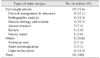
The majority of the study designs from full-length publications in both of the years were clinical management and outcomes related to TMJ (9 articles in 2014 and 2015), followed by articles on radiographic diagnosis and disease etiology and relation, with 4 and 3 articles, respectively.(Table 1)
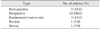
Most of the full-length articles were prospective studies, amounting to 60% of the articles, while the numbers of retrospective studies and randomized control trials (RCTs) were three.(Table 2) Other surveys and reviews accounted for approximately 10% of the articles. There was a considerable increase in the number of prospective studies compared to the review of 2011 to 2012, which contained 50% prospective studies.
IV. Full-Length Articles
1. Clinical management and outcomes
The TMJ has a very strategic location and prime function in the head and neck region. It is a very circuitous system with an assemblage of complex structures such as muscles, tendons, and bones. Generally, TMJ disorders fade and improve gradually with time, but serious difficulties demand proper interventions. We came across five articles dealing with the topic of management of TMJ disorders. Haq et al.3 tried a single stage instead of the usual two-stage approach for reconstruction of the joint following ankylosis in five patients. A virtual resection and reconstruction was carried out by a surgeon and an engineer via teleconference, and prosthetic implants were manufactured using computer aided design/computer aided manufacturing (CAD-CAM) technology in a period of six weeks, after which the surgeries were carried out. It was found that this approach is economical both in the sense of time and money. Jose et al.4 in their study of 62 gap arthroplasty cases in 35 patients over a period of six months demonstrate that using piezoelectric technology is superior to conventional drilling in terms of bleeding, postoperative complications, and amount of mouth opening, but is more time consuming. A one-year follow-up study by Zhou et al.5 of a modified prolotherapy of injecting lignocaine with 50% dextrose in the posterior pre-articular tissues, showed great results in 45 patients having non-neurogenic recurrent episodes of TMJ dislocation, suggesting no pain or difficulty associated with any nerve. Ninety-one percent of the patients had no recurrence, indicating that it is a simple, economical, and very effective line of treatment. In the 41 rehabilitated patients, 26 patients (63.4%) required a single injection, 11 patients (26.8%) had two treatments, and 4 patients (9.8%) needed a third injection. Abel et al.6 compared the conventional TMJ prosthesis to a customized condylar support prosthesis and found the latter to have a better physiologic form along with a better capacity to transfer load and reduce strain on the bone and screws. A retrospective comparison by Zhang et al.7 of the use of autogenous coronoid process grafts and costochondral grafts for the management of unilateral TMJ ankylosis patients yielded satisfactory results for both of the modalities on jaw deviation, mouth opening, and jaw movement parameters. Thus, they state that coronoid grafts can be a good treatment option.
The effectiveness of a management protocol is based on its outcome. It is very important to ascertain the potency of the treatment method, be it surgical or conservative, for its proper use and improved application. A total of four articles regarding the outcomes of different treatment techniques vis-à-vis TMJ were retrieved from these two years. Sipahi et al.8 carried out a placebo-controlled, double-blind study of 30 patients with symptomatic internal derangements of TMJ in order to ascertain the efficacy of intra-articular analgesics post arthrocentesis. Injections of 1 mL of 5% Ringer's lactate were given to one group, the second group received 1 mg morphine, and the third group was given 50 mg Tramadol intra-articularly immediately after the procedure. Pain relief was sustained for six months using morphine, while Tramadol, though equally effective, had a shorter duration. In a study on the outcome of joint replacement, Hussain et al.9 compared the outcomes of patients allergic to metals, using a titanium prosthesis in patients with a normal cobalt-chromium replacement. Based on mouth opening, diet, and disease and pain parameters, they found that there was no significant difference in the results of the groups. Based on similar parameters, Gruber et al.10 conducted an analysis of 84 patients and found that three and five years after TMJ replacement, patients in the United Kingdom had fewer complaints of pain and better functional morbidity, with very few patients showing complications such as weakness in the temporal branch of the facial nerve and prosthesis infections due to a localized head and neck infection. Enlightened by the holistic approach of multispecialty clinics, Ahmed et al.11 scrutinized the outcomes of patients visiting a multidisciplinary team for TMJ-related issues, consisting of specialists from the fields of maxillofacial surgery and prosthesis, oral medicine and psychiatry, and advocated that such types of clinics have a high impact on improving the overall quality of life of patients. They concluded that a multi-layer system and approach for dealing with a patient's complaint using all available resources should be inculcated into the system.
2. Radiographic analysis
In this modern era, technology plays a major role in all aspects of life. Technological advancements in the field of radiology and radiodiagnosis have made a huge impact and have a large role to play in planning, executing and determining the success of treatment regimens of different diseases. Four articles were discovered that comment on the diagnosis and treatment of TMJ with the aid of various radiological options. Li et al.12 executed a magnetic resonance imaging (MRI)-based study of 247 patients undergoing arthroscopic repositioning of anterior disc displacement. The patients were divided into two groups based on disc rupture, probable disc rupture and no rupture after repositioning. Analysis was conducted on the parameters of age, sex, symptom duration, mouth opening, unilateral or bilateral disc displacement, and Wilkes stage and concluded that teenagers and young adults have a high tendency for disc rupture. Furthermore, the Wilkes stage was more significant in the ruptured disc group compared to the other two groups. Describing the association between the anti-oxidant capacity of synovial fluid and intra-articular structures, Ishimaru et al.13 conducted diagnostic arthrograms, computed tomography (CT) scans, and chemiluminescence on the synovial fluid of 21 patients having TMJ disorders and measured them based on the constants of age, sex, and mouth opening visual analogue scale (VAS). Obtaining results for 11 patients with closed lock and 10 with no closed lock radiologically, they commented that there seems to be an association between synovial fluid oxidative stress and closed-lock in disorders of TMJ. Verifying the efficacy of the buccal fat pad in the gap arthroplasty procedure, Bansal et al.14 performed a volumetric analysis with ultrasonography at 15 days and 6 months following operation and found the shrinkage percentage of the fat pad to be 28%. With little or no heterotrophic calcification in the 6 month postoperative CT scan, it was deduced that the buccal fat pad is a viable, stable, and easily harvested option for management of TMJ ankylosis. The efficaciousness of cone-beam CT in the diagnosis and management of TMJ disorders was examined by de Boer et al.15 Patients were given a provisional diagnosis after obtaining their history, and following a clinical examination and orthopantomogram study. The patients then underwent CT scan, after which the degree of certainty was ascertained. In more than half of the cases of TMJ disorders, the diagnosis had to be changed following CT scans, indicating that they are an important component of proper identification of the disorder.
3. Disease etiology and relation
TMJ disorders very commonly are either associated with or a sequelae of some other condition. It is important to correctly diagnose the proper etiology of the disorder and to plan the treatment options, keeping in mind the postoperative complications and their impact on the TMJ. Xiang et al.16 investigated 492 cases of treated mandibular condylar fracture and found 16 cases of ankylosis recurrence during a follow-up period of 6 months to 10 years. Condylar head fractures are more likely to lead to ankylosis compared to condylar neck and subcondylar fractures. In addition, miniplates, wires, or removal of a fractured segment are more highly associated with ankylosis compared to fixation using long screws (bicortical screw). All of the postoperative ankylosed cases were associated with damaged articular discs. Matrix metalloproteins (MMPs) are supposedly a contributing factor to the loss of matrix and fibrocartilage inflammation of the TMJ. Taking this hypothesis into account and the fact that MMPs have increased activity in the serum of patients with polycystic ovary syndrome (PCOS), Soydan et al.17 tried to ascertain the prevalence of TMJ disorder in such patients. Comparing PCOS patients with a normal control group showed that, based on the symptoms of pain through VAS, the severity of TMJ disorders was greater in the former group, confirming the association between PCOS and TMJ disorder. Lund et al.18, by performing an extensive bacterial culture and serological tests on venous blood and synovial tissues from patients with reciprocal clicking and chronic closed lock, tried to determine the relation of reactive arthritis and internal derangements of TMJ. With no significant difference observed between the number of bacterial antibodies and the absence of antibodies for Chlamydia trachomatis and Campylobacter jejuni, it was confirmed that reactive arthritis does not play a role in the internal derangement of TMJ.
4. Animal research
A lot of research and experiments have been carried out on animals to determine the efficacy, limitations, and feasibility of application to humans. In a similar approach for visualizing the role of psychological stress on the TMJ, Huang et al.19 divided rats into three groups of 12, based on the duration of stress—3 weeks, 6 weeks, and more than 6 weeks. They compared them to a fourth control group. Based on the presence of MMP-3, a tissue inhibitor of metalloproteinase 3 (TIMP-3), thinning of articular cartilage, shedding of collagen fibers, cracks in articular discs, and other structural changes in the stained tissues of TMJ, it was concluded that psychological stress does have an impact on rats; however, further research is required for confirmation. On the other hand, to evaluate the effect of diazepam on TMJ, Figueroba et al.20, using iOVD, saline, and diazepam, studied approximately 40 male rats. A thorough examination was conducted on the thickness of the articular cartilage, articular area, and number of collagen fibers from the bilateral TMJs of the rats. Pro-inflammatory cytokines and tumor necrosis factor were also measured, and it was observed and confirmed that both iOVD and diazepam induced significant changes in the articular cartilages.
5. Review articles and survey
A single review article was published in these two years and reviewed all of the TMJ-related papers published between 2011 and 2012. It was written by Tahim et al.1 and found a total of 24 articles published in the given time frame.
A report on baseline data was published by Idle et al.21 It was prepared to establish the long-term collection of data on TMJ replacement from all centers in the United Kingdom. Data was collected and analyzed through Snap Survey, which accounted for 402 patients who had undergone replacements between 2011–2012. The main reasons for replacement were osteoarthritis, failed operation, ankylosis, and seronegative arthritis.
V. Other Articles
1. Short communications, technical notes, and letters to the editor
Two short communications were published during this period. Mustafa and Sidebottom22 assessed the risk of intraoperative dislocation of TMJ replacement as well as its management. Amongst 138 cases, 8% were identified as having a risk of intraoperative dislocation, which is associated primarily with coronoidectomy (30%) and inflammatory arthropathy (24%) and is mainly managed with intermaxillary elastics. Sidebottom and Mistry23 determined the incidences of metal allergies in prospective patients of total TMJ replacements and the efficacy of patch tests. It was observed that 39% of 101 patients had an allergy to one or more metals and were thus given a titanium prosthesis. No signs of allergy were seen six months following the operation; thus, it is advocated that all patients who are to undergo total joint replacement should be given patch tests to determine the need for a titanium prosthesis.
Two technical notes were published. Machon et al.24 suggested the use of mini instruments made from pre-tensioned stainless steel wire during visually guided arthroscopy of the TMJ. These instruments help with irrigation and permit the grading of chondromalacia and removal of adhesions. Ryba et al.25 wrote about the use of Arthrex Corkscrew for TMJ meniscopexy. In the publication, the strength, efficacy, and ease of use of the Arthrex system are discussed and compared to the Mitek system.
Four letters to the editor were published from 2014 to 2015. Kain et al.26 described the misdiagnosis of TMJ synostosis as ankylosis and it being treated as such. Bonte27 tried to cover the omitted part of the article by de Boer et al.15, in which the analysis of the axial view of the CT scan was not carried out. The other two letters were responses to previously published articles262728.
VI. Discussion
Table 2 indicates that there was a predominance of prospective studies. Experimental research contributed to a very minor portion of the publications. Hence, there seems to be a requirement to diversify the resources and workflow as well as to test the strength of imagination with the appropriate knowledge and cutting-edge technology of the field to revolutionize the work culture.
Though the number of full-length articles has increased compared to previous years, it is still low compared to other areas of the field such as oncology and salivary gland diseases. We need to give more attention to this field of specialty and focus on proper channeling of the knowledge and resources for expansion.
 XML Download