

 PDF
PDF ePub
ePub Citation
 Print
Print


I. Introduction
Although much controversy surrounds the treatment of intracapsular condylar fracture in adults, open reduction is considered superior to closed reduction in regard to restoration of vertical dimensions, condylar mobility, and articular disc positions1. In addition, open reduction and internal fixation have been reported to result in less crepitus, lateral deviation, occlusal disturbances, and disparity in ramus height than closed reduction2. However, even if open reduction is successful, destruction of the disc-ligament system can result in irregular reduction in the range of mandibular movement, with a prevalence of up to 40%2.
Conventional closed reduction for the treatment of intracapsular condyle fracture involves induction to maximum intercuspation, followed by maxillomandibular fixation (MMF) for a set period3. However, premature occlusal contact and deviation on the affected side during mouth opening can occur with such treatment. In cases of bilateral comminuted condylar fracture, patients might experience limited mouth opening (≤35 mm) and discomfort associated with a limited protrusive movement4567.
To resolve these problems associated with conventional closed reduction, we designed a novel method for closed reduction through protrusive MMF.
II. Cases Report
We established inclusion criteria. Among the cases of intracapsular fractures according to the Loukota classification8, the patients who were not contraindicated9 for MMF with comminuted fracture in which the fracture segment was displaced medially.
According to our criteria, two adult male patients were included in this study. A 37-year-old man was diagnosed with symphysis and bilateral intracapsular condylar fracture after a traffic accident. Also, a 42-year-old man injured in a fall was diagnosed with symphysis and right intracapsular condylar comminuted fracture with displacement.
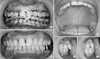
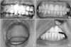
An Erich arch bar was applied under general anesthesia, and open reduction and internal fixation were performed for the symphysis fracture in both patients. After confirming proper occlusion, the mandible was protruded forward to form an edge-to-edge bite or crossbite, and MMF was performed using wires in the Class II direction in order to prevent setback after confirming disclusion of the posterior teeth.(Fig. 1, 2) All procedures were performed by the authors.
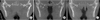
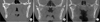
The patients were discharged four days after surgery with no notable events, and the protrusive MMF was removed two weeks after surgery. The patients were recommended to avoid mastication until one month after surgery and to open their mouths only to perform mouth opening exercise. The posterior teeth did not achieve a maximal intercuspal position spontaneously on the day of MMF removal, but stable posterior occlusion was observed three weeks after surgery and maintained at the six-month follow-up visit.(Fig. 1, 2) Mouth opening was ≤10 mm on the day of MMF removal but increased with time. Panoramic and computed tomography images were obtained at the six-month follow-up, and all data were analyzed by the authors. The mandibular condyle maintained its shape, which was close to normal in appearance, while exhibiting slight narrowing of the articular space.(Fig. 3, 4) Posterior ramus height (distance from the condylion to gonion) at pre-operation and postoperation six-month follow-up was measured with OnDemand 3D Application (Cybermed Inc., Seoul, Korea). The measurements of posterior ramus height are given in Table 1. Clinically, occlusion was favorable without premature contact of the posterior teeth, and mandibular movement was not limited including lateral excursion and protrusion without deviation in both patients. The measurements of mandibular movement are given in Table 2.
Due to the retrospective nature of this study, it was granted a written exemption for patient consent by the Institutional Review Board of Wonkwang University Dental Hospital (WKDIRB201607-02).
III. Discussion
Clinical limitations following closed reduction of an intracapsular condylar fracture include a decrease in maximum mouth opening, reduced mandibular range of motion including anterior guidance, and reduced occlusal stability. These problems are thought to be due to structural changes in the osseo-discoligamentous complex510. When an intracapsular condylar fracture occurs, the medial condyle fragment undergoes anteromedial and inferior displacement due to traction by the lateral pterygoid muscle, which creates a gap between the medial and lateral condylar stumps4101112. Such traction can induce bone overgrowth due to distraction osteogenesis between the medial and lateral condylar stumps, causing structural changes in the condyle412.
When MMF is performed in the maximum intercuspal position, persistent interdental contact causes masticatory muscle hyperactivity, even after the MMF is removed, leading to a decrease in the vertical dimension and premature posterior teeth contact1113. However, in growing patients, limitation in mandibular movement or decreased ramus height on the affected side rarely occurs as a result of a comminuted fracture14. One reason could be that the volume and activity level of the masticatory muscles become excessive with aging15. Injection of botulinum toxin has been reported to be useful in decreasing masticatory muscle hyperactivity in condylar fracture patients, but the attempts at treatment had been limited to cases of subcondylar and condylar neck fractures16.
Accordingly, we performed protrusive MMF in cases involving anteromedial and inferior displacement of the medial condyle fragment in intracapsular condylar fractures. This novel method has two main advantages. First, in the protrusive position, the lateral condylar fragment can be deviated in the anteromedial and inferior directions closer to the medial fragment, minimizing bone formation between the two fragments and structural changes4. Second, in the protrusive position, posterior disclusion is achieved, preventing masticatory muscle hyperactivity and subsequent gradual decrease in ramus height111317.
Compared to conventional MMF, change in the protrusive position can cause functional and aesthetic discomfort to the patient; thus, it is necessary to explain this possibility prior to surgery or protrusive MMF. The protrusive MMF is maintained for two weeks to allow formation of soft calluses 2 to 3 weeks after the fracture and that are thought to possess enough strength to partially resist a decrease in ramus height caused by increased masticatory muscle activity after MMF removal18. We have experienced cases of premature posterior contact from a decrease in ramus height after MMF removal in patients who underwent protrusive MMF for only one week; therefore, the duration of protrusive MMF was changed to two weeks. Posterior occlusion revealed a state of disclusion immediately after MMF removal, but as the activity of the masticatory muscles increased, the posterior teeth returned to the proper occlusion in both patients. The possibility of tooth extrusion and concomitant malocclusion during protrusive disclusion should be considered; however, we think that a two-week period is too short to produce tooth extrusion.
The results suggest that protrusive MMF can be a useful alternative to reduce the occurrence of posttreatment complications in patients with intracapsular condylar fracture.


 XML Download