

 PDF
PDF ePub
ePub Citation
 Print
Print


I. Introduction
Square faces due to prominent mandibular angles of varying degrees are often treated with plastic surgery in modern societies where oval faces with symmetrical and small overall facial outlines are considered esthetically pleasing.
Since brachycephalic or mesocephalic faces are typical of Asians, whereas dolichocephalic faces are typical of Caucasians, reduction of mandibular angles and zygomas resulting in oval face contours is widespread among Asians1.
Although factors leading to a prominent mandibular angle can be subdivided into 2 groups, i.e., congenital and developmental (e.g., bruxism), no exact cause has been identified so far, despite several studies2.
Numerous studies have been conducted on the cause, since the terms, masseter muscle hypertrophy or benign masseteric hypertrophy, were coined for a prominent unilateral or bilateral mandibular angle34, and improvement in surgical techniques have been made. Surgical corrections have been carried out since the late 1980s5. Techniques for the reduction of a mandibular angle are classified as masseter muscle reduction and mandibular bone reduction; the latter is preferred to the former by surgeons due to its many advantages56. While curved ostectomy of the mandibular angle is by far the most widely used technique, lateral cortical ostectomy of the mandibular angle and ramus is recently gaining popularity7. Indications, pros and cons as well as various procedures for both techniques have been described in detail.
In spite of numerous reports on the surgical technique for mandibular angle reduction, only a few studies have been conducted to evaluate patient postoperative satisfaction and esthetic improvements. Since there is a lack of established standards for the esthetic mandibular angle, the patient's request is the primary determinant in the operative plan. In other words, postoperative satisfaction and the demands of each patient are the most critical criteria in mandibular angle reduction. However, unclear understanding between the surgeon and the patients' esthetic demands may bring about unexpected outcomes. Moreover, a patient's misperception of self may lead to disappointment with the result of the treatment. Lack of standardized preoperative and postoperative assessment methods may result in such situations. Thus, an objective and systematic manner of evaluation and diagnosis is required.
Therefore, this study was conducted to test Han's ratio as an objective and quantitative method obtained from pre- and postoperative data in patients requesting mandibular angle reduction. Considering that the outline of oval shaped mandibles resembles an inverted triangle, favorable soft tissue landmarks were determined and quantified in order to compare the amount of reduction in patients who have undergone mandibular reduction surgery8.
II. Materials and Methods
1. Patients
Thirty patients, including 12 men and 18 women were selected from 175 patients who came to the Department of Oral and Maxillofacial Surgery, Dankook University Dental Hospital (Cheonan, Korea) from January 2007 to March 2012 with chief complaints of skeletal mandibular prognathism and prominent mandibular angle. Subjects were carefully chosen with the following criteria: (1) patients who had undergone both orthodontic and orthognathic surgery and (2) patients capable of 1 month presurgical and 3 to 6 months postsurgical follow-up. Due to the retrospective nature of this study, it was granted written exemption by the Dankook University Dental Hospital's Institutional Review Board. The written informed consent was obtained from all patients.
Mandibular angle reduction along with bilateral sagittal split ramus osteotomy with or without genioplasty in the mandible and advancement of the maxilla with or without Le Fort I osteotomy was conducted. Subjects were classified into 3 groups according to the types of surgical procedures involved. Group A consisted of 9 patients who had mandibular angle reduction and mandibular setback was achieved only with mandibular angle resection and bilateral sagittal split osteotomy; Group B consisted of 12 patients with mandibular angle resection, bilateral sagittal split ramus osteotomy and genioplasty; and Group C consisted of 9 patients with mandibular angle resection, bilateral sagittal split ramus osteotomy, Le Fort I osteotomy and genioplasty.
Moreover, in order to establish an unbiased standard, 82 Korean females who ranked high in beauty pageants from 2002 to 2011 were chosen as the control group.
2. Surgical technique
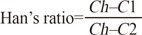
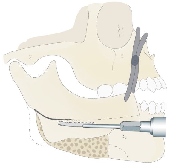
Curved ostectomy or lateral cortical ostectomy was performed for mandibular angle resection. Curved ostectomy involved bilateral sagittal splitting ramus osteotomy in which the proximal section was anteriorly displaced, followed by a gently curved ostectomy with a reciprocation saw starting from the posterior border of the mandible to the lower border of the mandibular body. At this point, the modified method for mandibular angle reduction was done. The line of resection was extended below the mental foramen for a shallower look in the lower lateral face.(Fig. 1, 2) Sharp resection margins were subsequently rounded with a round bur5. For lateral cortical ostectomy, the mandible was cut transversely at the mandibular occlusal level with the reciprocating saw and the cut extended antero-inferiorly along the lateral cortical bone to reach the first molar and at that point the mandible was vertically split. Segments were separated with a chisel and sharp margins were also smoothed with the round bur.
3. Analysis method for frontal facial photographs
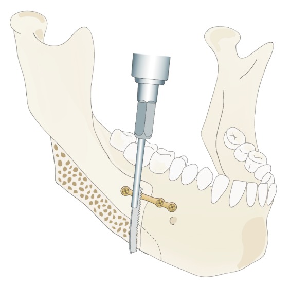
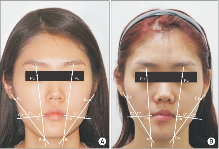
Frontal facial photos of patients were taken within 1 week before and 3 to 6 months after surgical correction. Subjects were instructed to stay still with their Frankfort horizontal plane (FH plane) parallel to the ground with both lips relaxed and closed naturally in centric occlusion. Then, photos were taken in a usual manner and saved in JPEG format. Utilizing the Adobe Photoshop ver. 14.0 program (Adobe Systems, Mountain View, CA, USA), the distance between each selected landmark was measured and ratios were obtained. The amount of discrepancy was assessed by cross-comparison of the ratios representative of each phase. As shown in Fig. 3 and 4, landmarks and measurements used in the present study were as follows.
-
1) Landmarks and dimensions
(1) Pu: midpoint of pupil
(2) Ch: most lateral point between upper and lower lip
(3) Bo: mandibular outline
(4) Ea: intersection of outlines of face and earlobe
(5) C1: intersection of a line drawn from Ch perpendicular to Po-Bo and a line between Ba-Bo
(6) C2: intersection of a line drawn from Ch perpendicular to Po-Bo and mandibular outline
-
2) Linear measurements
4. Statistical analysis
Preoperative and postoperative means and standard deviations were obtained from each patient group and the Wilcoxon signed rank test was performed to verify significance. In addition, the Mann-Whitney test at a significance level of 5% was conducted to verify significant differences between groups. PASW Statistics ver. 18.0 (IBM Co., Armonk, NY, USA) was used. Average values and standard deviations were calculated for beauty pageant winners.
III. Results
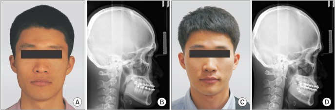
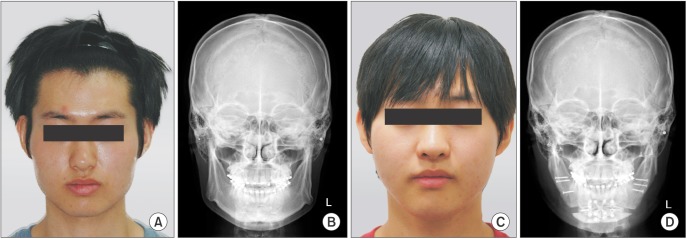
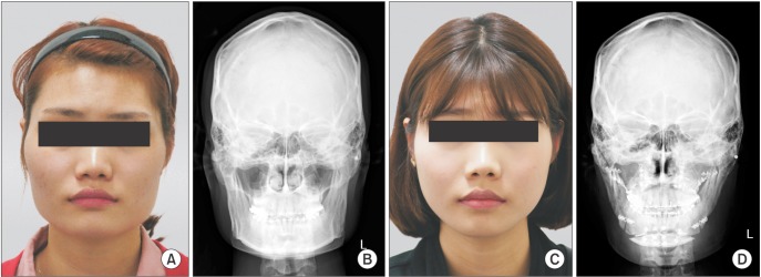
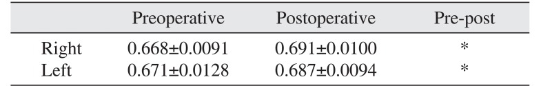
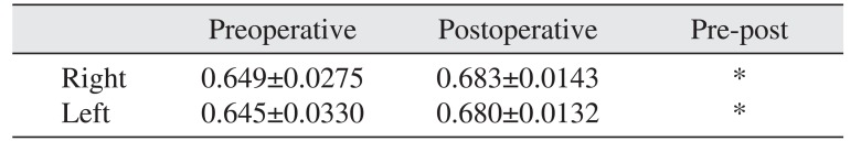
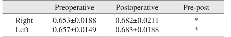
Postoperative Han's ratio was significantly increased compared to the preoperative Han's ratio in groups, A, B, and C.(Tables 1, 2, 3) The magnitude of the increase in the Han's ratio was not statistically different between groups (P>0.05). Representative cases of frontal facial photos and cephalometric radiographs of each group are shown in Fig. 5, 6, 7.
The average and standard deviation of Han's ratio acquired from 82 Korean beauty pageant winners were 0.688±0.06 on the right and 0.696±0.05 on the left.
IV. Discussion
A recent worldwide trend in maxillofacial reconstruction focuses on cosmetic surgeries primarily aimed at esthetic enhancement of individuals, unlike the past when the mainstream of treatment was reconstruction of facial deformities induced by malignant cancer or trauma. This is not a transient phenomenon but rather a transition owing to the demands of contemporary esthetically-minded patients. In fact, the range of surgery has been gradually broadened to meet the changing needs of patients more specifically, such as jaw and facial contouring surgery, predominantly including the correction of mandibular prognathism.
A face with a prominent mandibular angle makes a strong and masculine impression on others, so it is not preferred among Asians, including Koreans. Moreover, for genetic and racial reasons, Asians tend to have a wider lower face than Caucasians and as a result, mandibular angle reduction is more common in the East than in the West9.
The average bigonial distance of Korean women is 117.8 to 125.25 mm whereas that of Caucasian women is 105 to 109 mm10. Thus, a prominent mandibular angle is not a pathologic phenomenon but instead a problem that merely requires an esthetic manner of approach. Standards of beauty have historically been established respectively in western and eastern society. However, since modern times, the spread of the western centered culture worldwide has resulted in western standards of beauty dominating the East as well. When faces of 72 Korean-American women were scored by 10 other Korean-Americans using a visual analogue scale (VAS) of appearance, the highest rated were more similar to Western faces than that of the average Korean11. In the same sense, Asians with wide a interangular dimension consider narrower bigonal distance, typical of Caucasians, charming and desire mandibular angle reduction.
Baek et al.5 who first suggested the modern concept of mandibular angle reduction, concluded that a prominent mandibular angle is a relatively common esthetic problem and the cause lies in inferoposterior projection of the angular portion of the mandible rather than muscle hypertrophy. In addition, Legg12 reported that the prominent angles are attributed to racial differences, with hypertrophy of the masseter muscle predominant in Caucasians and bony projection in Asians.
Improvement in surgical techniques and research of causes have continued since the introduction of terms including prominent unilateral of bilateral angle, masseter muscle hypertrophy or benign masseteric hypertrophy.
Surgical correction by an extraoral approach was initially performed by Gurney13 with excision of the lateral portion of the hypertrophic masticatory muscle, followed by Adams14 with skin incision and finally the first intraoral approach by Converse15. Popular current techniques include curved ostectomy and lateral cortical ostectomy, originally introduced by Baek et al.516 in 1989 and 1994, respectively.
In spite of many reports on surgical techniques for mandibular angle reduction, studies on preoperative diagnosis and postoperative assessment are relatively scarce.
The gonial angle is the most critical part in the profile, especially in curved ostectomy, since patients with an initially large gonial angle may show an even larger angle and thus an abnormal and unnatural contour after the procedure7. The mean mandibular angle in Koreans is 128.71°±3.87° and the mandibular plane-sella nasion (MP-SN) is 32.69°±6.11°1718. Meanwhile, Jin7 insisted that mandibular ramus to body length ratio should also be considered important for profile analysis. When the length of the body is longer than that of the ramus, only a slight increase in mandibular angle brings about a very awkward appearance. Thus, enlargement of the mandibular angle is not favorable in either the case of mandibular prognathism with a long ramus length or mandibular recession with a short body length.
Since Baek et al.16 pointed out that the frontal as well as the profile view are crucial for the diagnosis and assessment of mandibular angle, diagnosis drawn from both sides has become a general procedure19.
Han and Kim9 measured bitemporal, bizygomatic and bigonial distance and calculated their ratios. He reported that ideally the bitemporal and bigonial distance should be equal and narrower than the bizygomatic distance by 10%. However, this is merely a concept of linear measurement and carries some disadvantages. Ambiguous criteria for nonbiased assessment of esthetic enhancement and an inability to evaluate facial asymmetry are examples of shortcomings in the evaluation of treatment results.
Barnett and Whitaker20 proposed a three-dimensional (3D) method for midface and lower face evaluation. Six landmarks were chosen and connected with lines and eventually constituted planes. However, this method is quite complicated and difficult to use in the clinical setting. Recently, computer-aided surgical simulation improved presurgical work-up efficiency. It also provided an opportunity to illustrate multidimensional correction at the skeletal and soft tissue levels. CT scanning images are needed for this, but due to economical and ethical issues, CT scanning images are not used every time. Instead, two-dimensional films are commonly used for post-orthognathic surgery follow-up visits. In clinical practice, there are limits to the 3D analysis of patient faces before and after operations. Therefore, in this study we suggested a simple and fast method to quantitatively analyze the differences in the pre- and postoperative mandibular angles using frontal facial photographs only.
The evaluation of facial symmetry should be carried out on the facial soft tissue of actual patients and not radiographs. Radiographic shape of bones cannot reflect real facial structure because of variation in the thickness of the masseter muscle. Moreover, it should be noted that a shorter than average bigonial distance may make a face look more beautiful7.
As to the assessment of patient satisfaction level after treatment, only a few studies have adopted objective methods such as the VAS to estimate patient aesthetic satisfaction. Choi et al.21 conducted a survey with 20 questions and reported that 97% of the patients who underwent mandibular reduction surgery were satisfied with the results.
In most studies, esthetic satisfaction was assessed in a subjective manner, for example satisfactory or unsatisfactory. The results revealed that a considerable number of patients were satisfied with the esthetic outcome of their operation. However, such studies may have been biased, since most were retrospective studies on the patient group and no established criteria for radiographic or clinical success existed.
In the present study, Han's ratio was obtained and compared to evaluate postoperative esthetic enhancement in exact numerical values, rather than patient subjective satisfaction level. A Han's ratio of approximately 1.0 indicates a more oval shaped lower face line. Although the standard of beauty is obviously a subjective notion, 82 winners of Korean beauty pageants from 2002 to 2011 can be presumed to represent the recent standard of beauty in Korea. Han's ratio was measured in their frontal facial photos and the mean values were 0.688 on the right and 0.696 on the left.
All 3 groups, i.e., Group A (mandibular angle reduction and mandibular setback osteotomy), Group B (mandibular angle resection, bilateral sagittal split ramus osteotomy and genioplasty), and Group C (mandibular angle resection, bilateral sagittal split ramus osteotomy, Le Fort I osteotomy and genioplasty), demonstrated an increase in Han's ratio. This data supported Han and Kim9 who reported a postoperative decrease of bigonial distance by an average of 12.3 mm in 12 patients after mandibular angle ostectomy, as compared to preoperative data.
There were no significant differences in preoperative and postoperative Han's ratio for each group i.e., control, Group A, and the experimental, Group B or Group C. Thus, we assumed that using a frontal view for preoperatively planned anteroposterior movement of the maxilla and the chin played little role in postoperative esthetic improvement resulting from a reduced mandibular angle.
Han's ratio proposed in the present study can be easily calculated by placing landmarks on frontal facial images. Moreover, it can be obtained separately on the right and left to analyze the degree of correction of facial asymmetry between preoperative diagnosis and postoperative evaluation. Meanwhile, since only frontal evaluations are possible with this method, supplementary lateral assessments of angle are required.
Whitaker17 pointed out that applying a plain and consistent method in every case is incorrect because of the ambiguity inherent in the evaluation of facial esthetics. In fact, enhancement of Han's ratio does not necessarily lead to esthetic improvement. In addition, overall facial balance and harmony with the nose, eyes and adjacent structures must be taken into consideration.
 XML Download