

 PDF
PDF ePub
ePub Citation
 Print
Print


I. Introduction
Imatinib mesylate is a member of a new class of chemotherapic agents that inhibit tyrosine kinase, a protein which belongs to a family of ubiquitous enzymes having a strategic role in signal transduction pathways and which influence gene transcription and/or DNA synthesis. Cell studies demonstrate that imatinib specifically inhibits proliferation of myeloid cell lines that express the BCR-ABL fusion proteins associated with chronic myeloid leukemia (CML). So, imatinib mesylate is used to treat CML, acute lymphoblastic leukemia and gastrointestinal stromal tumors (GISTs)1.
Osteonecrosis of the jaw (ONJ), commonly described as an adverse effect of the use of bisphosphonates, is the progressive destruction and death of bone that affects the mandible or maxilla of patients exposed to treatment with nitrogen-containing bisphosphonates, in the absence of previous radiation treatment2.
A few cases of ONJ associated with tyrosine kinase inhibitors (sunitinib, imatinib) have been reported in the literature and usually ONJ occurred in patients simultaneously treated with bisphosphonates. There are a lot of common side-effects of imatinib (nausea, vomiting, weakness, muscle cramps, edema especially periorbital and of the ankles, diarrhoea, exanthema, hypo-hyperpigmentation of the palate or oral mucosa) but no case of ONJ related to imatinib has been previously reported.
II. Case Report
A 72-year-old Caucasian male came to the Emergency Unit of Siena University Hospital (Siena, Italy) complaining of submandibular and right laterocervical pain with onset several days earlier. Medical history revealed that the patient had CD117-positive GISTs with a c-Kit genetic mutation since 2012. Since January 2013, he had been on imatinib at doses of 400 mg/day for 3 months followed by 600 mg/day for 4 months and then 800 mg/day. He had never taken bisphosphonates or undergone radiotherapy in the head and neck region. Moreover, he was not taking any other medication.
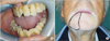
Examination, initially conducted by an ENT (ear-nose-throat) specialist, showed slight swelling at the right mandibular angle, multiple laterocervical and right submandibular lymphadenopathies, and hot reddened skin without signs of fistulas. Oral examination showed exposed bone in the right retromolar triangle, halitosis and sialorrhea.(Fig. 1. A) Rhino-fibrolaryngoscopic examination did not detect irregularities or pathological processes in the pharyngeal and laryngeal regions. The ENT specialist referred the patient for dental examination.
Medical history included surgical removal of the distal root of the first lower molar 10 years earlier. On April 2014 the patient went to his dentist complaining of lower right quadrant toothache. The dentist confirmed the finding of the ENT specialist and also found mobility of the mandibular right first molar and anaesthesia/hypoesthesia of the right half of the lower lip, suggesting homolateral mandibular nerve compression.(Fig. 1. B) The oral mucosa was normal. Since the patient did not recall exactly what dental work had been done, he consented to our contacting his dentist, who confirmed having performed a dental X-ray and tooth extraction (#47) because the tooth had fractured vertically and could not be saved.(Fig. 2. A) The extraction was performed under block anaesthesia with articaine 1:100.000 (1.8 mL) and suture hemostatic control. The patient was prescribed 1 g amoxicillin and clavulanic acid every 12 hours for 6 days. When the stitches were removed on day 7, the wound appeared to have healed. Five weeks after the extraction, the patient had pain in the same region and halitosis but did not seek medical advice, preferring to take nonsteroidal anti-inflammatory drugs and the antibiotic again (1 g amoxicillin and clavulanic acid, every 12 hours). Since the pain did not resolve, a week later he presented at the emergency unit where ENT examination was carried out. The specialist ordered an X-ray of the dental arches (Fig. 2. B) which showed sequestration of the right mandibular bone involving the retromolar triangle. An oral swab was taken and after disinfecting the oral cavity with 0.2% chlorexidine with anti-discoloration system, a bone fragment measuring 2.5×1.5 cm was removed together with underlying gingival tissue. The bone was sent to the pathology lab for examination. Cone-beam computed tomography (CBCT) was requested. The patient was prescribed antibiotics (3 g/day amoxicillin and clavulanic acid and 500 mg/day levofloxacin) and discharged.
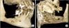
The pathology results available 72 hours later indicated positivity for Staphylococcus aureus, Candida albicans, Escherichia coli, and Enterococcus faecalis. Since the antibiogram showed sensitivity to levofloxacin, the patient continued the therapy already prescribed, to which fluconazole was added for C. albicans. CBCT showed a large area of osteonecrosis of the right hemimandibular body and angle with erosion of the vestibular cortex and complete destruction of the lingual cortex involving the mylohyoid line and the mandibular canal.(Fig. 3) Microscope examination of the tissues confirmed the clinical diagnosis of diffuse osteonecrosis and absence of neoplastic cells, therefore secondary localisations from GIST were excluded.(Fig. 4) Employing the Naranjo adverse drug reaction probability scale to determine the association of imatinib with osteonecrosis, the score revealed a probable adverse drug reaction.
III. Discussion
Imatinib is used to treat adults with GISTs that cannot be removed with surgery or have spread to other parts of the body, and adults who are at risk of GISTs coming back after surgical removal. The dose depends on the disease being treated, the age and condition of the patient, and the response to treatment, but it should not exceed 800 mg a day. The patient had been treated only with imatinib for 22 months at doses of 400 mg/day for 3 months followed by 600 mg/day for 4 months and then 800 mg/day and never with bisphosphonates or radiotherapy. In line with indications in the literature34, the oncologist did not suspend imatinib therapy because the GISTs were metastases. Clinical course after the tooth extraction included apparent healing in the first weeks followed by rapid local worsening leading to bone sequestration with pain, swelling, halitosis and partial hypoesthesia of the lower lip. This evolution largely resembles the clinical course of osteonecrosis due to bisphosphonates2. Although the patient had never been treated with bisphosphonates or radiotherapy of the head or neck, his clinical picture was identical to osteonecrosis stage 2 subclass b due to bisphosphonates. Therapy with imatinib and the concomitant dental extraction seem likely causes of the resulting osteonecrosis.
Only one case of osteonecrosis involving fracture of the tibia5 has been reported in a patient on imatinib. Various cases of osteonecrosis of the mandible have been reported in patients taking imatinib and bisphosphonates6. In our case, the patient was taking only imatinib.
Other tyrosine-kinase inhibitors, usually sunitinib, have been associated with an increased risk for ONJ in patients receiving concomitant treatment with intravenous bisphosphonates7. Cases of ONJ have also been reported in patients with renal cell carcinoma treated with sunitinib monotherapy8. Sunitinib is a vascular endothelial growth factor (VEGF) receptor 1-3 antagonist: inhibition of VEGF-dependent angiogenesis may play a major role in the onset of sunitinib-associated ONJ9. The present case of ONJ, however, cannot be due solely to a similar mechanism, since imatinib does not block VEGF receptors, although it could partly reduce VEGF expression through inhibition of the platelet-derived growth factor receptor (PDGFR) or c-Kit1011.
Some evidence suggests that imatinib can directly target skeletal cells, leading to dysregulation of bone remodelling12. Inhibition of c-Fms and c-Kit signaling by imatinib may decrease osteoclast number and activity. Imatinib also inhibits the proton-generating activity of carbonic anhydrase II, preventing dissolution of minerals from bone and hence inhibiting bone resorption. Furthermore, inhibition of PDGFR signalling on osteoblasts can reduce the production of osteoclastogenic cytokines, including macrophage-colony stimulating factor (M-CSF) and receptor activator of nuclear factor-κB ligand (RANKL), and inhibit osteoblastic cell proliferation.
A few cases of ONJ associated with tyrosine kinase inhibitors have been reported in the literature and they always occurred in patients simultaneously treated with bisphosphonates.
Considering the clinical and pharmacological history of our patient, ONJ could have been caused by imatinib treatment. Finally, it would be a good practice to avoid tooth extraction during imatinib therapy.


 XML Download