

 PDF
PDF ePub
ePub Citation
 Print
Print


I. Introduction
Human bite injuries to the face are extremely rare occurrences, with only a handful of cases reported to date12. The eyelids in particular are complex structures, and repair of eyelid injuries can be extremely difficult, with the risk of ptosis when the levator muscle is involved or of unsatisfactory surgical outcome due to improper technique34. Although human bite injury to the eyelid has been reported in several cases, the simultaneous involvement of both eyelids is an extremely rare occurrence; to the best of our knowledge, no cases have been reported. In most reported cases on human bites to the eyelids, the avulsed tissue was viable and available for reconstruction2.
Herein, we report a successful reconstruction of an extremely rare case of human bite injury with involvement of both the upper and lower eyelids of the right eye where the avulsed tissue was not available for reconstruction.
Go to : 
II. Case Report
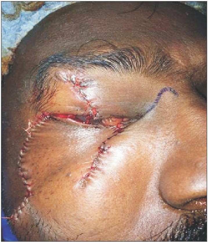
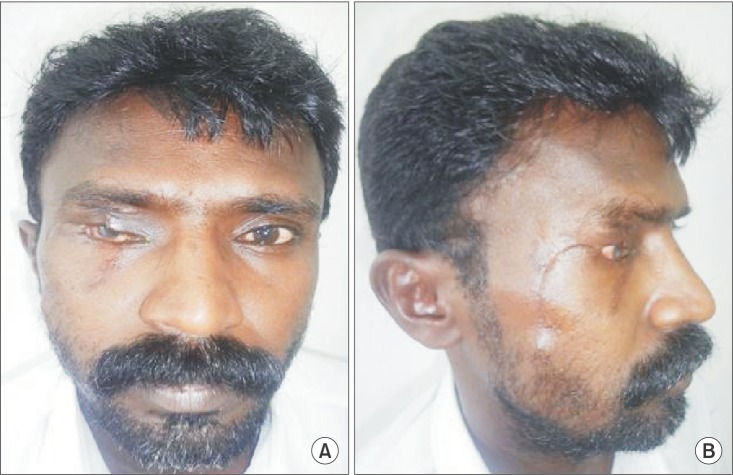
A 43-year-old male patient was referred to our unit 5 hours after he sustained human bite injury to the right eye with eyelid involvement. On clinical examination, approximately 60% full thickness loss of the upper eyelid up to the orbital septum was observed, extending from the lateral canthus to approximately 1 cm from the medial canthus. The defect extended approximately 12 mm vertically from the lid margin, and 80% to 90% full thickness loss of the lower eyelid was observed, extending from the lid margin up to the infraorbital margin.(Fig. 1) Computed tomography of the right orbit revealed no bony injuries. Ophthalmic examination of the right eye showed injury to the underlying globe, but the ocular motility and anterior and posterior segment examinations were within normal limits. The wound was thoroughly irrigated with normal saline, and tetanus toxoid was administered to the patient because he was unaware of his immunization status. Broad spectrum parenteral antibiotics were administered by injection (ceftriaxone 1 g and metronidazole 500 mg) preoperatively and continued for 7 days postoperatively. Since the avulsed tissue was not available at the time of presentation, we decided to reconstruct the defect immediately using a local cheek flap. After hematological investigations and obtaining informed written consent, the patient was scheduled for surgery under general anesthesia. Following standard draping and skin preparations, copious irrigation of the wound was performed again as human bite wounds are regarded as dirty, contaminated wounds with increased risk of infection5. Next, markings for the flap were performed with a marking pen, and the surgical site was infiltrated with 2% lignocaine and 1:80,000 adrenaline. The incision was planned in an inverted U manner over the right cheek below the lower lid, and an inverted U-shaped cheek flap was released and advanced to cover the defect of the lower eyelid and to reconstruct the lost lateral and middle portions of the upper eyelid. The wound was closed in layers using 5-0 and 6-0 vicryl rapide sutures.(Fig. 2) A few superficial punctures through the tarsus were performed to prevent retraction of the cheek skin and to ensure that the suture knots were facing outward in order to prevent corneal irritation. Neosporin ointment and a dressing were applied for 3 days, and the patient was regularly followed up to assess the healing progress.(Fig. 3) At the initial follow-up, postoperative edema and mild photophobia were evident but resolved uneventfully on subsequent follow-up visits. The patient was satisfied with the overall outcome of the procedure, although diminished vision in the right eye was reported as a consequence of the bite injury.

The written consent for the publication of the photographs was obtained from the patient.
Go to :
III. Discussion
Human bites are serious injuries plagued with infection, loss of function, and gross disfigurement. Human bite to the eyelids is an extremely rare injury. Although the involvement of the upper eyelid is slightly more common, the simultaneous involvement of both eyelids is an extremely rare presentation; to the best of the authors' knowledge, no cases have been reported in the literature.
The challenges of reconstruction depend on the fullness and extent of tissue loss and whether the avulsed tissue is available for reconstruction6. The goal of the reconstructive procedure should include adequate closure, restoring the anatomical landmarks, and minimizing surgical revision and psychological trauma7. The greater is the percentage of full thickness eyelid missing, the more complex is the reconstruction. If the avulsed tissues are not available at the time of reconstruction, various closure techniques can be attempted, from primary closure to the use of free flaps. Reconstruction of the missing defect should be attempted with consideration of the functional and cosmetic outcomes.
We reconstructed the avulsed defect of the eyelids using a local advancement cheek flap. A significant advantage of this technique is that the tissue is similar to the missing skin in color and texture and hence provides an optimal color match. Additionally, these flaps are less time-consuming and have excellent vascularity8.
In addition to repair of the primary injury, a major consideration in the management of human bites is the prevention of infection, most often caused by gram-positive or gram-negative organisms in the saliva9. No infection was observed in our case, probably due to copious saline irrigation and parenteral broad spectrum antibiotics (cephalosporin) and metronidazole targeting the anaerobes. The adequacy of this management protocol has been reported in several earlier surveys on management of human bites with tissue loss10111213.
Functionally, our patient reported good results as the orbital septum was not involved, and the levator function was intact. No development of ectropion was observed at the regular follow-up visits. Cosmetically, the upper eyelid appeared short due to full thickness loss of approximately 1.5 cm of tissue.
In conclusion, human bite injury to the eyelids with subsequent tissue loss presents both cosmetic and functional reconstructive challenges. These injuries should be repaired with proper technique and appropriate suture material to avoid the unwanted sequelae of kinked tarsal plate, notched lid margin, and long-term friction injury to the cornea14. Copious saline irrigations, appropriate antibiotic prophylaxis, and meticulous layered and principled surgical closure with close follow-up are instrumental for the management of such injuries and ensure good outcomes both cosmetically and functionally.
Go to :
 XML Download