Abstract
Dental infections and maxillary sinusitis are the main causes of osteomyelitis. Osteomyelitis can occur in all age groups, and is more frequently found in the lower jaw than in the upper jaw. Systemic conditions that can alter the patient's resistance to infection including diabetes mellitus, anemia, and autoimmune disorders are predisposing factors for osteomyelitis. We report a case of uncommon broad maxillary osteonecrosis precipitated by uncontrolled type 2 diabetes mellitus and chronic maxillary sinusitis in a female patient in her seventies with no history of bisphosphonate or radiation treatment.
Macbeth offered three classifications for the causes of osteomyelitis occurring in the maxilla: traumatic, rhinogenic, and odontogenic1. Osteomyelitis of the maxilla occurs primarily in infants, but rarely in adults2. Osteomyelitis tends to occur more frequently in the mandible than in the maxilla, because the maxilla has an extensive blood supply, thin cortical bones, and bone marrow with struts3. Since the advent of antibiotic therapy, osteomyelitis of the jaw has decreased24.
Contributing factors for infection include the virulence and count of the infectious microbe, and patient characteristics including immunocompromised status, such as seen in compromised healing related to diabetes.
A female patient in her seventies presented with signs of swelling and pain in the palate around the second molar region of the right maxilla. There were signs of drainage from an oroantral fistula and there was mobility of the tooth, but the patient exhibited no percussion reaction or any other abnormal findings.
Osteomyelitis was suspected based on radiographic exam. (Fig. 1) About 14 months before her visit for the treatment of maxillary osteonecrosis, the patient had been hospitalized for uncontrolled diabetes.
After incision and drainage, she was administered amoxicillin/clavulanic acid (Amocla Tab. 375 mg; Kuhnil Pharm., Seoul, Korea), decongestants, and mucolytics to reduce maxillary sinusitis symptoms for two weeks following treatment. Her blood glucose level was appropriately regulated.
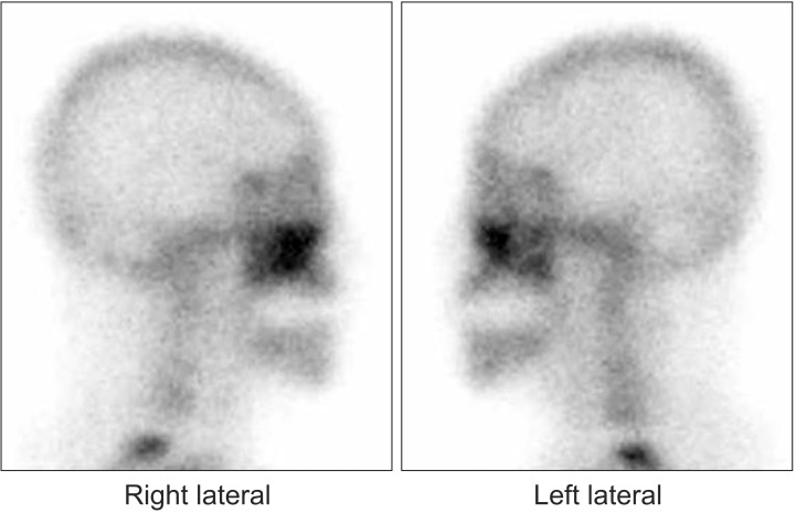
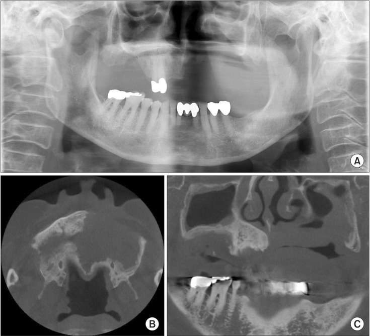
The patient returned 14 months later chiefly complaining of swelling in the left maxilla molar, buccal, and palatal areas, and an oroantral fistula.(Fig. 2) Panoramic radiograph and cone-beam computed tomography were taken, and the osteonecrosis now appeared broader than previously seen.(Fig. 3) A bone scan of the lesion also showed increased intake for both sides of the maxilla relative to the prior scan.(Fig. 4) She was therefore diagnosed with extensive osteomyelitis of the maxilla and surgery was planned.
When the patient had been hospitalized for treatment of uncontrolled diabetes mellitus, her blood glucose level was estimated to be about 309 mg/dL, given that her glycosylated hemoglobin A (HbA1c) was approximately 12.4. During the 14 months when the patient was not seen, her blood glucose level was estimated to be about 200 mg/dL, given that her HbA1c was approximately 8 on follow-up in the Department of Endocrinology. After the patient was admitted to the Department of Endocrinology, her blood glucose level was controlled (HbA1c, 5.7; blood glucose level, 111 mg/dL) and surgery was performed.
Under general anesthesia, partial maxillectomy was performed to remove lesions in the canine and molar regions on the left maxilla. Sequestrectomy of the lesions in the right molar region was also performed. The defect site was covered with a buccal fat pad flap. From the removed tissue, widespread necrosis and sequestrum extending to palate area was found and a sequestrum was also observed on the right side.(Fig. 5)
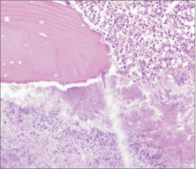
The histopathologic lesions on the upper right and left maxilla were diagnosed as acute and chronic osteomyelitis with actinomycosis.(Fig. 6)
Predisposing factors for osteomyelitis on the maxilla include dental infections, maxillary sinusitis, trauma, radiation therapy, and bisphosphonate treatment. Dental infections and sinusitis are the main causes of maxillary osteomyelitis, followed by trauma5.
Osteomyelitis caused by sinusitis occurs frequently in the frontal bone and rarely in the maxilla as the maxilla has relatively well-developed vascularity and a thin bone structure6. However, these features can allow the lesion to spread to the surrounding soft tissue and the sinuses78.
In this case, necrosis of the maxilla caused by bisphosphonate-related osteonecrosis of the jaws (BRONJ) was strongly suspected because the number of general osteomyelitis cases caused by infections has decreased and BRONJ has become more common. However, we ruled out BRONJ because the patient had no history of bisphosphonate treatment. The patient also had no history of radiation treatment.
Patients with diabetes, anemia or immunodeficiency, and patients who have been prescribed bisphosphonate9 are at higher risk of osteomyelitis. Diabetes has well-known effects on patients' immune systems. According to Peravali et al.10, 20% of mandibular osteomyelitis cases are related to diabetes, and 68% cases of osteomyelitis of the maxilla are related to diabetes. This is because high blood glucose levels reduce immune system efficiency by altering the blood flow distribution of the lesion, and thereby may contribute to the onset of osteomyelitis.
It is difficult to find particular odontogenic causes, and it is thought that a history of uncontrolled diabetes is an important etiological factor. Therefore, we suspect that the widespread maxilla necrosis was due to the spread of preexisting chronic maxillary sinusitis in the context of impeded blood flow and a weakened immune system due to infection. At the same time, the patient underwent amputation of the lower extremities due to diabetic foot complications, indicating poor glycemic control, and that she was susceptible to infection due to a weakened immune system.
There are many treatment methods for osteomyelitis ranging from non-invasive approaches to radical, invasive surgery11. The use of antibiotics in combination with surgery is known to be effective in the treatment of osteomyelitis of the jaw. In this case, because there was widespread necrosis and compromised blood supply, effective penetration with antibiotics was unlikely5. The thick and well-vascularized palatal mucosa remained intact even with maxillary necrosis spreading to the palate.
Therefore, in combination with antibiotics, sequestrectomy and partial maxillectomy were performed. No signs of recurrence have been observed over the course of 13 months of follow-up.
In this case, the patient's immunocompromised state due to uncontrolled diabetes is believed to have worsened the chronic maxillary sinusitis, allowing spread into the maxillary bone. Proper control of the underlying conditions that can hinder healing is mandatory to prevent such devastating infections.
References
2. Indrizzi E, Terenzi V, Renzi G, Bonamini M, Bartolazzi A, Fini G. The rare condition of maxillary osteomyelitis. J Craniofac Surg. 2005; 16:861–864. PMID: 16192871.

3. Hudson JW. Osteomyelitis of the jaws: a 50-year perspective. J Oral Maxillofac Surg. 1993; 51:1294–1301. PMID: 8229407.

4. Schenck HP. Osteomyelitis of the frontal bone. Ann Otol Rhinol Laryngol. 1959; 68:336–345. PMID: 13661805.

5. Prasad KC, Prasad SC, Mouli N, Agarwal S. Osteomyelitis in the head and neck. Acta Otolaryngol. 2007; 127:194–205. PMID: 17364352.
6. Koorbusch GF, Fotos P, Goll KT. Retrospective assessment of osteomyelitis. Etiology, demographics, risk factors, and management in 35 cases. Oral Surg Oral Med Oral Pathol. 1992; 74:149–154. PMID: 1508521.
7. Adekeye EO, Cornah J. Osteomyelitis of the jaws: a review of 141 cases. Br J Oral Maxillofac Surg. 1985; 23:24–35. PMID: 3156622.
8. Borle RM, Borle SR. Osteomyelitis of the zygomatic bone: a case report. J Oral Maxillofac Surg. 1992; 50:296–298. PMID: 1542073.

9. Reid IR. Osteonecrosis of the jaw: who gets it, and why? Bone. 2009; 44:4–10. PMID: 18948230.
10. Peravali RK, Jayade B, Joshi A, Shirganvi M, Bhasker Rao C, Gopalkrishnan K. Osteomyelitis of maxilla in poorly controlled diabetics in a rural Indian population. J Maxillofac Oral Surg. 2012; 11:57–66. PMID: 23449555.

11. Patel V, Harwood A, McGurk M. Osteomyelitis presenting in two patients: a challenging disease to manage. Br Dent J. 2010; 209:393–396. PMID: 20966998.

Fig. 1
Radiographic exams at the first visit show relatively well defined bone destructive lesion (sequestrum) on right maxillary molar area with elevation of sinus floor. A. Panoramic radiograph. B, C. Cone-beam computed tomography.
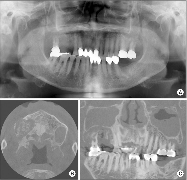
Fig. 2
Intra-oral photographs show oro-antral fistula (palate; A) and bony exposure (buccal; B) on left maxilla after 14 months since the first visit.
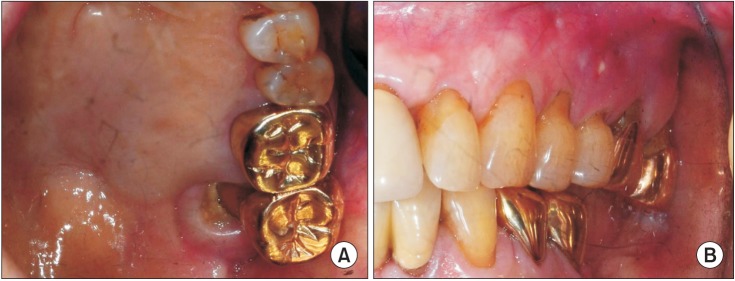
Fig. 3
Radiographic exams show severe bony destructive lesions on both maxilla and mucosal thickening on both maxillary sinuses after 14 months since the first visit. A. Panoramic radiograph. B, C. Cone-beam computed tomography.
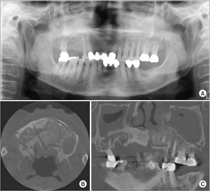
Fig. 5
Clinical photographs were taken during operation (A) extensive necrotic destruction of the both maxilla (B) excised specimen of left maxilla and palate (C) reconstruction with buccal fat pad flap (D) primary wound closure.
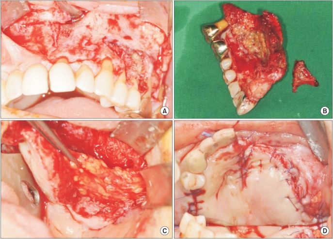
Fig. 6
Histopathologically, bony tissues show bony necrosis, marrow fibrosis and numerous sulfur granules with acute and chronic inflammatory cell infiltration (H&E staining, ×200).


 PDF
PDF ePub
ePub Citation
 Print
Print



 XML Download