

 PDF
PDF ePub
ePub Citation
 Print
Print


I. Introduction
The etiology of supernumerary teeth is unclear. The most frequent type of supernumerary tooth is the mesiodens, located between the central incisors in the anterior maxilla12.
Mesiodenses are more common in males than in females. A mesiodens, which is often located on the palatal side of the root of a deciduous tooth, can interfere with the eruption of the permanent incisor or can cause malocclusion such as rotation of a permanent incisor and diastema. Rarely, it is associated with a dentigerous cyst3.
In this study, we investigated the characteristics of recent mesiodens cases and compared them with mesiodens research by Kim and Lee4 in 2003.
Go to : 
II. Materials and Methods
This research was conducted on patients who underwent mesiodens extraction at Chosun University Dental Hospital between January 2009 and September 2013. All patients were examined clinically and radiologically and were assessed in terms of gender, age, main reason for the visit to the hospital, effects on permanent incisors, shape of the supernumerary tooth, and presence of dentigerous cysts. In all patients, mesiodenses were extracted surgically. A comparative analysis was carried out between these data and the results of research published in 20034.
Go to :
III. Results
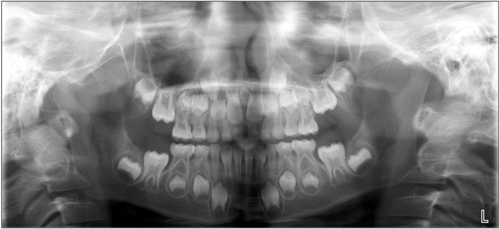
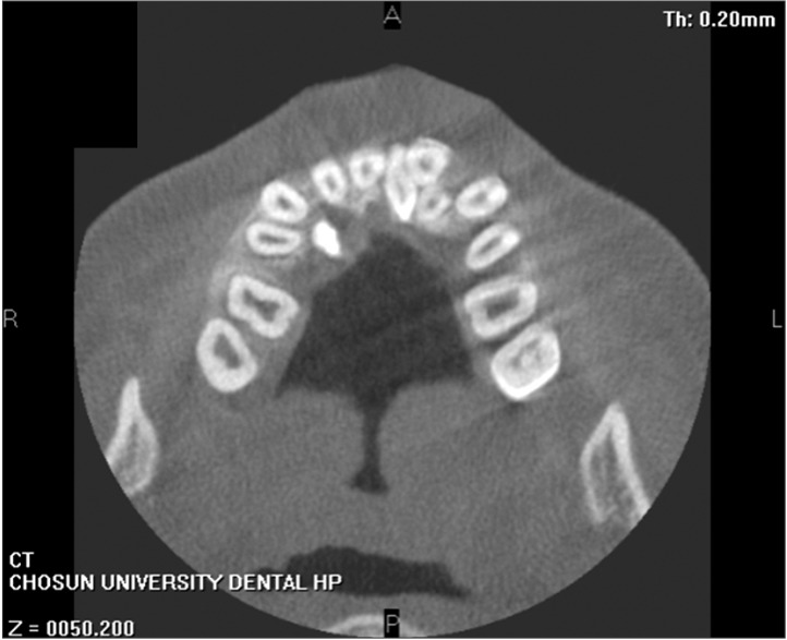
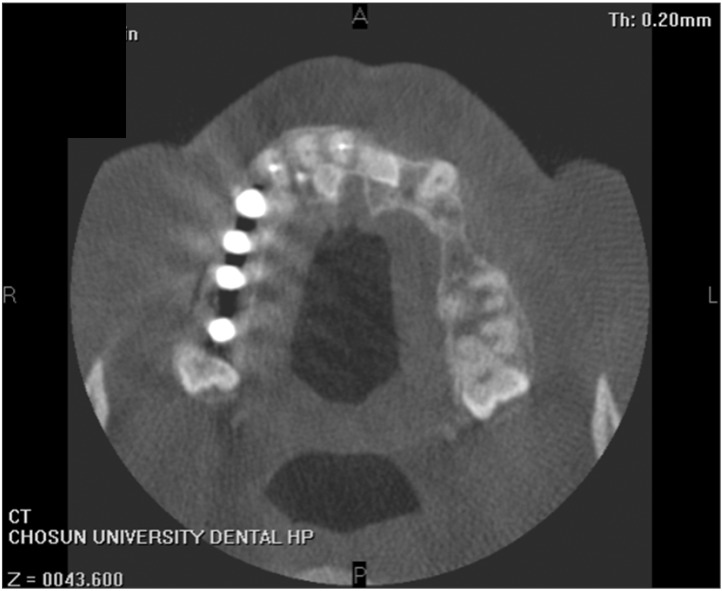
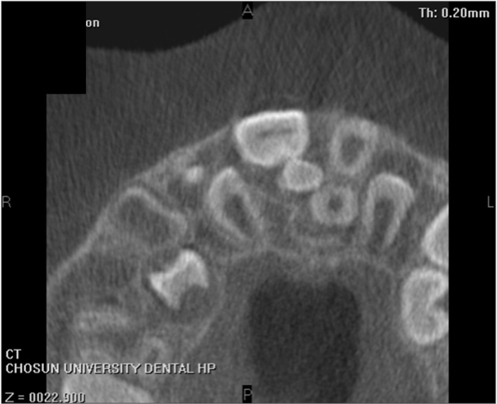
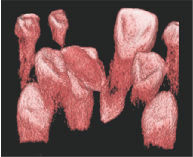
In total, 378 patients visited the hospital with mesiodens between January 2009 and September 2013.(Fig. 1,2,3,4,5) There were 298 males and 80 females, showing a male-to-female ratio of 3.7:1. Their ages varied from 3 to 75 years, with a mean age of 8.4 years.
Of the patients, 265 patients (70.1%) had one supernumerary tooth, 112 patients (29.6%) had two supernumerary teeth, and one patient (0.3%) had three supernumerary teeth. In patients with more than one supernumerary tooth, all supernumerary teeth were extracted at the same visit.
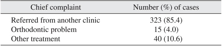
Patients were analyzed for motivation to visit to the hospital. Of the 378 enrolled patients, 323 patients (85.4%) were referred from other hospitals specifi cally for removal of the supernumerary tooth. In 15 patients (4.0%) cases, an orthodontic problem was caused by the mesiodens; in 40 patients (10.6%) cases, the mesiodens was found by chance during their visit to the hospital for another treatment.(Table 1)
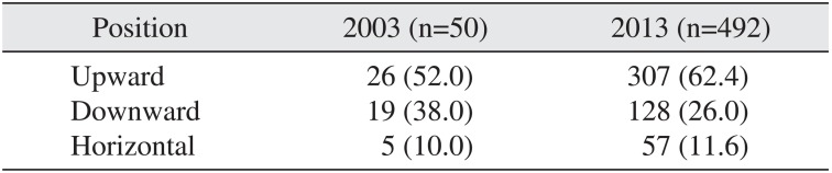
We radiologically examined 492 supernumerary teeth for impaction type and found that 307 teeth (62.4%) were impacted in the opposite direction, 128 teeth (26.0%) were impacted in the normal direction, and 57 teeth (11.6%) were impacted horizontally.(Table 2)
On the basis of clinical examinations, 200 patients (52.9%) had no specifi c symptoms due to the presence of mesiodens, 94 patients (24.9%) had diastema, 59 patients (15.6%) had delayed eruption of a permanent incisor, and 22 patients (5.8%) had an angled incisor due to the mesiodens. Infl ammation caused by the mesiodens was observed in three (0.8%) patients.(Table 3)
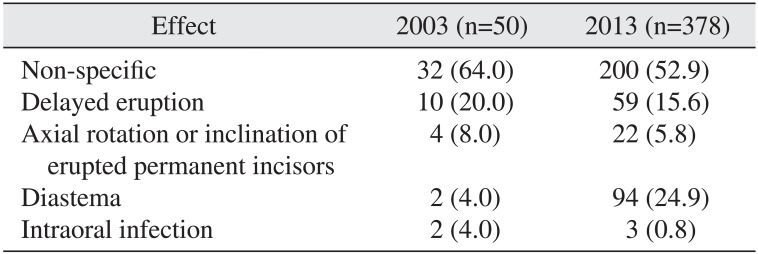
Table 3
Effects on permanent maxillary incisors according to year
![]()
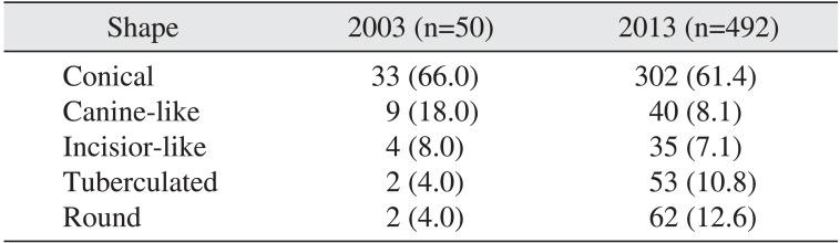
Following extraction, the crown shape was analyzed. A conical shape was found in 302 crowns (61.4%), a round shape in 62 crowns (12.6%), a tubercular type in 53 crowns (10.8%), a shape similar to a canine in 40 crowns (8.1%), and a shape similar to an incisor in 35 crowns (7.1%).(Table 4) In two patients (0.5%), dentigerous cyst was found radiologically.
Go to :
IV. Discussion
In many cases, a mesiodens is a single impacted tooth. Comparing results of our previous study with the present study, we found that 75.0% and 70.1% patients had only one mesiodens, and 25.0% and 29.6%, respectively4, had two mesiodenses. Orhan et al.5 reported that 76% to 86% of patients had only one mesiodens, 12% to 23% had two, and fewer than 1% had three or more.
Ray et al.6 observed that mesiodenses appeared twice as frequently in males than females, and Rajab and Hamdan7 reported a male-to-female ratio of 2.2:1 in Jordanian children. Both our previous and present results suggest a higher male-to-female ratio. We found 4:1 in our previous study and 3.7:1 in this study.
Morphologically, mesiodenses can have various shapes8. The most common shapes are conical, incisor-like, and tubercular types. The conical type was seen in both our previous study and in the present study, at frequencies of 66.0% and 61.4%, respectively4.
In some patients, the mesiodens erupts 'normally' but is mostly impacted or is pointed in the opposite direction. This is probably because the supernumerary tooth has an abnormal eruption path or erupts ectopically910. An impacted mesiodens can interfere with the eruption of the adjacent permanent teeth or can cause malocclusion11. Comparing impaction direction between our previous study and this study showed that 52.0% and 62.4% were impacted in the opposite direction, 38.0% and 26.0% were in the normal direction, and 10.0% and 11.6% were impacted horizontally, respectively4. No specific effect on permanent incisors was consistently found in either the previous study or in this study (64.0% and 52.9%, respectively). Delayed eruption was found in 20.0% and 15.6% of cases, diastema in 4% and 24.9%, and rotation of a permanent incisor in 8.0% and 5.8% between the previous study and this study, respectively. Malocclusion of a permanent incisor was found in 32.0% and 46.3% of previous and current patients, respectively, and infl ammation was observed in 4% and 0.8% of cases4.
Only two patients (0.5%) in the present study were radiologically observed to have dentigerous cyst associated with a mesiodens, and Dinkar et al.3 have reported a correlation between dentigerous cyst and a mesiodens, although such a fi nding is rare.
Go to :
V. Conclusion
It is important to determine both the number and location of mesiodenses in a patient. It is also necessary to carefully examine the impaction direction, the effects on adjacent teeth, and the formation of dentigerous cyst. When a pathologic condition is found related to the presence of a mesiodens, extraction of the mesiodens should be considered.
Go to :
 XML Download