

 PDF
PDF ePub
ePub Citation
 Print
Print


I. Introduction
Mandibular fractures in infants (younger than 1 year) are uncommon. In the 0-1 year age group, the frequency has been reported to range from 0.9% to 2.6%1. In terms of treatment, pediatric mandibular fractures are treated with a wide variety of fixation methods. Although conservative treatment such as closed reduction is preferable because displaced fractures have relatively lower incidence in pediatric patients and tooth buds can be damaged, the open reduction-internal fixation method is possible for unfavorably displaced fractures2,3. We report a case of an 11-month-old infant whose displaced fractured mandible was treated with an open surgery method.
Go to : 
II. Case Report
On June 11, 2010, an 11-month-old infant was brought
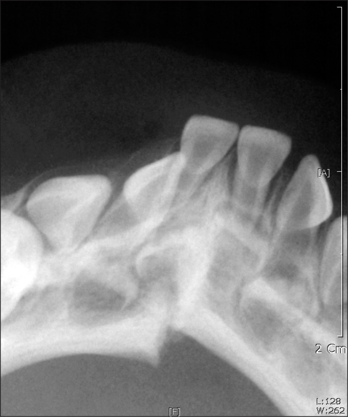
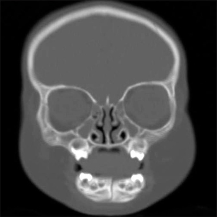
to the Department of Oral and Maxillofacial Surgery, Ajou university hospital after falling down from a baby carriage. The examination revealed extra-oral swelling and ecchymosis around the area of symphysis. The intraoral examination and standard radiography showed bony displacement between the lower right first and second primary incisor teeth.(Fig. 1) Diagnostic computed tomography was performed under sedation, confirming the fracture.(Fig. 2) Specifically, the fracture was visibly displaced, and fragments were freely movable.
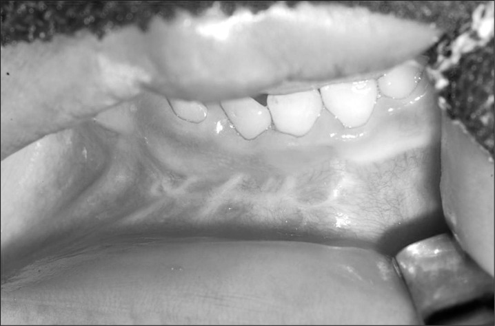
A decision was made to treat the fracture with a combination of open reduction and internal fixation with microplate. Surgery was performed under general anesthesia with sevoflurane, thiopental sodium, fentanyl citrate. After inducement, the patient was intubated with conventional endotracheal tube with 4.0 s cuff via the oral route without difficulty. Lidocaine 2% with 1:100,000 epinephrine was infused in the mandibular anterior vestibule. Horizontal incision was made in the anterior labial vestibule, approximately 3 mm inferior to the mucogingival junction. A mucoperiosteal flap was elevated to expose the fracture line, which ran obliquely from the right second primary incisor to the inferior border of the mandible with step on the buccal cortex and extended to the symphyseal region on the lingual side.(Fig. 3) After gentle reduction of the displaced parasymphyseal fracture by interdental wiring, a four-hole L-shaped microplate (overall length of 13 mm, thickness of 0.6 mm; JEIL Medical Co., Seoul, Korea) was adapted to the buccal side of the mandible close to the inferior border to avoid damaging the unerupted primary teeth and permanent tooth germs. After careful drilling, the plate was secured with four 3 mm, low-profile screws.(Fig. 4) The short, low-profile screw was selected to avoid damaging the developing teeth and dental follicles. The incision was sutured with 5-0 chromic. The day after the operation, breastfeeding commenced and continued with good acceptance. Penicillin was administered twice daily at a dose of 50 mg/kg to prevent infection. The patient was discharged on the fourth postoperative day, with the interdental wiring removed about a week after the operation. In the follow-up period, the healing of the fracture was satisfactory.(Fig. 5) No deformities, functional restrictions, or complications such as mouth opening limitation were recorded. Nine weeks after surgery, the microplate was removed under general anesthesia. The fractured wound had healed, and there was evidence of bone growth over the margins of the plate. The lower right primary canine had erupted normally.(Fig. 6)


Total follow-up period was 3 months from the first surgery; further follow-up was impossible since the patient's family had immigrated to the United States.
Go to :
III. Discussion
Mandibular fractures are rare in infants. This has been attributed to the relatively small size of the mandible, resilient nature of the bones, relatively low tooth to bone ratio, and protected environment. For these reasons, facial fractures in infants occur mostly in the form of incomplete fractures or simple fractures without significant displacement4. Mandibular fractures in infants may result from the trauma of short falls. Fractured mandible in infants is diagnosed clinically based on step deformity, hematoma in the floor of the mouth, and mobility of the fractured segments.
The treatment of infant fractures is divided into conservative treatment and surgical treatment. Minor greenstick fractures of the mandible in infants generally do not require immobilization. For more complex fractures, open and closed reduction methods have been used.
The debate on open treatment versus closed treatment of pediatric mandibular fractures remains. Note, however, that recent literature5-8 shows a change in using open reduction and internal fixation (ORIF) in fracture stabilization. The risks of facial growth disturbance in ORIF have not been investigated9. In contrast, non-treatment of unrecognized mandibular fractures leads to high incidence of orthognathic surgery and craniofacial treatment10. The potential damage to tooth roots and follicles can be minimized with a careful technique, which involves placing the fixation screws in the lower mandibular border to avoid the dental follicle.
Closed reduction with splints fixed with circummandibular wires is the commonly recommended treatment3. Cir-cummandibular wiring is advantageous since it neither damages the tooth germs nor requires intermaxillary fixation (IMF). Moreover, this method does not interfere with condylar growth, but it has limited indications, i.e., it is useful only for anterior mandibular fractures without significant displacement when sufficient eruption of the teeth for fixation is completed. There are difficulties in IMF-such as the Arch bar-when this method is applied to infants due to the potential risk of condylar growth and imbalanced nutrition during fixation. Such conservative treatments alone do little to control a significantly displaced fracture of the symphysis11. On the other hand, severely displaced fractures and mobile fragments are common indications for open reduction and internal fixation12. Nonetheless, this method carries the risk of interfering with the eruption of teeth. The developing permanent incisors are located lingual to the primary teeth. Care should be taken to prevent damage to the tooth germs when a minimally traumatic technique is used in this region with microplates and low-profile screws and when plates are placed at the lower border13.
The titanium microplate and screws were subsequently removed after 9 weeks. According to previous research, plates and screws should be removed as early as 2-3 months after placement14-16. Others maintained rigid fixation for 3 weeks17 or 10 weeks18. The timing for plate removal is still not well-established. The use of an absorbable plate was considered, but the smallest absorbable plate would have been too large and bulky considering the small size of the mandible. The fracture site was mobile and displaced, requiring rigid
fixation. On the other hand, IMF was contraindicated due to the absence of erupted dentition to support the fixation.
Bone quality and thickness as well as the degree of mobility between the fracture ends, erupted dentition, and possible hindrances to future growth are important considerations that affect the choice of treatment. In such cases, the surgeon should choose the least traumatic procedure. Nevertheless, the mandibular function has important influence on future facial development19. The restoration of mandibular continuity after a fracture is important not only for the immediate function but also for future mandibular development20.
Displaced symphyseal fractures of the mandible in infants may be treated with open reduction when careful positioning of the plates at the lower border of the mandible is performed.
This case with limited follow-up showed no facial growth or tooth eruption problems. Nonetheless, further follow-up would be necessary to observe other problems such as the normal eruption of permanent canine as well.
Go to :
 XML Download