

 PDF
PDF ePub
ePub Citation
 Print
Print


I. Introduction
An implant with a small diameter and a short length may be useful in orthodontic treatment1. However, the stability of a mini-implant is uncertain because mini-implants with a small diameter can be easily loosened by low removal torque2,3. A previous study reported that the failure rate of orthodontic mini-implants ranges from 10% to 30%4. Therefore, further efforts to improve the clinical survival rate of mini-implants are needed. A number of studies on the shape2,5, soft tissue contact surface6, and insertion method7 of mini-implants have been conducted to enhance success rates. Various methods, such as the modification of the shape of mini-implants, have been suggested to increase mechanical stability. Sullivan et al.8 showed that the removal torque of a screw-shaped implant with a 3.74 mm diameter T-chamber was greater than 10 Ncm, despite a lack of osseointegration. A vertical bump on a mini-implant provides vertical space for bone growth and could improve stability when rotational movement is applied9. The stability of implants with an anti-rotational vertical bump may be increased through bone remodeling and bone proliferation.
Stability in the form of resistance to rotational movement can be analyzed by measuring the removal torque. The mobility test, resonance frequency analysis and torque analysis are techniques to evaluate the stability of implants10. Although insertion torque analysis was developed as a method to measure the stability and supportive capacity of implants11, insertion torque may have a weak relationship with stability, and removal torque may be more useful to test the mechanical stability of implants5. Torque ratio (TR) can show the increase in stability from the insertion stage to the final stage2. Angular momentum, which is defined as the torque integrated during a certain period, represents the stability of the implant12, and removal angular momentum (RAM) describes the resistance to removal rotational movement13.
This study was performed to evaluate the stability of orthodontic mini-implants with an anti-rotational vertical bump in rabbits through the mechanical analysis of maximum insertion torque (MIT), maximum removal torque (MRT), TR and RAM at two and four weeks after insertion.
II. Materials and Methods
1. Implants and subjects
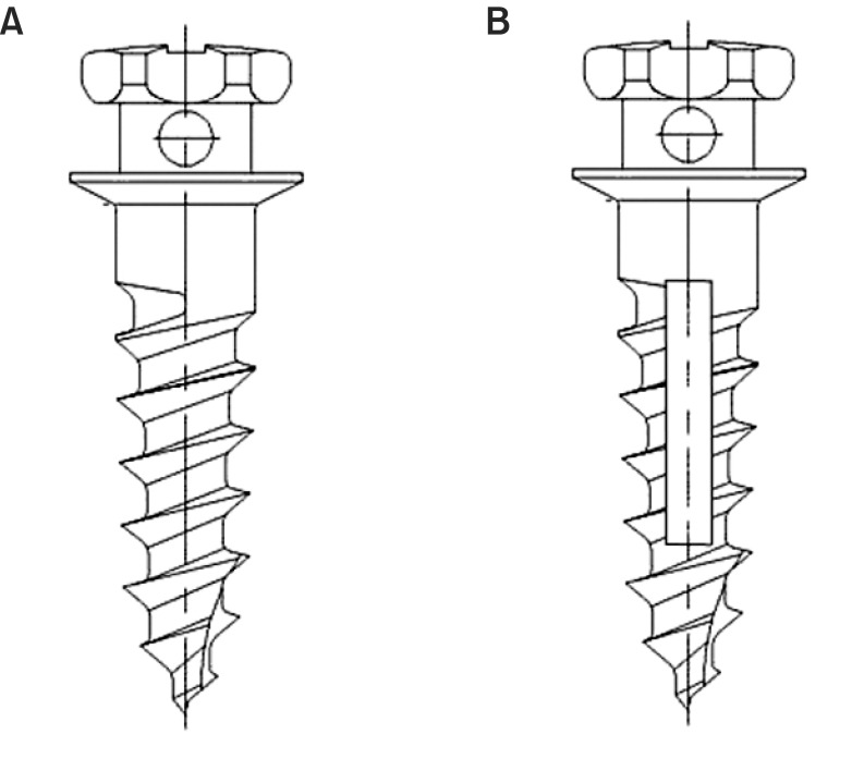
A total of 64 titanium orthodontic mini-implants (length 6.0 mm, diameter 1.6 mm, Dual-Top; Jeil Medical Corporation, Seoul, Korea) were included in the present study: 32 in the control group and 32 in the rotation bump (RB) group.(Fig. 1) This study was approved by the Ethics Committee for Research on Animals (Seoul National University Ethical Board, SNU 120308-2). Sixteen three-month-old New Zealand white rabbits (mean weight was 3.5 to 4.0 kg, Dooyeol Biotech, Seoul, Korea) were used in this study. This study was done from March 2011 to February 2012 in Dental Research Institute of Seoul National University (Seoul, Korea).
2. Surgical procedures
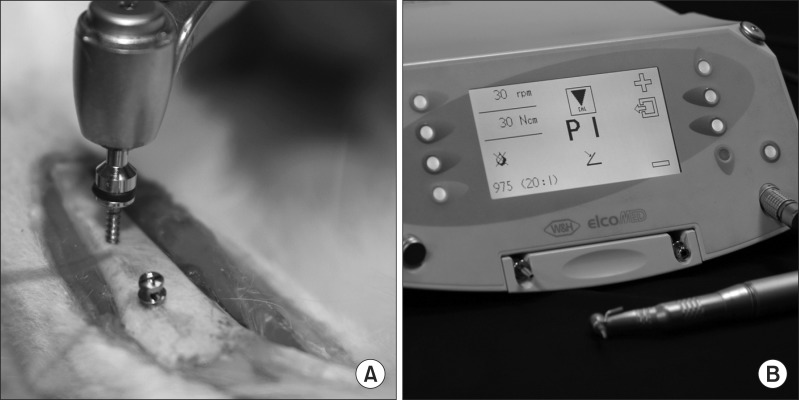
The rabbits were anesthetized with an intravenous injection of Zoletil 50 (7.5 mg/kg; Virbac Korea, Seoul, Korea) and Rompun 2% (0.15 mL/kg; Bayer Korea, Seoul, Korea). The internal surface of the tibia was further blocked with 0.5 mL of 2% lidocaine. The metaphyses of both end sides of the tibia were surgically exposed by scalpel incision to the periosteum and blunt dissection. The mini-implant was inserted into the metaphysis of the tibia with a surgical implant engine (Elcomed SA200C; W&H Dentalwerk, Bürmoos, Austria) after pre-drilling (Ø 1.0 mm) under saline irrigation.(Fig. 2. A)
3. Mechanical analysis
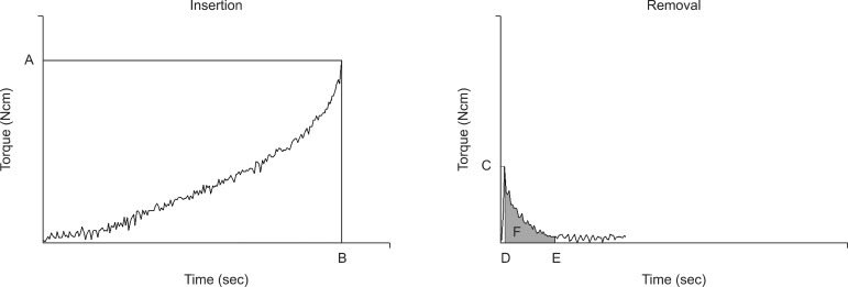
The mini-implants were removed at two weeks and four weeks after implantation, with the torque recorded by the surgical engine (Fig. 2. B), which had a rotational speed of 20 rpm. Impdat software (Kea Software GmbH, Poecking, Germany) was used for the readout of the removal torque value. The torque was recorded eight times per second. MIT, MRT and TR of MRT to MIT were measured to analyze mechanical stability.(Fig. 3) To analyze the energy exerted to remove the mini-implant from the bone, the RAM (Ncms) was calculated by integrating the torque during a half turn, which took 1.5 seconds under 20 rpm after MRT.
III. Results
No failures of mini-implants occurred at two weeks. However, two mini-implants in each group failed at four weeks, resulting in identical failure rates.
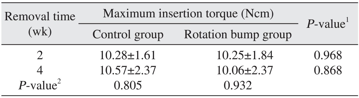
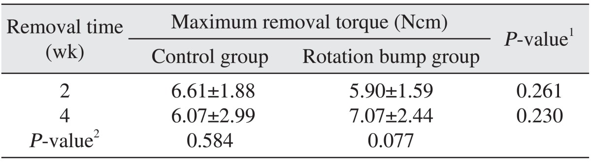
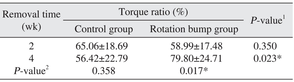
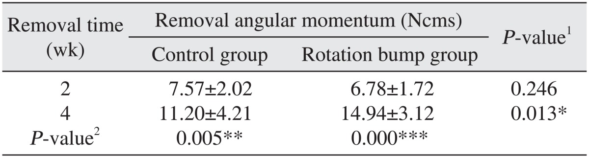
There were no significant differences between the two groups with regard to MIT and MRT at two or four 4 weeks.(Tables 1, 2) However, the TR of the RB group (79.80%) was significantly higher than that of the control group (56.42%) at four weeks (P<0.05).(Table 3) The RAM of the RB group (14.94 Ncms) was significantly higher (P<0.05) than that of the control group (11.20 Ncms).(Table 4)
TR and RAM at four weeks in the RB group were significantly higher than in the control group (P<0.05). The TR of the RB group significantly increased from 58.99% at two weeks to 79.80% at four weeks (P<0.05). In both groups, RAM at four weeks was significantly higher than at two weeks (P<0.05). RAM increased from 7.57 to 11.20 in the control group and from 6.78 to 14.94 in the RB group.
IV. Discussion
Mini-implants have low stability in bone because their surface area is small. The removal torque of an implant is proportional to its surface area, and the osseointegrated area is proportional to the square of the implant radius7 under the same osseointegration ratio condition. This indicates that mini-implants with a small radius have low removal torque requirements and can be easily removed without an additional operation. A previous study reported that mini-implants could be loosened with application of orthodontic loading, which can induce a rotational moment14.
A vertical rotation bump can provide vertical space for bone proliferation. The bone growth into the rotation bump space can resist the rotational movement of mini-implants. In the present study, there were no significant differences between the control group and RB group at two or four weeks.(Tables 1, 2) However, RAM at four weeks in the RB group was significantly higher than that of the control group.(Table 4) These results imply that the mini-implants in the RB group are more resistant to removal rotation. Because RAM was defined as the integrated torque value during a half turn, high RAM might indicate high resistance to removal rotation. The removal torque suddenly decreased after MRT. High RAM might indicate that a high torque value was maintained after MRT15. When rotational moments were applied to the mini-implant, high RAM might have made the mini-implant more resistant to the loosening moment. In both groups, RAM at four weeks was significantly higher than at two weeks. This might indicate that the osseointegration at four weeks was better than at two weeks. The higher RAM of the RB group at four weeks compared to the control group might mean that the bone growth and the osseointegration in the vertical rotation bump space improve resistance to removal torque16.
TR was significantly increased at four weeks in the RB group and was significantly higher than that of the control group.(Table 3) Increased TR may indicate an increase in final stability compared to initial stability2. The MRT of the RB group was increased at four weeks, although it did not show a statistically significant difference.
Although the stabilities of both groups did not differ, there was an increase in the resistance to the removal rotational movement in the RB group. This might suggest that the vertical rotation bump did not enhance the stability of the mini-implant at the initial stage. If the mini-implant remains stable during initial bone remodeling, the vertical rotation bump could improve the resistance of a small mini-implant to rotational movement, which could occur under clinical orthodontic loading. Although conventional orthodontic force is not a rotational movement, food irritation and practices in the orthodontic environment could cause rotation force around the mini-implants during a long treatment period. A rotation bump could support resistance to some unexpected rotational movements.
A previous study suggested that immediate or early orthodontic loading did not affect mini-implant stability because similar histomorphometric results were observed for immediate loading, early loading and delayed loading groups17. However, the histological results did not analyze the intensity or change in stability over time. The results of the present study suggest that RAM changes with time, and that a vertical rotation bump can reinforce the stability of mini-implants.
When orthodontic loading is applied to the mini-implant immediately after insertion, a vertical rotation bump does not enhance mini-implant stability. However, a mini-implant with a rotation bump may have greater stability than a mini-implant without such a bump, as long as the implant is kept stable at the initial stage.
Further studies of histological changes and clinical observations are needed to improve stability and to decrease tissue stress.
V. Conclusion
The rotation bump group did not show significant differences compared to the control group in MIT and MRT at two or four weeks after insertion of mini-implants. However, the TR and RAM of the rotation bump group were significantly higher than the control group at four weeks. RAM was significantly increased at four weeks in both groups. This suggests that the mini-implants were securely osseointegrated at four weeks, and bone growth at the rotation bump space may increase resistance to removal rotation movements.
 XML Download