

 PDF
PDF ePub
ePub Citation
 Print
Print


I. Introduction
Odontogenic keratocyst (OKC) was first introduced in the 1950s to describe keratin containing jaw cysts1. This cyst is considered an important odontogenic cyst because of its aggressive behavior, high recurrence rate, and specific histopathologic features, and it accounts for about 5-15% of all odontogenic cysts. In 2005, the World Health Organization reclassified OKC as "keratocystic odontogenic tumor" (KCOT)2. Recently, this tumor has been re-classified as a benign neoplasm of odontogenic origin and not as cyst; it is defined as a "benign uni- or multi-cystic odontogenic intraosseous tumor, with lining of parakeratinised stratified squamous epithelium and aggressive behavior"1-3. The cystic lumen is filled with creamy proteinaceous material or clear yellowish fluid, which is also an important diagnostic marker for KCOT1.
Several investigators agreed that KCOT arises from cell rests of dental lamina, and that its growth is related to enzymatic activity or unknown factors in the fibrous cystic wall4,5. Clinically, the posterior body of the mandible and ascending ramus especially related to impacted third molar were the prevalent occurring site of KCOT, showing slightly male predilection with peak incidence in patients between 10 and 40 years of age6,7. Radiologically, differently sized radiolucent lesions with displacement of impacted or erupted teeth, root resorption, or root displacement were easily detected in the jaw bones7. The treatment strongly recommended for KCOT consists of complete enucleation and curettage of the lesion to reduce the recurrence rate, although more conservative methods including marsupializa tion and decompression have also been tried in many cases. Interestingly, several recent studies observed the increased expressions of cell proliferation and anti-apoptosis related proteins such as MKI67 (Ki-67), tumor protein 53 (p53), tumor protein 63 (p63), B-cell lymphoma 2 (BCL2), and cyclooxygenase-2 (COX-2) in the basal cell layer of KCOT specimens, indicating that these proteins could be used as biological markers to diagnose KCOT5,8-14.
When odontogenic cysts or tumors including KCOT, calcifying odontogenic cyst, and dentigerous cyst are involved in the maxillary sinus, the cysts or tumors could asymptomatically expand and result in a large expanded cyst involving the whole maxilla and maxillary antrum15-19. To achieve total excision of expanded lesions and complete hemostasis in the anatomically delicate areas of the maxilla, a more extensive approach, such as Weber-Ferguson incision, would be needed. Here, we report two cases of large expanded maxillary KCOT, which occupied the entire maxilla and maxillary sinus. These expansile benign cystic tumors could be completely excised via wide facial approach through Weber-Ferguson incision. In addition, the tumor specimens were evaluated for the expression of BCL2, BCL2-associated X capital letter (BAX), Ki-67, and p53 and p63 proteins by immunohistochemistry to analyze their characteristics.
II. Cases Report
1. Case 1
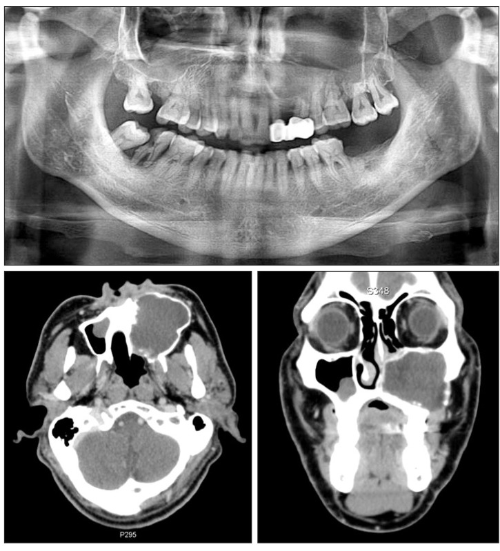
A 57-year-old man was referred to the clinic for the evaluation of facial swelling and asymmetry. The panoramic view showed a large cystic lesion from the anterior maxilla to the entire left side of the maxilla and maxillary sinus. The teeth involved showed no deviation, but slightly resorbed roots were detected. In the computed tomography (CT) views, the cystic tumor was expanded to the buccal and palatal bony wall with partial disruption of cortical bone.(Fig. 1) The lesion was tentatively diagnosed as KCOT because of its aggressive expansion with homogenous fluid-filled lumen. The patient underwent endodontic treatment for all teeth involved, and the expansile maxillary tumor was radically excised with Weber-Ferguson incision, showing no recurrent sign for more than 5 years' follow-up period.(Fig. 2)
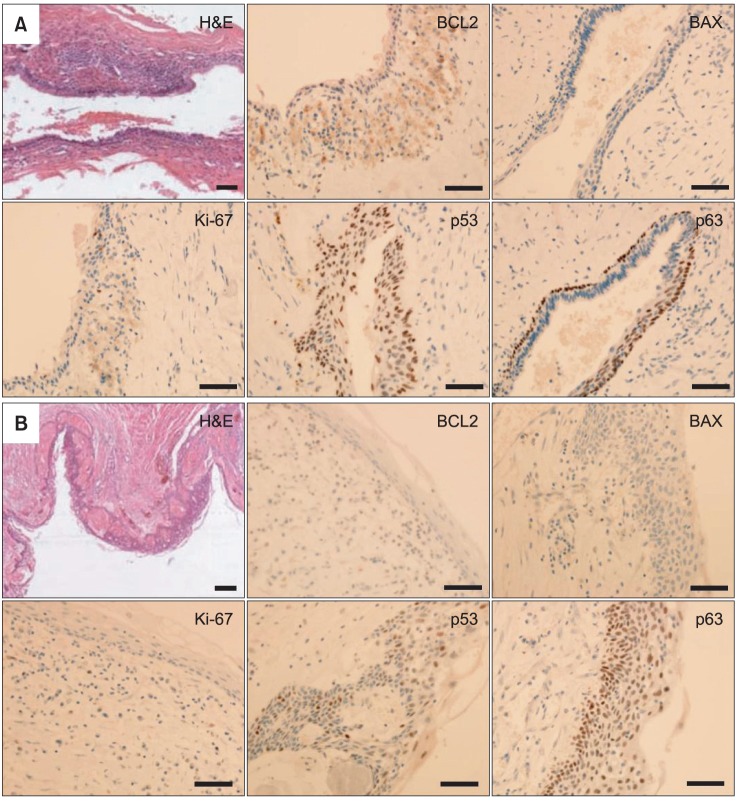
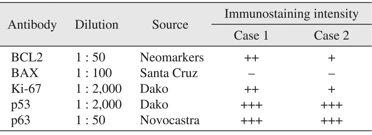
In the histopathological features of H&E-stained slides, thin lining of stratified squamous epithelium and thin parakeratotic surface on the lumen side of the cyst were observed, indicating KCOT. To characterize this tumor, specimens were immunostained with BCL2, BAX, Ki-67, and p53 and p63 antibodies. For the immunohistochemical analysis, tumor specimens were embedded in paraffin blocks, and then cut into 4-µm sections and mounted on silane-coated glass slides. Sections were maintained at room temperature for 12 hours, and then deparaffinized; after hydration they were immunostained using an automated immunostainer (BenchMark XT; Roche Korea, Seoul, Korea). The primary antibodies used and immunohistochemical staining results are summarized in Table 1. Positive immunostaining intensities were graded as +++, ++, +, and - for strong, moderate, weak, and negative staining, respectively.
2. Case 2
A 54-year-old man visited for evaluation after sustaining unilateral facial swelling. Following the radiological examination, large expanded intrabony cystic lesion was found in the left maxilla and maxillary sinus. The lesion occupied the entire unilateral maxilla and maxillary sinus with the expansion and erosion of cortical bone. Tooth displacement was not detected, but some root resorption was observed in the teeth involved.(Fig. 3) Benign cystic lesion much like KCOT was diagnosed due to its radiological and clinical features and was completely excised with Weber-Ferguson incision. The patient had been followed up for 2 years without any evidence of recurrence.(Fig. 4)
The histopathological feature of H&E-stained slide was similar to that of case 1, with thin-lining epithelium and parakeratinized luminal surface as demonstrating features of KCOT. Immunohistochemical analysis of the tumor specimen was performed as described previously with case 1. Strong expression of p53 and p63, positive but weak detections of BCL2 and Ki-67, and negative detection of BAX were observed, and these expression patterns were similar to those of case 1.(Fig. 5. B, Table 1)
III. Discussion
Multidirectional studies for KCOTs using various biomarkers of cell proliferation and apoptosis have been performed because KCOT is an aggressive benign neoplasm with high recurrence rate and extensive local invasion1. Higher proliferation rate and inhibition of apoptosis of cells are regarded as one of the most important steps in tumor formation, allowing the survival of genetically unstable cells and accumulating mutations that lead to neoplasm10,13. In previous studies, KCOT showed higher expression of cell proliferation markers particularly Ki-67, COX-2, proliferating cell nuclear antigen (PCNA) and p53, and epithelial stem cell factor p638-14. It also showed increased expression of BCL2, an anti-apoptosis marker, whereas the expression of BAX, a pro-apoptotic marker, in KCOT decreased13,20.
The p53 protein is expressed in the G1 phase of the cell cycle to allow repair of damaged DNA and to arrest cell cycle progression to the S phase. Some investigators reported that the overexpression of p53 in odontogenic tumors and cysts might be associated with increased cell proliferation12. The p63 protein has a role in epithelial development, stem cell biology, proliferation of limb and craniofacial-structures, and carcinogenesis. It may also have an important role in the progression of odontogenic cystic lesions because it is essentially of epithelial origin8,11. Ki-67 expression increased with cell cycle progression, reaching its peak during the G2 and M phases; thus suggesting that it can be used as cell proliferation marker12. In several studies, Ki-67 was expressed higher in the epithelium of KCOT than in the radicular cyst or oral mucosa, indicating that the epithelium of KCOT has higher cell proliferation rate9,10,12,13. Apoptosis plays an important role in the maintenance of cell homeostasis with programmed cell death for senescent or damaged cells. This mechanism has been regulated with pro-apoptosis and anti-apoptosis proteins. In previous studies, BCL2, one of the anti-apoptosis markers, showed higher expression level in the epithelium of KCOT specimens compared with radicular cyst13,20. Note, however, that BAX, the pro-apoptosis marker, was weakly expressed in KCOT13. These results demonstrate that the epithelium of KCOT has decreased apoptotic activity, allowing increased cell survival activity and invasive growth.
When odontogenic cysts or tumors formed in the maxilla, especially near or in the maxillary sinus, the lesion usually expanded to a large size15-19. For the complete excision of the expansile tumor and achievement of hemostasis in the maxilla particularly in anatomical fragile portions such as posterior maxillary wall and lateral nasal space and orbital floor, more extensive approaches are needed even though a lesion has no malignant evidences. Anatomically less dense structure, such as maxillary sinus, could allow the rapid growth of the lesion and tolerate the tumor's occupancy in the entire maxilla within a short period of time16,18,19. In this study, two cases of KCOT were developed and expanded in the unilateral entire maxilla and maxillary sinus, invading the opposite site of the maxilla. The anatomical structure, loose bone density of the maxilla, and empty space of the maxillary sinus could be considered one of the contributing factors for the development of expansile tumor in the present cases. Note, however, that the aggressive characteristics of KCOT in previous immunohistochemical studies enhanced cell proliferation rate and increased anti-apoptotic activity, possibly serving as other putative factors for enlarging tumor size.
In this report, the p53 and Ki-67 showed increased expre-ssion, although Ki-67 was weakly detected in the specimen in case 2, suggesting that the KCOTs of the present cases have higher cell proliferation rate. In addition, the anti-apoptotic factor, BCL2, showed moderate and weak expression in cases 1 and 2, whereas the pro-apoptotic factor, BAX, was almost negatively expressed in both cases. This result suggests that the present KCOT cases have decreased apoptosis activity and inhibit spontaneous cell death, leading to abnormal tumor growth. Moreover, the epithelial stem cell marker, p63 protein, showed high expression level in the basal layer and suprabasal layer of the tumor epithelium, indicating that KCOT is a tumor originating in epithelial tissue. Taken together, these properties of KCOT can contribute to tumor enlargement, aggressive behavior, and high recurrence rate.


 XML Download