

 PDF
PDF ePub
ePub Citation
 Print
Print


I. Introduction
There has been increasing interest in the change of tongue, hyoid bone, and pharyngeal airway space after bimaxillary surgery1-5. Many authors have studied the change in airway space according to the movement direction of the maxilla and mandible and have shown different results. In particular, the impact of mandibular posterior movement on airway was controversial, as one with the airway space reduced and the other with no change2,4-13.
The majority of previous studies on pharyngeal airway change of skeletal class III patients after bimaxillary surgery have been conducted mainly by means of twodimensional analysis using lateral cephalometric radiograph1,6-14. Few studies on threedimensional analysis using computed tomograpy (CT) reported that the pharyngeal airway space had insignificant change or even had increased after mandibular setback surgery2-4. Such disagreement was believed to have resulted from the difference in the corres ponding movement direction of the maxilla.
The main purpose this retrospective study is to evaluate the changes of pharyngeal airway volume and minimal axial area in skeletal class III patients undergoing mandibular setback and maxillary setback or posterosuperior movement surgery, which is expected to decrease the pharyngeal airway space. Additionally, the effect of genioplasty advancement on pharyngeal airway was evaluated.
II. Materials and Methods
1. Materials
The study sample consisted of 18 skeletal class III Korean patients that had undergone maxillary setback or posterosuperior movement and mandibular bilateral sagittal split osteotomy (BSSRO) setback surgery from May 2010 to August 2010 at the Department of Oral and Maxillofacial Surgery, Seoul St. Mary's Hospital, the Catholic University of Korea (8 males and 10 females, mean age of 28.7; range 1743).
2. Methods
This study was conducted with the approval of the Institutional Review Board in the Catholic University of Korea (Grant No.: KC11RISI0437).
Cone beam CT (CBCT) images of 0.4 mm axial thickness were taken 1 month before (T0) and 6 months after (T1) ortho gnathic surgery using i-CAT (Imaging Sciences International Inc., Hatfield, PA, USA) CBCT scanner. Patients were instructed to sit upright in natural head position with their jaws at maximum intercuspation and the lips and tongue in a resting position.
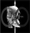
The axial images were reconstructed into three-dimensional images using InVivo Dental software (version 5.1; Anatomage, San Jose, CA, USA). The threedimensional images were reoriented by using Frankfort horizontal (FH) plane. The FH plane was constructed from the right and left porions located in the most superior point of the external auditory meatus and the left orbitale.(Fig. 1)
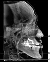
The airway space was divided into nasopharyngeal space, oropharyngeal space, and laryngopharyngeal space. Three horizontal pharyngeal cross sections were parallel to the FH plane and each passed through posterior nasal spine (PNS), epiglottis tip, and the most anteroinferior point of the body of 4th cervical spine (C4). The anterior limit of the nasopharyngeal space was defined by the coronal plane perpendicular to the FH plane and sagittal plane that passes through PNS. Oro pharyngeal space was defined as the airway space between the planes passing through PNS and the epiglottis tip. Laryngopharyngeal space was defined as the airway space between the plane passing through the epiglottis tip and the most anteroinferior point of the body of C4.(Fig. 2)
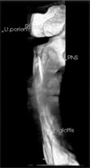
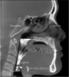
Since the airway is an empty space surrounded with hard and soft tissue, the Inversion function of InVivo Dental software was used. The space of interest was separated by removing the unnecessary structures through freehand sculpture function.(Fig. 3) To standardize the measured values and to minimize the errors, the voxels within the range of -1,024 HU and -600 HU were calculated. The volume was then measured through the volume measurement function of the software. The airway measurement function was used to measure the minimum crosssectional area of each airway space.(Fig. 4)
All statistical analyses of the obtained data were performed using the SPSS program (version 18.0: SPSS Inc., Chicago, IL, USA). The differences of the airway space volume and the minimum crosssectional area between T0 and T1 were evaluated through a Wilcoxonsigned rank test. The pharyngeal airway changes of the genioplasty advancement group (7 patients) and the other without genioplasty (11 patients) at T0 and T1 were analyzed by MannWhitney U test. P<0.05 was regarded as significant.
III. Results
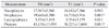
From the analysis of the CBCT data, a significant reduction in oropharyngeal space volume was observed (P<0.05). (Table 1) The total pharyngeal space volume which includes nasopharyngeal space, oropharyngeal space, and laryngopharyngeal space significantly decreased from an average of 43,130±13,595 mm3 preoperatively to an average of 38,227±13,688 mm3 postoperatively. There was no significant difference in nasopharyngeal and laryngopharyngeal space volume. The change of oropharyngeal space volume was from an average of 19,648±8,300 mm3 at T0 to an average of 14,710±5,916 mm3 at T1.(Table 1)
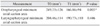
The minimum crosssectional area of oropharyngeal space decreased from an average of 249.33±126 mm2 at T0 to an average of 166.86±94 mm2 at T1 (P<0.05). The minimum crosssectional area of laryngopharyngeal space decreased from an average of 204±114 mm2 at T0 to an average of 190.73±110 mm2 at T1. However, the difference was not statistically significant.(Table 2)
For the genioplasty advancement group, the oropharyngeal space volume decreased by an average of 4,373±2,488 mm3, and the laryngopharyngeal space volume decreased by an average of 496±2,242 mm3. In the group without genioplasty, the average oropharyngeal space volume showed a small decrease, whereas the laryngopharyngeal space volume was slightly increased. However, the changes in both groups were not statistically significant.(Table 3)
IV. Discussion
Pharyngeal airway change corresponding to soft and hard tissue change from orthognathic surgery have been thought to be important in functional aspect, and numerous previous studies have evaluated it1-15. Until recently, cephalo metry was mainly used in studies with purpose of finding out relationship between orthognathic surgery and pharyngeal airway space. In a series of studies on mandibular setback surgery, the distance between tongue base and pharyngeal wall in the lateral cephalometry has been shown to decrease significantly6,8,10,11. In contrast, Saitoh9 reported that oropharyngeal area first showed significant decrease in 3-6 months after surgery and after 2 years the change was not significant anymore. On the other hand, Kawakami et al.7 reported that there first was no change in the distance between tongue base and pharyngeal wall up to 1 month. One year after, the significant reduction of the distance followed with the superior movement of hyoid bone.
There also was controversy for the bimaxillary surgery. Chen et al.14 reported that for mandibular single jaw setback surgery, airway distance was significantly decreased both in oropharynx and laryngopharynx. However, for bimaxillary surgery with maxillary advancement, airway distance first decreased until 3-6 months postoperatively and after 2 years, there was no significant change. Marşan et al.15 and Pereira-Filho et al.1 also reported similar results. In other studies with two dimensional analysis conducted on CT, anteroposterior distance and width of airway space at the level of tongue base was shown to decrease postoperatively12,13. Mattos et al.5 con ducted a meta analysis and reported that in mandibular single jaw setback surgery, anteroposterior airway distance decreased at the level of soft palate and tongue base. For the bimaxillary surgery with maxillary advancement, the anteroposterior airway distance at the level of soft palate and tongue base still decreased and the distance increased only at the level of posterior nasal spine. They also reported that whether significant change of cross sectional area of airway before and after mandibular setback or bimaxillary surgery is there or not is yet controversial.
However, these two dimensional studies based on lateral cephalometry or CT have innate limitations to reflect three dimensional changes of airway after orthognathic surgery. Because of this limitation, the study results of three dimensional changes of airway on CT or CBCT are showing different results2-4. Park et al.2 reported that there was no significant change of nasopharyngeal and oropharyngeal airway volume after mandibular setback surgery. They suggested that the sagittal compression of the pharyngeal airway was compensated by physiologic deformation such as lateral expansion. Jakobsone et al.3 reported that after bimaxillary surgery with maxillary advancement and mandibular setback, oropharyngeal and hypopharyngeal airway volume increased. They assumed that the difference of their result from the previous works was from the movement of the tongue to the space created by advancement of maxilla on an average of 4.6 mm. Hong et al.4 observed significant decrease of airway volume in both mandibular setback group and bimaxillary surgery group on CBCT. However, the direction of maxillary movement was not specified in their study. The present study showed significant decrease of oropharyngeal airway volume which was similar to the result of Hong et al.4. Until present, there was no study in the literature that evaluated the impact of maxillary posterior or posterior superior movement on the pharyngeal airway space in combination with BSSRO setback. In this study, there was no significant change of nasopharyngeal space volume. Whether oropharyngeal space will be affected by maxillary posterior movement alone is uncertain and requires further study.
From the results of previous studies on nasal airway resistance after maxillary movement, the resistance has been shown to follow the change of nares which has the smallest cross sectional area of the nasal airway16,17. From this point of view, evaluating the least cross sectional area of each airway space would reflect the functional aspect, such as airway resistance. In this study, we evaluated the change of the minimum cross sectional area of oropharyngeal and laryngopharyngeal space. The oropharyngeal space showed significant decrease, whereas the laryngopharyngeal space did not.
Genioglossus advancement is one of the surgical therapies for obstructive sleep apnea syndrome18. Similarly, we expected genioplasty advancement to have effect on the position of the hyoid bone and tongue which would then have effect on the pharyngeal airway space. We compared the laryngopharyngeal airway volume between one group that had genioplasty advancement and the other that had not. Althought there was difference in mean laryngopharyngeal airway volume, it was not statistically significant. It might have been due to the small amount of advancement, which was an average of 3.42 mm.
The present study had limitations such as relatively short follow up period of 6 months and small sample size. In future studies, longer follow up period and larger sample size would help us to have better knowledge of the pharyngeal airway changes.
V. Conclusion
In the study of 18 Korean patients that had received bimaxillary surgery with mandibular BSSRO setback and maxillary posterior or posterosuperior movement and had had the follow up period of 6 months, the following results were obtained.
Nasopharyngeal and laryngopharyngeal airway volume did not have significant change whereas oropharyngeal volume decreased significantly.
The minimum cross sectional area of oropharyngeal space decreased after surgery, and laryngopharyngeal space did not show significant change.
The genioplasty advance did not have significant effect on the oropharyngeal and laryngopharyngeal airway volume.

 XML Download