

 PDF
PDF ePub
ePub Citation
 Print
Print


I. Introduction
The cystic lesion occurring on the mandible is one of most frequent osseous lesions in the clinical fields of dentistry and oral surgery. Since such lesion is due to odontogenic causes, and it may influence the adjacent bones, enucleation is recommended as treatment on the whole1,2. In the mandible, there are the inferior alveolar nerve canal and vital teeth. In many cases, the surgery itself may pose some difficulties in case access to cystic lesions is made using general methods. When the cystic lesion in the mandible is removed surgically, the method of surgery should be decided considering the area, size, and pathological characteristics. The method of surgery may differ depending on the individual clinician, and preservative treatment and aggressive surgery may be applied3. Preservative treatments may include enucleation, curettage, marsupialization, and decompression4. Aggressive treatments include enucleation accompanied by chemical curettage5 or mandibulectomy6.
Williams and Connor6 and Bramley7 introduced the excision of bone with cysts and bone grafting as a surgical method for odontogenic keratocysts. Such method may minimize the possibility of recurrence, but its widened scope of surgery may become a disadvantage. Since minimally invasive surgery has a disadvantage of narrow visibility, and it may cause some difficulties in terms of exactly accessing lesions or iatrogenic damage in the surrounding tissues, minimally invasive methods with good visibility using an endoscope have been utilized recently3,8. Aside from those methods, the method of gaining access after cutting out the buccal or lingual side of the bone in the lesion to access the cystic lesion in the mandible, method of repositioning the relevant cortical bone after excising and storing the cortical bone only and removing the inside lesions, and method of repositioning after performing Le Fort osteotomy9 and removing the lesions are available in the clinical fields.
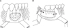
When removing cystic lesions, the authors generally utilize the method of foregoing repositioning after cutting out the buccal cortical bone plate (Fig. 1. A) and the method of repositioning after cutting out and storing the cortical bone plate and removing the lesions.(Fig. 1. B) Such method was applied in a few cases wherein the defects of soft tissue are judged to be noticeable due to the excessive bone defects when the entire buccal cortical bone plate was removed and the normal anatomical structures were likely to have been damaged. Reports on the occurrences of complications or bone healing in connection with this method have not been sufficiently made. Thus, in this article, the authors tried to report the occurrence of complications and bone healing by comparing the method of repositioning the buccal cortical bone plate (reposition, R) and foregoing repositioning (no reposition, NR) after cutting out the cortical bone.
II. Materials and Methods
1. Research subjects
The subjects of this research were patients for which retrospective review of their medical records was possible among the patients who underwent surgery for the treatment of cystic lesions in the mandible at the Department of Oral and Maxillofacial Surgery of Samsung Medical Center from 2007 to March 2010 and whose buccal monocortical bone plate was removed among the patients for whom panoramic radiographs (Planmeca ProMax; Planmeca Oy, Helsinki, Finland) were taken for over 6 months. Among the patients concerned, those patients whose lingual bicortical bone plate - not the buccal monocortical bone plate - in the mandible was removed but bone healing was not observed in the relevant regions during the process observation period or for which the boundary of bone defects was not well-defined in the panoramic radiographs for the process observation were excluded. The radiographs utilized in this study were panoramic radiographs. A total of 60 subjects - 22 men and 38 women - were considered, with the R group consisting of 34 persons and the NR group having 26 persons.
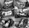
A total of 5 clinicians performed the surgeries, and they utilized the R method at their own discretion in case extensive bone removal was needed to access the lesions or the defects of soft tissue were judged to be noticeable when the buccal cortical bone plate was removed. The tools for fixing after repositioning the buccal cortical bone plate with plate, wire, suture materials, and their combinations.(Fig. 2) The selection of method for fixing the buccal cortical bone plate was made based on the criterion that there should be no mobility in the bone fragments when using them. The observation of process was carried out for 6 months for 60 persons, for 12 months for 41 persons (R: 24 persons, NR: 17 persons), and for 24 months for 18 persons (R: 11 persons, NR: 7 persons). The average age was 38.5 years old; 13 patients underwent bone grafting using xenogenic bones (Bio-oss; Geistlich Pharma AG, Wolhusen, Switzerland) or autogenous bones, 29 patients had the insertion of materials such as Teru-plug (Olympus Terumo Biomaterial, Tokyo, Japan) and fibrin sealant (TISSEEL; Baxter International Inc., Deerfield, IL, USA) done, and 40 patients had the drainage tube inserted.
2. Research method
1) Measurement of bone healing
Bone healing aspects were evaluated by measuring the width of the radiolucent lesion area in the radiographs taken in the 6th, 12th, and 24th month after the surgery of patients who met the aforesaid criteria. The sizes of the lesions in the digitalized radiographs were measured by pixel unit using the polygonal selection of Image J (National Institutes of Health, Bethesda, MD, USA). The bone defect site projected in the panoramic radiograph included the surrounding bones when the lesion was removed but excluded the adjacent normal sponge bones. The reference area whose boundary was well-defined in the relevant radiograph was established.
This reference area was set as the size of tooth crown, which was not changed during the observation period but was adjacent to the lesion. To minimize the error of enlargement and reduction that the radiograph itself had, the ratio between the lesion size and the reference area was calculated for all panoramic radiophotos. Bone healing was evaluated in the proportion of the lesion size measured during the observation of the process against the lesion size in the panoramic radiograph taken just after the surgery. Out of a total of 60 patients who met the aforesaid criteria, there were extensive differences in width (4,419-535,129 pixel dimension). Accordingly, the bone healing aspects of 20 persons in the R group and 20 persons in the NR group and who had a certain level of lesion sizes were also evaluated at each period.
2) Evaluation of occurring complications
Through the retrospective study using medical records, whether complications occurred was determined; the after-effects were defined based on continuous discomfort, edema, dysgeusia, location change of bony window, infection, and recurrence. Whether the surgical method (R or NR), gender, case with or without grafting materials, case with or without bone grafting, and case with or without the drain tube meaningfully influenced the occurrence of complications was assessed.
III. Results
1. Measurement of bone healing
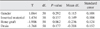
In the panoramic radiograph of 60 patients with 4,419-535,129 pixel dimension in the 6th, 12th, and 24th month, the statistical analysis result for each group as to the difference in how much the radio opacity of lesions increased between the R group (n=34) and the NR group (n=26) showed no statistically meaningful difference. Neither was there any meaningful difference between the R group (n=20) and the NR group (n=20) with 23,000-80,000 pixel dimension.(Table 1)
2. Evaluation of complication occurrences
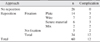
For the 26 patients in the NR group, there was no complication defined in the research method. Nonetheless, complications occurred in 12 out of the 34 patients in the R group (Table 2). Two out of these twelve patients complained of continuous discomfort, but such symptoms disappeared after the wire was removed from the relevant site. One person complained of dysgeusia, but the symptoms disappeared after the plate was removed. Four patients were infected, and incision, drainage of abscess, and sequestrectomy were performed. Recurrent aspects were observed in 3 patients, and re-surgery was performed. Curettage was done since bone grafting for one patient failed. Since abnormality of the bony window location was noted in one patient, the bony window was removed.
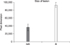
According to the result of the analysis on whether there was a difference in the size of the existing lesion based on the method of surgery, the average of the NR group was 37,024 (pixel dimension, 1.55×1.55 cm), and that of the R group was 92,863 (pixel dimension, 2.44×2.44 cm); thus implying that the former method was used for statistically smaller lesions.(Fig. 3) Whether complications occurred depending on gender as well as the kinds of bone grafting materials, case with or without bone grafting, and case with or without the insertion of drainage tube did not show any statistically meaningful connection.(Table 3)
IV. Discussion
Partsch10,11 announced the application of marsupialization (Partsch I) and enucleation (Partsch II) for the treatment of cyst in the jaw bones in 1892 and 1910. Today, Gold et al.12 divide the surgical methods of removing the cysts generated in the jaw bones into four levels and refer to them as follows: enucleation, curettage, marsupialization, and resection with or without breakage. Enucleation means completely separating the lesion from the adjacent bone and removing it. Curettage involves raking out the lesion together with part of the adjacent bone (generally, 1-2 mm) using mechanical, physical, and chemical materials.
Ghali and Connor2 used the terms curettage and peripheral ostectomy, taking the former to mean the use of hand tools and defining the latter as the case of using rotary cutting instruments. Curettage and peripheral ostectomy are fundamental surgical methods of removing the lesions of aggressive cysts in the mandible. Marsupialization is a method of creating an opening from the inside of cyst into the oral cavity, maxillary sinus, or nasal cavity by making a bony window on the wall of the cyst. This method would minimize - or even later forego the need for - enucleation by reducing the size of the cyst, decreasing the pressure inside the cyst, and allowing the bone to fill the cyst. As mentioned earlier, in marsupialization, the cyst wall and its adjacent tissues are sutured, unlike in the method wherein decompression is done simply by inserting the drainage tube.
As the most aggressive treatment, osseous resection may be divided into two methods. The first method is resection without loss of jaw continuity. According to some authors, the satellite cysts that may cause later recurrence could be removed using this method. The method of resection with loss of jaw continuity may be applicable in case pathological fracture is likely to occur due to cystic lesion, there is a recurring, widespread odontogenic cyst, or the lesion includes the mandibular condyle or there is a carcinomatous or ameloblastomatous degeneration within a keratocyst.
The odontogenic keratocyst has the characteristics of expanding through the bony wall and penetrating deep into the structures. Due to such characteristics, some authors support the execution of aggressive surgery in case there is a large cyst in the mandible6. Others prefer the preservative method, however4. Sometimes, the preservative method may be carried out first, and cyst enucleation may then be executed. It is very similar to performing marsupialization first followed by cyst enucleation13. The wall of the odontogenic keratocyst is known to undergo histological deformation after decompression. The wall of the cyst becomes thicker14 and truly orthokeratinized further15, getting deformed into one similar to the normal mucous membrane of the oral cavity sans its uniquely aggressive characteristics (inhibition of interleukin [IL]-1a, loss of cytokeratin-10 production)16,17. If marsupialization followed by cystectomy is applied considering such characteristics, it may be a safe method that can protect the surrounding structures cultivating the lesion itself. In a few cases, completely resecting out the odontogenic cyst may be very risky and not advisable. In such case, the main surgery process may be performed to a minimal extent, and the adjunctive procedure (liquid nitrogen cryotherapy, Carnoy's solution, peripheral ostectomy) may be executed. These three adjunctive procedures may generally reduce the risk of recurrence (epithelial remnants or satellite cysts)18 in the adjacent peripheral bone after performing the enucleation surgery.
The aforesaid adjunctive therapy may be chosen in case the lesion is difficult to remove completely through surgery or access to the lesion may damage the adjacent normal structures. A typical one is inferior alveolar nerve injury, one of the most frequent and likely complications in the oral surgery field. It may occur in minor surgery such as 3rd molar tooth extraction and implants installation; in major surgery performed in the mandible, however, it is reported as a case with high probability19. Since inferior alveolar nerve injury may cause discomfort for patients and legal problems20, surgery should ideally proceed by making best efforts to avoid such injury.
The complete removal of cystic lesions is well-known to be able to lower the recurrence rate and sometimes eliminate recurrence21,22. The cystic lesion that recurs in the mandible occurs in connection with the remained teeth. Often, it may not be easy to remove the lesion completely since visibility is not secured around the surrounding area of the remained teeth.
The authors judged that such situation occurred since it was difficult to access the lesion, and the cyst could not be removed completely. The remnant cyst that was not completely removed may form a new cyst23. Some researchers reported that the cystic lesion in the mandibular angle and ramus showed higher recurrence rate24, probably because it was difficult to have access to the relevant region and it was not easy to secure visibility. For a multilocular lesion, access should be performed through the septum and locule. Note, however, that the general method of accessing cystic lesions has the disadvantage of resecting an excessive amount of bones. The possibility of pathologic fracture occurrence in the mandible is known to be low25. In using the general method to have access to cystic lesions, however, the excessive volume of bone may have to be excised. In case the remnant bone is too small, the probability of morbid fracture may be higher; in such case, the partial excision of the mandible and additional surgery to reconstruct it using vasculized free flap may be needed26. In case efforts to reduce the amount of bone to be excised are made, the complete resection of cysts may not be possible due to limited visibility and access path.
The R method utilized by the authors was a method that had been applied with expectations of higher bone healing as well as better visibility for the lesions in the lingual regions and the lesions behind the tooth root. To be able to remove the lesion, identifying it with good visibility may become an advantage in dissecting the lesion safely from the nerves. In this study, however, R group (35.3%) showed more complications than NR group (0%), presumably due to the fact that the former was a method used in the larger lesion and the repositioned bone fragment worked as free bone grafting. Moose et al. attempted to retain the cortical bone plate even after surgery by including it in the mucoperiosteal flap instead of removing it27. The author discarded the bone if the buccal cortical bone plate came off from the mucoperiosteal flap. Tay28 performed buccal corticotomy to access the impacted 3rd molar and proceeded with the surgery by maintaining the lower margin parts of the mandible without breakage without removing the buccal cortical bone completely. In this study, the reason for the higher complication occurrence as seen in the R group was that the free bone made after being separated from the mucoperiosteal flap was repositioned without the application of the aforesaid method.
The dislocation of the bone fragments - one of the typical complications occurring in free bone grafting - was observed in a patient. Inflammation was noted in 4 patients. In addition, considering the fact that the symptoms improved after removing the wires from the two patients in whom the bone fragments were repositioned by fixing the fragments with wires, using the plate was judged to be more desirable than the wire, which may cause mobility when the bone plate is repositioned.
The retention of the buccal cortical bone plate in the alveolar bone area has been studied, and the transplantation and retention of the relevant bone plate may keep the contour of soft tissues29. Rattan and Sethi30 utilized for the patient with arteriovenous malformation in the mandible a method wherein they fixed the buccal cortical bone plate using the plate after removing and repositioning the buccal cortical bone plate to access the lesion and reported that such method was more effective than the existing method wherein lesions were accessed after excising the whole bone. They were able to obtain an esthetic result without the defects of soft tissue.
Dysgeusia was reported to be likely to be caused by trauma, drugs, tumor. In most cases, however, its cause was unknown31. In this study, dysgeusia was reported in one patient from the R group. The patient, who underwent removal of lesion in the posterior teeth of the mandible and iliac bone grafting, complained of intermittent dysgeusia, which disappeared when the plate was removed 17 months after the surgery. Since there was no finding of inflammation after the surgery, and no defects were observed in the lingual nerve during surgery, the case was judged as one of unknown cause as mentioned in the previous study.
The R group had higher complication rate compared to the NR group, but statistics showed that the former was used in case of large lesions.(Fig. 3) The bone healing aspects of bone defects may be determined through the panoramic radiographs and were calculated as the proportion of bone defect density against that of adjacent bone32. In this study, an examiner measured the ratio of bone defect against the unaffected structures during the process observation period, but there was no statistical difference between the R group and the NR group in the 6th, 12th, and 24th month.(Table 1) Considering this point, the R group was judged not to have a merit over the NR group in aspect of bone healing.
Securing visibility is important in parts of surgical approach. When good visibility is secured, iatrogenic damage to the tooth root or inferior alveolar nerve canal - which should not be damaged - may be prevented. As a merit of the R method, it prevents iatrogenic damage to the inferior alveolar nerves in the posterior teeth since the buccal cortical bone plate in the R method is not grind but split, unlike in the NR method. Thus, the R method may be considered a surgical method that can be applied selectively in case the inferior alveolar nerve canal has to be included in the surgical area to access the lesion.
Although the objects were limited to those patients whose radiographs definitely showed the boundary of bone defects, in this study, 2-dimensional assessment was performed for the 3-dimensional bone defects, and there were probably some limitations in the research. Therefore, we believe a study based on 3-dimensional assessment should follow hereafter.
V. Conclusion
The authors considered applying the R method and the NR method to the multilocular cystic lesion in the mandible. The reposition method was selected to facilitate bone healing after removing and repositioning the bone and securing good visibility. Since the R method did not have a merit compared to the NR method but showed statistically more complications, however, there was no advantage at all in terms of the improvement of visibility during surgery and bone healing as initially intended. Therefore, limited application is deemed advisable in case the loss of soft tissue contour is highly likely.

 XML Download