Journal List > J Korean Assoc Oral Maxillofac Surg > v.38(3) > 1091661

Jang, Choi, Park, Kim, Kim, and Kim: Clinical evaluation of temporomandibular joint disorder after orthognathic surgery in skeletal class II malocclusion patients

Abstract

Objectives

This study was performed in order to evaluate the occurrence of temporomandibular joint disorder after surgical correction of skeletal class II malocclusion.

Materials and Methods

This study included 21 patients who underwent orthognathic surgery for the correction of dentofacial deformities by a single surgeon at Mokdong Hospital, Ewha Womans University from 2000 to 2010. They underwent bilateral sagittal split ramus osteotomy for the treatment of undesirable mandibular advancement. The temporomandibular disorder (TMD) symptoms prior to surgery were recorded and the radiographic evaluation (panorama, bone scan, and magnetic resonance imaging [MRI]) of the post-surgery temporomandibular joint (TMJ) were assessed in order to evaluate condylar resorption, remodeling and disc displacement. The minimum follow-up period, including orthodontic treatment, was 12 months. Orthognathic procedures included 1-jaw surgery (n=8 patients) and 2-jaw surgery (n=13 patients). The monocortical plate was used for bilateral sagittal split ramus osteotomy fixation.

Results

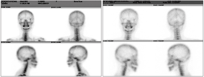
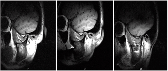
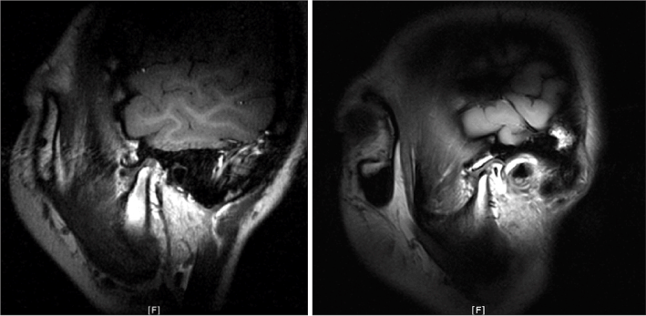
Among class II malocclusion patients with TMD symptom, clicking improved in 29.1%, and maximum mouth opening increased from 34.5±2.1 mm to 37.2±3.5 mm. The differences were not statistically significant, however. Radiographic changes in bone scan improved slightly based on the report by radiologist but not in TMJ dynamic MRI.

Conclusion

No particular improvements were found in patients with joint sound only. Patients with limitation of mouth opening showed an increase in the degree of opening, but the difference was not statistically significant (P>0.05).

REFERENCES

1. Wisth PJ. Mandibular function and dysfunction in patients with mandibular prognathism. Am J Orthod. 1984; 85:193–198.
crossref
2. Magnusson T, Ahlborg G, Svartz K. Function of the masticatory system in 20 patients with mandibular hypo- or hyperplasia after correction by a sagittal split osteotomy. Int J Oral Maxillofac Surg. 1990; 19:289–293.
crossref
3. O'Ryan F, Epker BN. Surgical orthodontics and the temporomandibular joint. I. Superior repositioning of the maxilla. Am J Orthod. 1983; 83:408–417.
4. Sanders B, Kaminishi R, Buoncristiani R, Davis C. Arthroscopic surgery for treatment of temporomandibular joint hypomobility after mandibular sagittal osteotomy. Oral Surg Oral Med Oral Pathol. 1990; 69:539–541.
crossref
5. Lee JY, Kim SG, Seung JH, Ahn JM. Alterations of temporomandibular joint symptom after orthognathic surgery. J Korean Assoc Maxillofac Plast Reconstr Surg. 2003; 25:448–451.
6. White CS, Dolwick MF. Prevalence and variance of temporomandibular dysfunction in orthognathic surgery patients. Int J Adult Orthodon Orthognath Surg. 1992; 7:7–14.
7. Kerstens HC, Tuinzing DB, van der Kwast WA. Temporomandibular joint symptoms in orthognathic surgery. J Craniomaxillofac Surg. 1989; 17:215–218.
crossref
8. Wolford LM, Reiche-Fischel O, Mehra P. Changes in temporomandibular joint dysfunction after orthognathic surgery. J Oral Maxillofac Surg. 2003; 61:655–660.
crossref
9. Proffit WR, Phillips C, Dann C 4th, Turvey TA. Stability after surgical-orthodontic correction of skeletal Class III malocclusion I. Mandibular setback. Int J Adult Orthodon Orthognath Surg. 1991; 6:7–18.
10. Panula K, Somppi M, Finne K, Oikarinen K. Effects of orthognathic surgery on temporomandibular joint dysfunction. A controlled prospective 4-year follow-up study. Int J Oral Maxillofac Surg. 2000; 29:183–187.
crossref
11. Aoyama S, Kino K, Kobayashi J, Yoshimasu H, Amagasa T. Clinical evaluation of the temporomandibular joint following orthognathic surgery--multiple logistic regression analysis. J Med Dent Sci. 2005; 52:109–114.
12. Farella M, Michelotti A, Bocchino T, Cimino R, Laino A, Steenks MH. Effects of orthognathic surgery for class III malocclusion on signs and symptoms of temporomandibular disorders and on pressure pain thresholds of the jaw muscles. Int J Oral Maxillofac Surg. 2007; 36:583–587.
crossref
13. Dervis E, Tuncer E. Long-term evaluations of temporomandibular disorders in patients undergoing orthognathic surgery compared with a control group. Oral Surg Oral Med Oral Pathol Oral Radiol Endod. 2002; 94:554–560.
crossref
14. Pahkala RH, Kellokoski JK. Surgical-orthodontic treatment and patients' functional and psychosocial well-being. Am J Orthod Dentofacial Orthop. 2007; 132:158–164.
crossref
15. Lindenmeyer A, Sutcliffe P, Eghtessad M, Goulden R, Speculand B, Harris M. Oral and maxillofacial surgery and chronic painful temporomandibular disorders--a systematic review. J Oral Maxillofac Surg. 2010; 68:2755–2764.
crossref

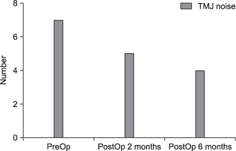
Fig. 1.
Changes in TMJ noise after orthognathic surgery in group 1. (TMJ: temporomandibular joint, PreOp: preoperative, PostOp: postoperative)
jkaoms-38-139f1.tif
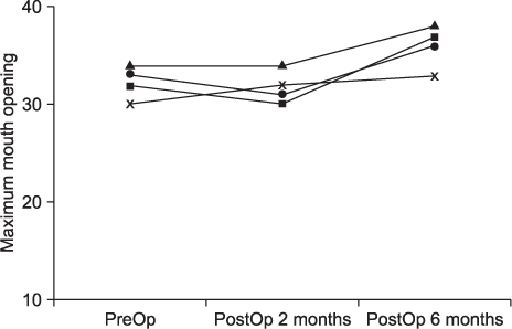
Fig. 2.
Changes after operation in limitation of mouth opening only (group 2). (PreOp: preoperative, PostOp: postoperative)
jkaoms-38-139f2.tif
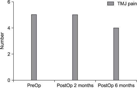
Fig. 3.
Changes after the operation in group 3 patient (group 3). (TMJ: temporomandibular joint, PreOp: preoperative, PostOp: postoperative)
jkaoms-38-139f3.tif
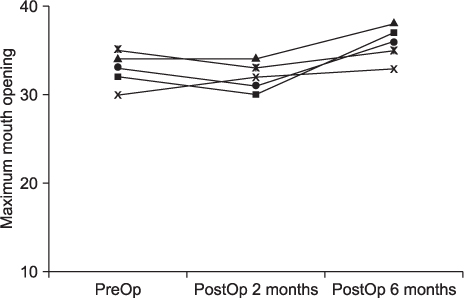
Fig. 4.
Changes of mouth opening after the operation in limited mouth opening with TMJ pain (group 4). (TMJ: temporomandibular joint, PreOp: preoperative, PostOp: postoperative)
jkaoms-38-139f4.tif
Fig. 5.
Changes of bone scan after orthognathic surgery (right: 2 years before, left: 1 year after orthognathic surgery).
jkaoms-38-139f5.tif
Fig. 6.
Changes of magnetic resonance imaging after orthognathic surgery. 2 years, 1 month, and 1 year after orthognathic surgery. Anterior disc displacement has not changed (patient 1, 2-jaw). (LT: left)
jkaoms-38-139f6.tif
Fig. 7.
Changes of magnetic resonance imaging after orthognathic surgery. 2 years before, and 1 year after orthognathic surgery. Anterior disc displacement has not changed (patient 2). (LT: left)
jkaoms-38-139f7.tif
Table 1.
Classification of patients G4 PreOp and PostOp MMO
Patient No. of patients TMJ noise LOM (<35 mm) TMJ pain
G1 n=7 + - -
G2 n=4 - + -
G3 n=5 + - +
G4 n=5 - + +

(PreOp: preoperative, PostOp: postoperative, MMO: maximum mouth opening, TMJ: temporomandibular joint, LOM: limitation of mouth opening, G1: group 1, G2: group 2, G3: group 3, G4: group 4)

Table 2.
PreOp and PostOp MMO of G4
Patient PreOp PostOp 6 months
Patient 1 32.00 35.00
Patient 2 33.00 36.00
Patient 3 34.00 40.00
Patient 4 35.00 46.00
Patient 5 32.00 45.00

(PreOp: preoperative, PostOp: postoperative, MMO: maximum mouth opening, G4: group 4)

Table 3.
Paired t-test of group 4
Paired samples test1
Paired differences
T df Sig. (2-tailed)
Mean Std. deviation Std. error mean 95% confidence interval of the difference
Lower Upper
Pair 1 PreOp–PostOp 6 months -7.20000 4.60435 2.05913 -12.91705 -1.48295 -3.497 4 0.025

(Std.: standard, df: degree of freedom, Sig.: significant)

1 No statistics are computed for one or more split files.

Table 4.
Changes after the operation in bone scan findings
Patient PreOp TMJ bone scan findings PostOp (minimum 6 month after) TMJ bone scan findings
Patient 1 Suggestive arthritis Mild uptake
Patient 2 Mild uptake No activity on TMJ
Patient 3 Increased uptake Mild uptake
Patient 4 Mild uptake Increased uptake
Patient 5 Mild uptake Mild uptake

(PreOp: preoperative, PostOp: postoperative, TMJ: temporomandibular joint)

Table 5.
MRI changes in 2 patients
PreOp MRI PostOp MRI (minimum 6 month after)
Patient 1 ADD without reduction ADD without reduction, no change from PreOp MRI finding
Patient 2 ADD without reduction ADD without reduction, no change from PreOp MRI finding

(PreOp: preoperative, PostOp: postoperative, MRI: magnetic resonance imaging, ADD: anterior disc displacement)

TOOLS
Similar articles