Journal List > Ann Lab Med > v.37(6) > 1091555

Choi, Kim, Yoon, Choi, Park, Seong, Kim, Hwang, Choi, Chae, Kim, Kim, Lee, Nam, and Lim: A Unique Mutational Spectrum of MLC1 in Korean Patients With Megalencephalic Leukoencephalopathy With Subcortical Cysts: p.Ala275Asp Founder Mutation and Maternal Uniparental Disomy of Chromosome 22

Abstract

Background

Megalencephalic leukoencephalopathy with subcortical cysts (MLC) is a rare inherited disorder characterized by infantile-onset macrocephaly, slow neurologic deterioration, and seizures. Mutations in the causative gene, MLC1, are found in approximately 75% of patients and are inherited in an autosomal recessive manner. We analyzed MLC1 mutations in five unrelated Korean patients with MLC.

Methods

Direct Sanger sequencing was used to identify MLC1 mutations. A founder effect of the p.Ala275Asp variant was demonstrated by haplotype analysis using single-nucleotide polymorphic (SNP) markers. Multiple ligation-dependent probe amplification (MLPA) and comparative genomic hybridization plus SNP array were used to detect exonic deletions or uniparental disomy (UPD).

Results

The most prevalent pathogenic variant was c.824C>A (p.Ala275Asp) found in 7/10 (70%) alleles. Two pathogenic frameshift variants were found: c.135delC (p.Cys46Alafs*12) and c.337_353delinsG (p.Ile113Glyfs*4). Haplotype analysis suggested that the Korean patients with MLC harbored a founder mutation in p.Ala275Asp. The p.(Ile113Glyfs*4) was identified in a homozygous state, and a family study revealed that only the mother was heterozygous for this variant. Further analysis of MLPA and SNP arrays for this patient demonstrated loss of heterozygosity of chromosome 22 without any deletion, indicating UPD. The maternal origin of both chromosomes 22 was demonstrated by haplotype analysis.

Conclusions

This study is the first to describe the mutational spectrum of Korean patients with MLC, demonstrating a founder effect of the p.Ala275Asp variant. This study also broadens our understanding of the mutational spectrum of MLC1 by demonstrating a homozygous p.(Ile113Glyfs*4) variant resulting from UPD of chromosome 22.

INTRODUCTION

Megalencephalic leukoencephalopathy with subcortical cysts (MLC; MIM 604004) is an autosomal recessive leukodystrophy characterized by infantile-onset macrocephaly and gradual motor deterioration. Infants with MLC are healthy at birth, but macrocephaly develops during the first year of life. Early development is usually normal, but motor disability in the form of spasticity and ataxia manifests slowly [12]. Many individuals with this condition experience epileptic seizures, which are easily controlled with medication. Cognitive function is relatively well preserved. Magnetic resonance imaging (MRI) shows diffuse signal abnormality in cerebral white matter and subcortical cysts in the anterior temporal region.
A causative gene, MLC1, was first identified in 2001 and maps to chromosome 22q13.33 [34]. Approximately 75% of MLC patients have MLC1 mutations, which are inherited in an autosomal recessive manner. Although MLC patients with MLC1 pathogenic variants may show clinical variability, no evidence of genotype-phenotype correlation has been reported [5]. More than 90 different MLC1 mutations have been identified [6789]. There are several reports that suggest founder effects. An ancestral mutation has also been described in Indian Agarwal [10], Jewish [11], Egyptian [12], and Japanese patients [13]. To date, there has been no study on the mutational spectrum of MLC1 in Korean MLC patients.
We performed a comprehensive mutational analysis of MLC1 in five Korean patients with MLC. We found a unique mutational spectrum of MLC1 in these patients by demonstrating a founder mutation. We also presented a case of MLC caused by maternal uniparental disomy (UPD) of chromosome 22.

METHODS

1. Clinical features

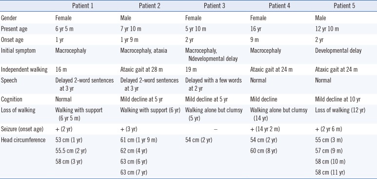
We studied five patients with MLC from unrelated Korean families (Table 1). All five MLC patients were born to nonconsanguineous Korean parents. Initial presentations were macrocephaly and/or motor developmental delay. Macrocephaly was usually detected before 18 months of age (range, 9 months to 2 yr). All patients could walk independently (range, 16–28 months), although most of them showed gait ataxia. Three of the patients exhibited delayed language development and mild declines in cognitive function during the follow-up period, for a median of 8 yr (range, 3–15 yr). Only one patient (Patient 5) was wheelchair bound at the age of 12 yr. Four of the five patients (80%) experienced epileptic seizures, which were easily controlled with antiepileptic medications. All patients had typical MRI findings showing diffuse signal abnormalities in cerebral white matter and the presence of subcortical cysts in the anterior temporal regions (Supplemental Data Fig. S1).

2. Mutational analysis

The study was approved by the institutional review board at Seoul National University Hospital, Korea (IRB No. 1605-133-765). Written informed consent was obtained from all participants. Genomic DNA was obtained from blood samples for each patient and their parents. Direct Sanger sequencing of the entire coding region of the MLC1 (NM_015166.3) was performed. To further evaluate the mutation of Patient 1, we applied multiple ligation-dependent probe amplification (MLPA) (SALSA MLPA P107; MRC-Holland, Amsterdam, The Netherlands) and used a SurePrint G human genome comparative genome hybridization (CGH) plus single nucleotide polymorphism (SNP) 400K microarray (Agilent Technologies, Santa Clara, CA, USA). All procedures were performed according to the manufacturers' protocols. For variant interpretation, we followed the international guidelines of the American College of Medical Genetics and Genomics and the Association for Molecular Pathology (ACMG/AMP) [14].

3. Genotyping and haplotype reconstruction

We selected 10 SNPs with a minor allele frequency of 0.39–0.50 in this population around the MLC1 region spanning 170 kb as follows: rs36168477, rs5771069, rs137855, rs5771305, rs2010453, rs932330, rs138208, rs731857, rs138244, and rs6010148. Genotyping of 39 unrelated normal controls and five patient families was done by direct sequencing using specific primers for each polymorphic marker. Hardy–Weinberg equilibrium was calculated by using Arlequin (version 3.5.2.2) [15]. Haplotypes were reconstructed by using PHASE (version 2.1) [16].

RESULTS

1. Mutational analysis

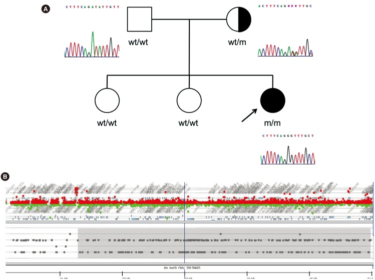
Direct sequencing of the MLC1 in all five patients and available family members revealed three pathogenic variants (Table 2). The p.Ala275Asp variants in exon 10 were observed in seven of 10 alleles (70%). This missense variant was heterozygous in both parents and was inherited in an autosomal recessive manner. Two frameshift variants p.(Cys46Alafs*12) and p.(Ile113Glyfs*4) were also identified. All three variants mentioned were considered pathogenic according to the international guidelines of the ACMG/AMP (Supplemental Data Table S1). Patient 1 was homozygous for the p.(Ile113Glyfs*4) variant. Family studies were performed for this female patient. Only her mother was heterozygous for the p.(Ile113Glyfs*4) variant (Fig. 1A). MLPA did not reveal multiple exon deletions of MLC1. By using the CGH plus SNP microarray analyses, loss-of-heterozygosity of chromosome 22q11.22-q13.33 including MLC1 was predicted. However, no evidence of any large deletion involving the same chromosomal region was identified (Fig. 1B). Therefore, we concluded that a UPD spanning the region of chromosome 22q11.22-q13.33 including the MLC1 was the most plausible mechanism for the homozygous state of the p.(Ile113Glyfs*4) variant in Patient 1.
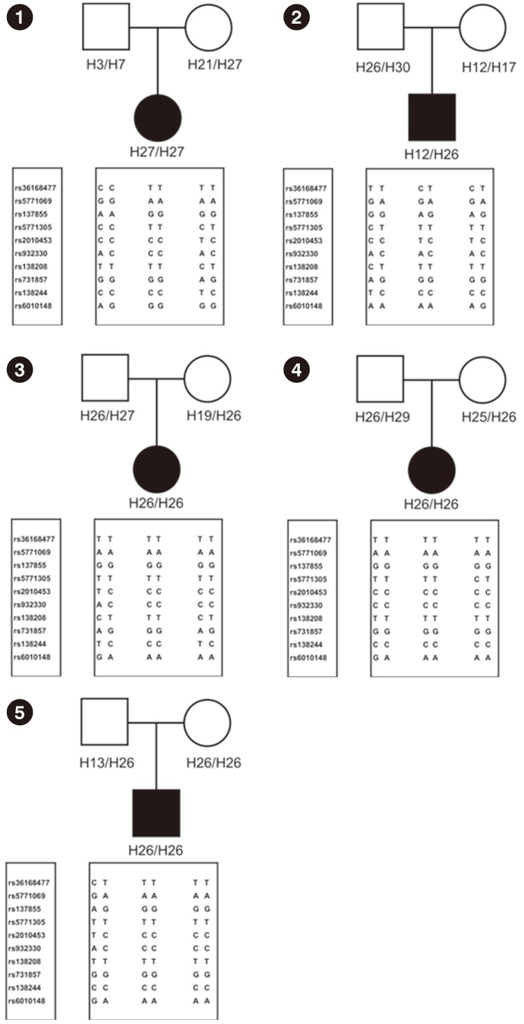
In addition, two chromosomes 22 harboring the MLC1 region were of maternal origin in this patient as shown by haplotype analysis using 10 SNP markers (Fig. 2).

2. Founder effect

Thirty haplotypes were obtained from our genotype data (Supplemental Data Table S2). Haplotype H26 was found in three patients (Patients 3, 4, and 5) with a homozygous p.Ala275Asp variant, and was also found in one patient (Patient 2) along with a heterozygous p.Ala275Asp variant (Fig. 2). Haplotype H26 was segregated within each family, respectively, suggesting that the p.Ala275Asp variant had a common origin.

DISCUSSION

We reported three different MLC1 pathogenic variants from five MLC patients. Seven alleles contained the p.Ala275Asp variant in exon 10. Two frameshift variants p.(Cys46Alafs*12) and p.(Ile113Glyfs*4) were also identified. Using the ACMG/AMP sequence interpretation guidelines [14], we classified all three variants as pathogenic.
The p.Ala275Asp variant was the most prevalent, most common ancestral mutation in the Korean population. Since it was first reported by Montagna et al [17], this variant has been reported in two Japanese MLC patients [1718]. This variant was regarded as the second most common variant in Japanese patients after the p.Ser93Leu variant, which accounts for approximately 80% of Japanese patients with MLC [19]. The p.(Ile113Glyfs*4) variant found in Patient 1 was also reported in one Japanese patient with MLC [12]. These findings suggest a shared genetic background between Japanese and Korean patients with MLC. However, there appears to be clear differences between these two ethnic groups, because the p.Ser93Leu variant was not found in our study.
Patient 1 had a homozygous frameshift variant caused by maternal UPD of chromosome 22. UPD can cause disease by meiotic nondisjunction followed by gamete complementation, trisomy rescue, monosomy duplication, and somatic chromosomal chiasmata [20]. Nondisjunction during meiosis I produces uniparental heterodisomy, in which both homologs of a part of a chromosome from a single parent are passed on to the offspring. By contrast, nondisjunction during meiosis II produces isodisomy, in which two genetically identical copies of a single parental chromosome are inherited [21]. Our results from MLC1 sequencing, MLPA, CGH plus SNP array, and haplotype analysis suggested that UPD of chromosome 22 including the MLC1 region caused MLC in this patient. UPD can cause autosomal recessive disorders, particularly when only one of the parents is a carrier [22]. UPD of chromosome 22 as a disease-causing mechanism has been documented in cases of metachromatic leukodystrophy [23], congenital methemoglobinemia [24], and PLA2G6-associated neurodegeneration [25]. Interestingly, Cao et al [26] recently reported the first case of MLC caused by maternal UPD of chromosome 22. There is no evidence that this could occur more frequently on chromosome 22 than on other chromosomes. However, our case could be an additional example, showing that UPD of chromosome 22 needs to be considered as a possible cause of MLC when conducting MLC1 mutational analysis.
This study is the first to present genetic analyses of Korean patients with MLC. From haplotype analysis, the p.Ala275Asp variant was proven to be an ancestral mutation. We also found that MLC could be caused by maternal UPD, which broadens the documented mutational spectrum of MLC1.

Acknowledgments

This research was supported by Basic Science Research Program through the National Research Foundation of Korea (NRF) funded by the Ministry of Science, ICT & Future Planning (2013-R1A1A1008888).

Notes

Authors' Disclosures of Potential Conflicts of Interest: No potential conflicts of interest relevant to this article were reported.

References

1. van der Knaap MS, Barth PG, Stroink H, van Nieuwenhuizen O, Arts WF, Hoogenraad F, et al. Leukoencephalopathy with swelling and a discrepantly mild clinical course in eight children. Ann Neurol. 1995; 37:324–334. PMID: 7695231.
2. Topcu M, Saatci I, Topcuoglu MA, Kose G, Kunak B. Megalencephaly and leukodystrophy with mild clinical course: a report on 12 new cases. Brain Dev. 1998; 20:142–153. PMID: 9628190.
3. Topçu M, Gartioux C, Ribierre F, Yalçinkaya C, Tokus E, Oztekin N, et al. Vacuoliting megalencephalic leukoencephalopathy with subcortical cysts, mapped to chromosome 22qtel. Am J Hum Genet. 2000; 66:733–739. PMID: 10677334.
4. Leegwater PA, Yuan BQ, van der Steen J, Mulders J, Könst AA, Boor PK, et al. Mutations of MLC1 (KIAA0027), encoding a putative membrane protein, cause megalencephalic leukoencephalopathy with subcortical cysts. Am J Hum Genet. 2001; 68:831–838. PMID: 11254442.
5. van der Knaap MS, Boor I, Estévez R. Megalencephalic leukoencephalopathy with subcortical cysts: chronic white matter oedema due to a defect in brain ion and water homoeostasis. Lancet Neurol. 2012; 11:973–985. PMID: 23079554.
6. López-Hernández T, Sirisi S, Capdevila-Nortes X, Montolio M, Fernández-Dueñas V, Scheper GC, et al. Molecular mechanisms of MLC1 and GLIALCAM mutations in megalencephalic leukoencephalopathy with subcortical cysts. Hum Mol Genet. 2011; 20:3266–3277. PMID: 21624973.
7. Leegwater PA, Boor PK, Yuan BQ, van der Steen J, Visser A, Könst AA, et al. Identification of novel mutations in MLC1 responsible for megalencephalic leukoencephalopathy with subcortical cysts. Hum Genet. 2002; 110:279–283. PMID: 11935341.
8. Ilja Boor PK, de Groot K, Mejaski-Bosnjak V, Brenner C, van der Knaap MS, Scheper GC, et al. Megalencephalic leukoencephalopathy with subcortical cysts: an update and extended mutation analysis of MLC1. Hum Mutat. 2006; 27:505–512. PMID: 16652334.
9. Stenson PD, Mort M, Ball EV, Shaw K, Phillips A, Cooper DN. The human gene mutation database: building a comprehensive mutation repository for clinical and molecular genetics, diagnostic testing and personalized genomic medicine. Hum Genet. 2014; 133:1–9. PMID: 24077912.
10. Gorospe JR, Singhal BS, Kainu T, Wu F, Stephan D, Trent J, et al. Indian Agarwal megalencephalic leukodystrophy with cysts is caused by a common MLC1 mutation. Neurology. 2004; 62:878–882. PMID: 15037685.
11. Ben-Zeev B, Levy-Nissenbaum E, Lahat H, Anikster Y, Shinar Y, Brand N, et al. Megalencephalic leukoencephalopathy with subcortical cysts; a founder effect in Israeli patients and a higher than expected carrier rate among Libyan Jews. Hum Genet. 2002; 111:214–218. PMID: 12189496.
12. Mahmoud IG, Mahmoud M, Refaat M, Girgis M, Waked N, El Badawy A, et al. Clinical, neuroimaging, and genetic characteristics of megalencephalic leukoencephalopathy with subcortical cysts in Egyptian patients. Pediatr Neurol. 2014; 50:140–148. PMID: 24315536.
13. Tsujino S, Kanazawa N, Yoneyama H, Shimono M, Kawakami A, Hatanaka Y, et al. A common mutation and a novel mutation in Japanese patients with van der Knaap disease. J Hum Genet. 2003; 48:605–608. PMID: 14615938.
14. Richards S, Aziz N, Bale S, Bick D, Das S, Gastier-Foster J, et al. Standards and guidelines for the interpretation of sequence variants: a joint consensus recommendation of the American College of Medical Genetics and Genomics and the Association for Molecular Pathology. Genet Med. 2015; 17:405–424. PMID: 25741868.
15. Schneider S, Roessli D, et al. Arlequin: a software for population genetics data analysis. User manual ver 2.000. Geneva: Genetics and Biometry Lab, Dept. of Anthropology, University of Geneva;2000.
16. Stephens M, Scheet P. Accounting for decay of linkage disequilibrium in haplotype inference and missing-data imputation. Am J Hum Genet. 2005; 76:449–462. PMID: 15700229.
17. Montagna G, Teijido O, Eymard-Pierre E, Muraki K, Cohen B, Loizzo A, et al. Vacuolating megalencephalic leukoencephalopathy with subcortical cysts: functional studies of novel variants in MLC1. Hum Mutat. 2006; 27:292.
18. Shimada S, Shimojima K, Masuda T, Nakayama Y, Kohji T, Tsukamoto H, et al. MLC1 mutations in Japanese patients with megalencephalic leukoencephalopathy with subcortical cysts. Hum Genome Var. 2014; 1:14019. PMID: 27081509.
19. Saijo H, Nakayama H, Ezoe T, Araki K, Sone S, Hamaguchi H, et al. A case of megalencephalic leukoencephalopathy with subcortical cysts (van der Knaap disease): molecular genetic study. Brain Dev. 2003; 25:362–366. PMID: 12850517.
20. Ledbetter DH, Engel E. Uniparental disomy in humans: development of an imprinting map and its implications for prenatal diagnosis. Hum Mol Genet. 1995; 4:1757–1764. PMID: 8541876.
21. Engel E, DeLozier-Blanchet CD. Uniparental disomy, isodisomy, and imprinting: probable effects in man and strategies for their detection. Am J Med Genet. 1991; 40:432–439. PMID: 1746607.
22. Zlotogora J. Parents of children with autosomal recessive diseases are not always carriers of the respective mutant alleles. Hum Genet. 2004; 114:521–526. PMID: 15024643.
23. Niida Y, Kuroda M, Mitani Y, Yokoi A, Ozaki M. Paternal uniparental isodisomy of chromosome 22 in a patient with metachromatic leukodystrophy. J Hum Genet. 2012; 57:687–690. PMID: 22854541.
24. Huang YH, Tai CL, Lu YH, Wu TJ, Chen HD, Niu DM. Recessive congenital methemoglobinemia caused by a rare mechanism: maternal uniparental heterodisomy with segmental isodisomy of a chromosome 22. Blood Cells Mol Dis. 2012; 49:114–117. PMID: 22658170.
25. Zhang P, Gao Z, Jiang Y, Wang J, Zhang F, Wang S, et al. Follow-up study of 25 Chinese children with PLA2G6-associated neurodegeneration. Eur J Neurol. 2013; 20:322–330. PMID: 22934738.
26. Cao B, Yan H, Guo M, Xie H, Wu Y, Gu Q, et al. Ten novel mutations in Chinese patients with megalencephalic leukoencephalopathy with subcortical cysts and a long-term follow-up research. PLoS One. 2016; 11:e0157258. PMID: 27322623.

SUPPLEMENTARY MATERIALS

Supplemental Data Fig. S1

Brain magnetic resonance images of the five patients in this study. All five patients showed diffuse high signal intensity in cerebral white matter on T2-weighted axial images (on the left panel). Subcortical cysts in the anterior temporal lobes were also present in T1-weighted axial images (on the right panel). (A, B) Patient 1 (6 yr), (C, D) Patient 2 (1 yr 9 months), (E, F) Patient 3 (2 yr), (G, H) Patient 4 (2 yr), and (I, J) Patient 5 (3 yr).
alm-37-516-s001.pdf

Supplemental Data Table S1

Interpretation of three putative sequence variants based on the American College of Medical Genetics and Genomics and the Association for Molecular Pathology criteria
alm-37-516-s002.pdf

Supplemental Data Table S2

MLC1 haplotypes reconstructed by using 10 single nucleotide polymorphisms
alm-37-516-s003.pdf
Fig. 1

Pedigree and MLC1 mutational analysis of Patient 1. (A) Pedigree of Patient 1 and the c.337_353del17insG mutation status in her family. Direct Sanger sequencing revealed that c.337_353 del17insG mutation was homozygous in the patient and heterozygous in the mother. The father did not harbor this mutation. (B) Loss of heterogeneity spanning chromosome 22q11.22-q13.33 (Chr22: 23279752-51103692, hg19) was predicted by the CGH plus SNP array.

Abbreviations: wt, wild type; m, mutant; CGH, comparative genomic hybridization; SNP, single nucleotide polymorphism.
alm-37-516-g001
Fig. 2

Results of haplotype analysis in the five families. Families of Patient 1-5 are presented as a number accordingly.

alm-37-516-g002
Table 1

Clinical features of five Korean patients with megalencephalic leukoencephalopathy with subcortical cysts

alm-37-516-i001
Patient 1 Patient 2 Patient 3 Patient 4 Patient 5
Gender Female Male Female Female Male
Present age 6 yr 5 m 7 yr 10 m 5 yr 10 m 16 yr 12 yr 10 m
Onset age 1 yr 1 yr 9 m 2 yr 9 m 2 yr
Initial symptom Macrocephaly Macrocephaly, ataxia Macrocephaly, Ndevelopmental delay Macrocephaly Developmental delay
Independent walking 16 m Ataxic gait at 28 m 19 m Ataxic gait at 24 m Ataxic gait at 24 m
Speech Delayed 2-word sentences at 3 yr Delayed 2-word sentences at 3 yr Delayed with a few words at 2 yr Normal Normal
Cognition Normal Mild decline at 5 yr Mild decline at 5 yr Mild decline Mild decline at 10 yr
Loss of walking Walking with support (6 yr 5 m) Walking with support (6 yr) Walking alone but clumsy (5 yr) Walking alone but clumsy (14 yr) Loss of walking (12 yr)
Seizure (onset age) + (2 yr) + (3 yr) + (14 yr 2 m) + (2 yr 6 m)
Head circumference 53 cm (1 yr) 61 cm (1 yr 9 m) 54 cm (2 yr) 54 cm (2 yr) 55 cm (3 m)
55.5 cm (2 yr) 62 cm (4 yr) 60 cm (8 yr) 57 cm (9 m)
58 cm (3 yr) 63 cm (6 yr) 58 cm (10 m)
63 cm (7 yr) 58 cm (11 yr)

Abbreviation: m, months.

Table 2

MLC1 (NM_015166.3) mutation status of five Korean patients with megalencephalic leukoencephalopathy with subcortical cysts

alm-37-516-i002
Patient Allele 1 Allele 2
Nucleotide change Amino acid change Nucleotide change Amino acid change
Patient 1 c.337_353delinsG p.(Ile113Glyfs*4) c.337_353delinsG p.(Ile113Glyfs*4)
Patient 2 c.135delC p.(Cys46Alafs*12) c.824C > A p.Ala275Asp
Patient 3 c.824C > A p.Ala275Asp c.824C > A p.Ala275Asp
Patient 4 c.824C > A p.Ala275Asp c.824C > A p.Ala275Asp
Patient 5 c.824C > A p.Ala275Asp c.824C > A p.Ala275Asp
TOOLS
Similar articles