Journal List > Ann Lab Med > v.37(3) > 1091501

Lee, Lee, Ng, Chiu, Tang, Loh, and Koay: Comparison of Luminex NxTAG Respiratory Pathogen Panel and xTAG Respiratory Viral Panel FAST Version 2 for the Detection of Respiratory Viruses

Abstract

Owing to advancements in molecular diagnostics, recent years have seen an increasing number of laboratories adopting respiratory viral panels to detect respiratory pathogens. In December 2015, the NxTAG respiratory pathogen panel (NxTAG RPP) was approved by the United States Food and Drug Administration. We compared the clinical performance of this new assay with that of the xTAG respiratory viral panel (xTAG RVP) FAST v2 using 142 clinical samples and 12 external quality assessment samples. Discordant results were resolved by using a laboratory-developed respiratory viral panel. The NxTAG RPP achieved 100% concordant negative results and 86.6% concordant positive results. It detected one coronavirus 229E and eight influenza A/H3N2 viruses that were missed by the xTAG RVP FAST v2. On the other hand, the NxTAG RPP missed one enterovirus/rhinovirus and one metapneumovirus that were detected by FAST v2. Both panels correctly identified all the pathogens in the 12 external quality assessment samples. Overall, the NxTAG RPP demonstrated good diagnostic performance. Of note, it was better able to subtype the influenza A/H3N2 viruses compared with the xTAG RVP FAST v2.

Respiratory tract infection is a leading cause of death worldwide [1]. Laboratory testing is required to identify the underlying etiologic agent of respiratory infections, as they commonly present with similar signs and symptoms [2]. The xTAG respiratory viral panel (xTAG RVP) FAST v2 is a multiplexed molecular assay for respiratory viral infections manufactured by Luminex Corp. (Austin, TX, USA) that has been routinely used in our clinical laboratory to detect respiratory viruses.
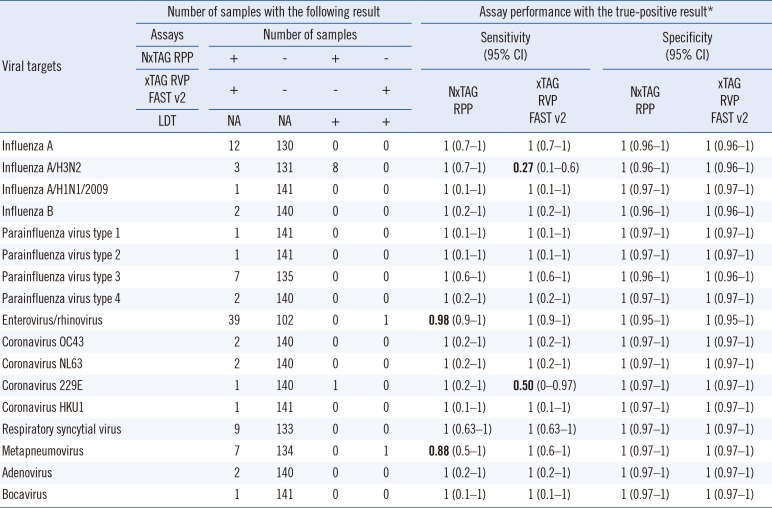
In December 2015, Luminex introduced the NxTAG respiratory pathogen panel (NxTAG RPP), following approval from United States Food and Drug Administration. Both the NxTAG RPP and xTAG RVP FAST v2 have the same number of viral targets, including influenza A virus (A/H3N2, A/H1N1, and A/H1N1/2009 strains), influenza B virus, parainfluenza virus types 1 to 4 (PIV 1-4), enterovirus/rhinovirus, coronaviruses (OC43, NL63, 229E, and HKU1), respiratory syncytial virus (RSV) A and B, metapneumovirus, adenovirus, and bocavirus. The NxTAG RPP has two additional atypical bacterial targets, namely Mycoplasma pneumoniae and Chlamydophila pneumoniae. Recent studies have compared the performance of the new NxTAG RPP with that of other respiratory panels such as the BioFire FilmArray RVP [34], RespiFinder-22 [5], Anyplex II RV16 [6], and xTAG RVP FAST v2 [7]. Overall, these reports demonstrated that the NxTAG RPP is at least comparable to, if not better than, some of the comparators. Here, we assessed the clinical performance of the NxTAG RPP versus the xTAG RVP FAST v2 in detecting respiratory viruses.
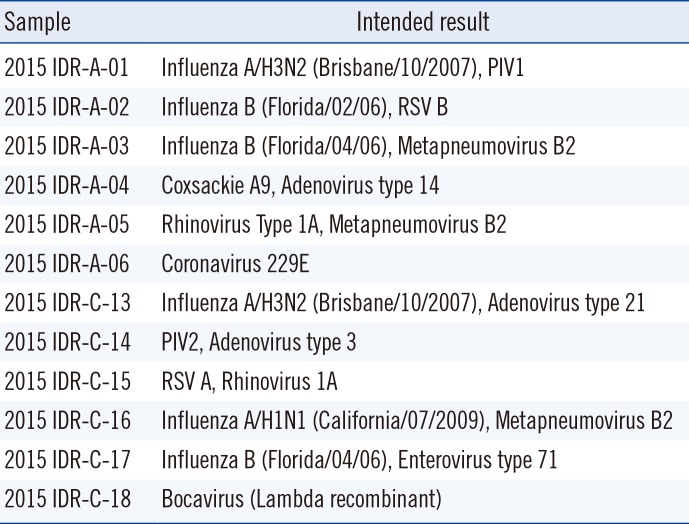
This study was approved by the local institutional ethics board (National Healthcare Group Domain-Specific Review Board A, reference: 2016/00044) and was performed between May and December 2015. Here, 142 de-identified clinical respiratory samples submitted to the Molecular Diagnosis Centre of the Singapore National University Hospital were included (see Table 1 for the list of viral pathogens included). Additionally, 12 external quality assessment (EQA) samples from the College of American Pathologists (CAP) infectious disease respiratory panel, received in year 2015, were tested (Table 2). Total nucleic acid was extracted with the Qiagen EZ1 Virus Mini Kit v2.0 on the BioRobot EZ1 extractor (Qiagen, Hilden, Germany).
All samples were initially tested with the xTAG RVP FAST v2 as part of our routine clinical service. In brief, the extracted nucleic acid (10 µL) was used for target amplification by multiplex reverse transcription PCR (RT-PCR). The PCR product (2 µL) was hybridized to a bead mix; next, reporter dye was added in a new reaction vessel, which was sealed and incubated. The amplification and hybridization/incubation were performed on the Applied Biosystems Veriti thermal cycler (Thermo Fisher Scientific, Wohlen, Switzerland), as per the manufacturer's recommendations. Signal acquisition was performed on the MAGPIX instrument (Luminex Corp). After testing, the extracted nucleic acids were immediately frozen at −80℃ until further testing.
Residual frozen archival samples were retrieved and tested with the NxTAG RPP, a closed-tube nucleic acid assay containing premixed lyophilized reagents for target amplification, PCR product hybridization/incubation, and detection. All procedures were carried out according to the manufacturer's instructions. The extracted nucleic acid (35 µL) was added to resuspend the preplated lyophilized bead reagents in the vessel. Multiplex RT-PCR, bead hybridization, and reporter dye incubation were performed on the Veriti thermal cycler, as per the manufacturer's recommendations. Finally, the vessel was placed onto the MAGPIX instrument for signal acquisition.
When discordant results were found between the two assays for a particular sample, a third method—a laboratory-developed, clinically validated RVP—was used for confirmation. The laboratory-developed RVP methodology is described in Supplemental file S1. In this scenario, the result concurrent between any two of the three methods was considered true. The concordance rate and Cohen's kappa coefficient of the two Luminex assays were determined by using GraphPad QuickCalcs (GraphPad, La Jolla, CA, USA).
Of the 142 clinical samples tested, 131 had concordant results, 60 and 71 of which were negative and positive, respectively. The 11 discordant samples containing metapneumovirus, enterovirus/rhinovirus, coronavirus 229E, and eight influenza A/H3N2 viruses tested positive in the laboratory-developed RVP assay and thus, were considered true positives (Table 1). The overall concordance rate between the two Luminex assays was 92.3% (131/142) with a Cohen's kappa coefficient of 0.85 (95% confidence interval [CI] 0.757–0.932), indicating a substantial degree of agreement. Of the discordant samples, the xTAG RVP FAST v2 missed eight influenza A/H3N2 viruses and one coronavirus 229E, while the NxTAG RPP missed one enterovirus/rhinovirus and one metapneumovirus. On the basis of the CAP results, both NxTAG RPP and xTAG RVP FAST v2 correctly identified all the pathogens in the samples tested.
The NxTAG RPP detected the presence of M. pneumoniae in one of the samples included in this study. This finding was confirmed by using a commercial real-time PCR assay for Mycoplasma, the Venor GeM qEP Mycoplasma detection kit (Minerva Biolabs GmbH, Berlin, Germany). Subsequent sensitivity testing using the lyophilized 10 colony-forming units (CFU) Sensitivity Standards (Minerva Biolabs GmbH) showed that NxTAG assay was capable of detecting M. pneumoniae strain down to 10 CFUs/PCR.
We assessed the clinical performance of the new NxTAG RPP against that of the xTAG RVP FAST v2 using a representative panel of viral pathogens and negatives. Notably, the seasonal influenza A/H1N1 virus was not detected in our local population, and was not included in this study. This strain appears to have been completely replaced by the pandemic influenza A/H1N1/2009 virus since 2009/2010 [8]. Overall, both assays showed comparable sensitivity and specificity for all viral targets, except for the influenza A/H3N2 virus. Notably, the xTAG RVP FAST v2 showed poor performance in influenza A/H3N2 subtyping, which may be due to primer mismatches. To the best of our knowledge, only one study has compared the performance of the NxTAG RPP with that of the xTAG RVP FAST v2; however, missed detection of influenza A/H3N2 was not reported [7]. It is unclear whether the missed detection by the xTAG RVP FAST v2 was related to the variant H3N2 virus reported by the Cen-ters for Disease Control and Prevention (Atlanta) recently [910]. Nonetheless, the inability to simultaneously detect and subtype these H3N2 viruses is a major hindrance for clinical laboratories to return test results within established turn-around-time. The influenza A/H3N2 virus is a clinically significant respiratory pathogen. Therefore, the ability to rapidly provide subtype information is important during an outbreak or in epidemiologic investigations. By contrast, the influenza A/H3N2 primers in the NxTAG RPP have been updated to detect these untypable strains. Our study suggests that existing xTAG RVP FAST v2 users should switch to the NxTAG RPP, which has better sensitivity for influenza A/H3N2, without a significant drop in sensitivity for the other respiratory viral targets.
Enterovirus/rhinovirus infections comprised 27.5% (39/142) of our study population. However, the inability of both Luminex assays to distinguish enterovirus from rhinovirus infections in patients lowers their overall clinical utility. This distinction is clinically important, particularly for septic workups in neonates and other vulnerable/immunocompromised patients, as enteroviruses can disseminate to cause systemic infection and involve multiple organs, whereas rhinoviruses generally do not [11]. The RVP assay can detect multiple viral targets simultaneously. Our study revealed nine cases of co-infections. Most of the co-infections involved enterovirus/rhinovirus (78%), consistent with results of previous studies [1213], and 43% of these cases involved enterovirus/rhinovirus and RSV.
A limitation of the current study is that the numbers per target were relatively low for influenza B, parainfluenza virus types 1, 2, and 4, coronaviruses, adenovirus, and bocavirus, and may not be sufficient to reflect the true diagnostic capability of the two assays. Such low detection rates of these viruses have been observed elsewhere [1213]. Additionally, we were unable to evaluate the performance of the bacterial panel in the NxTAG RPP, as the additional bacterial targets were not detectable by the xTAG RVP FAST v2 or the laboratory-developed RVP. Further-more, we detected only a single case of M. pneumoniae with the NxTAG RPP.
Experimentally, the xTAG RVP FAST v2 assay had a turnaround time of 5 hr for 48 samples. However, the need to manipulate post-amplification products presents an inherent risk for laboratory contamination. Moreover, the need to remove the seal from the vessel during the detection presents another potential source of sample cross-contamination, leading to false-positives. Besides cross-contamination, high background noise (Fig. 1) is another source of false-positives, which is commonly associated with the Luminex bead-based suspension array technology due to suboptimal hybridization conditions involving temperature divergences or operator variations. In contrast, the NxTAG RPP is a closed-tube, one-step system, which abolishes the need for post-amplification product manipulation and removal of the seal. The hands-on time is significantly reduced with the simplified workflow, alleviating process variations and giving a turnaround time of <4 hr for 48 samples. Overall, the streamlined workflow minimizes cross-contamination and background noise. However, initially, where the extracted nucleic acid is used to resuspend the preplated lyophilized bead reagents, the repeat pipetting can cause possible cross-over contamination. Finally, the NxTAG RPP can process between 1 and 96 samples per run, without wasting additional consumables or reagents. This flexible throughput can cater to the needs of laboratories with different and/or variable volume demands.
In conclusion, the two Luminex assays performed comparably for most pathogens, with the NxTAG RPP having the advantages of being able to detect atypical bacteria and having better diagnostic sensitivity for certain viruses.

Notes

Authors' Disclosures of Potential Conflicts of Interest: No potential conflicts of interest relevant to this article were reported.

References

1. World Health Organization. The top 10 causes of death. Updated on May 2014. Accessed on 27 October 2016. http://www.who.int/mediacentre/factsheets/fs310/en/.
2. Meissner HC. Uncertainty in the management of viral lower respiratory tract disease. Pediatrics. 2001; 108:1000–1003. PMID: 11581456.
3. Tang YW, Gonsalves S, Sun JY, Stiles J, Gilhuley KA, Mikhlina A, et al. Clinical evaluation of the Luminex NxTAG respiratory pathogen panel. J Clin Microbiol. 2016; 54:1912–1914. PMID: 27122378.
4. Chen JH, Lam HY, Yip CC, Wong SC, Chan JF, Ma ES, et al. Clinical evaluation of the new high-throughput Luminex NxTAG respiratory pathogen panel assay for multiplex respiratory pathogen detection. J Clin Microbiol. 2016; 54:1820–1825. PMID: 27122380.
5. Beckmann C, Hirsch HH. Comparing Luminex NxTAG-respiratory pathogen panel and RespiFinder-22 for multiplex detection of respiratory pathogens. J Med Virol. 2016; 88:1319–1324. PMID: 26856438.
6. Brotons P, Henares D, Latorre I, Cepillo A, Launes C, Muñoz-Almagro C. Comparison of NxTAG respiratory pathogen panel and Anyplex II RV16 tests for multiple detection of respiratory pathogens in hospitalized children. J Clin Microbiol. 2016; 54:2900–2904. PMID: 27629904.
7. Esposito S, Scala A, Bianchini S, Presicce ML, Mori A, Sciarrabba CS, et al. Partial comparison of the NxTAG respiratory pathogen panel assay with the Luminex xTAG respiratory panel fast assay V2 and singleplex real-time polymerase chain reaction for detection of respiratory pathogens. Diagn Microbiol Infect Dis. 2016; 86:53–57. PMID: 27401400.
8. Palese P, Wang TT. Why do influenza virus subtypes die out? A hypothesis. MBio. 2011; 2:pii:e00150-11.
9. Gray GC, Cao WC. Editorial commentary: variant Influenza A(H3N2) virus: looking through a glass, darkly. Clin Infect Dis. 2013; 57:1713–1714. PMID: 24065323.
10. Jhung MA, Epperson S, Biggerstaff M, Allen D, Balish A, Barnes N, et al. Outbreak of variant influenza A(H3N2) virus in the United States. Clin Infect Dis. 2013; 57:1703–1712. PMID: 24065322.
11. Rotbart HA. Antiviral therapy for enteroviruses and rhinoviruses. Antivir Chem Chemother. 2000; 11:261–271. PMID: 10950388.
12. Babady NE, Mead P, Stiles J, Brennan C, Li H, Shuptar S, et al. Comparison of the Luminex xTAG RVP Fast assay and the Idaho Technology FilmArray RP assay for detection of respiratory viruses in pediatric patients at a cancer hospital. J Clin Microbiol. 2012; 50:2282–2288. PMID: 22518855.
13. Gadsby NJ, Hardie A, Claas EC, Templeton KE. Comparison of the Luminex Respiratory Virus Panel fast assay with in-house real-time PCR for respiratory viral infection diagnosis. J Clin Microbiol. 2010; 48:2213–2216. PMID: 20357215.

Supplemental File S1

METHODS

1. Laboratory-developed respiratory viral panel
The laboratory-developed respiratory viral panel (RVP) is a capillary electrophoresis-based multiplex RT-PCR assay for the detection of 16 respiratory viral pathogens, including influenza A virus, influenza B virus, parainfluenza virus (PIV) types 1-4, enterovirus, rhinovirus, coronaviruses (OC43, NL63, 229E, and HKU1), respiratory syncytial virus (RSV) A and B, metapneumovirus, and adenovirus.
The RVP is comprised of 4 separate multiplex reactions. The first multiplex RT-PCR included enterovirus, rhinovirus, influenza A virus, influenza B virus, and metapneumovirus. The sequences of the primers were: enterovirus/rhinovirus, forward: 5′-GGC CCC TGA ATG YGG CTA A -3′, enterovirus-specific, reverse: 5′-HEX-GAA ACA CGG ACA CCC AAA GTA -3′, and rhinovirus-specific, reverse: 5′-HEX-CAA AGT AGT TGG TCC CAT CC -3′; influenza A, forward: 5′-FAM-GGA ATG GCT AAA GAC AAG ACC AAT -3′ and reverse: 5′-GGG CAT TTT GGA CAA AGC GTC TAC -3′; influenza B, forward: 5′-FAM-CCA GGG ATT GCA GAC ATT GA -3′ and reverse: 5′-ACA GGT GTT GCC ATA TTG TAA AGA G -3′; metapneumovirus, forward: 5′-CAT ATA AGC ATG CTA TAT TAA AAG AGT CTC -3′ and reverse: 5′-FAM-CCT ATT TCT GCA GCA TAT TTG TAA TCA G -3′. The second multiplex RT-PCR included RSV A/B, HKU1, OC43 and adenovirus. The sequences of the primers were: RSV, forward: 5′-GGA AAC ATA CGT GAA CAA RCT TCA -3′, reverse: 5′-FAM-TGG AAC ATR GGC ACC CAT ATT G -3′; HKU1, forward: 5′-HEX-CGT TCG TAC CGT CTA TCA G -3′ and reverse: 5′-ATT AAA CCA CAC TCA CAA GCA T -3′; OC43, forward: 5′-FAM-CGT GCA TCC CGC TTC A -3′ and reverse: 5′-AAA ATC TAC GCC CAC AAG CAT -3′; adenovirus, forward: 5′-FAM-GCC CCA GTG GTC TTA CAT GCA CAT C -3′ and reverse: 5′-GCC ACG GTG GGG TTT CTA AAC TT -3′. The third multiplex RT-PCR included PIV types 1-4. The sequences of the primers were: pan-PIV reverse primer: 5′-TGA TTG TCT CCT TGA ACC AT -3′; PIV type 1, forward: 5′-FAM-ACC TAT GAC ATC AAC GAC AAC AG -3′; PIV type 2, forward: 5′-FAM-CTT TCG ATC TAG ATA AAG TAT T -3′; PIV type 3, forward: 5′-FAM-TTA CAC CCT CGT CTT GAA G -3′; PIV type 4, forward: 5′-FAM-AAG ACA ATA CAA TTA CAC TTG A -3′. The final multiplex RT-PCR included 229E and NL63. The sequences of the primers were: 229E/NL63, forward: 5′-FAM-TTT RTT GTC MAT GCT GCT AAT G -3′, reverse: 5′-ACA CTC AAC CAT AAC TCC TG -3′. All RT-PCR reactions were carried out using the Qiagen OneStep RT-PCR kit (Qiagen, Hilden, Germany) on the Biometra T3000 thermocycler (Biometra GmbH, Gottingen, Germany). RT-PCR was initiated with an RT step at 50℃ for 30 min and a 15-min denaturation at 95℃, followed by 50 amplification cycles consisting of 30 sec at 94℃, 45 sec at 58℃ (for multiplex RT-PCRs 1 and 2) or 51℃ (for multiplex RT-PCRs 3 and 4), and 12 sec at 72℃.
After amplification, fragment-size analysis was performed on the Applied Biosystems 3130xl genetic analyzer (Thermo Fisher Scientific, Wohlen, Switzerland). The analytical sensitivity of the RVP was determined to be 20 copies/PCR using plasmids cloned from the respective viral targets. Clinical specificity and sensitivity were evaluated with 65 archived, pre-tested clinical samples using single-plex (RT-) PCRs, which demonstrated 100% result concordance.
alm-37-267-s001.pdf
Fig. 1

High background noise observed with the Luminex bead hybridization technology in a run. (A) Sample A initially tested positive for coronavirus HKU1 with the xTAG respiratory viral panel (RVP) FAST v2 (top left). Of note, the internal control signal intensity was higher than that in previous runs. After repeating the bead hybridization step, sample A was negative for all viral targets (false-positive) and the internal control signal intensity was within the expected range (bottom left). (B) Sample B initially tested positive for seasonal influenza A/H1N1 virus, influenza A/H1N1/2009 virus, and enterovirus/rhinovirus (top right). Again, the internal control signal intensity was higher than that in previous runs. After repeating the bead hybridization step, seasonal influenza A/H1N1 virus signal was found to be negative (false-positive), and the internal control signal intensity was within the expected range (bottom right). Subsequent investigation revealed that the high background is likely due to operator variations.

Abbreviations: Corona, coronavirus; RSV, respiratory syncytial virus; Para, parainfluenza virus; MFI, median fluorescence intensity.
alm-37-267-g001
Table 1

Summary of the performance of the NxTAG respiratory pathogen panel (NxTAG RPP) and the xTAG respiratory viral panel (xTAG RVP) FAST v2 for the detection of viral pathogens in 142 clinical samples

alm-37-267-i001
Viral targets Number of samples with the following result Assay performance with the true-positive result*
Assays Number of samples Sensitivity (95% CI) Specificity (95% CI)
NxTAG RPP + +
xTAG RVP
FAST v2
+ + NxTAG
RPP
xTAG
RVP
FAST v2
NxTAG
RPP
xTAG
RVP
FAST v2
LDT NA NA + +
Influenza A 12 130 0 0 1 (0.7–1) 1 (0.7–1) 1 (0.96–1) 1 (0.96–1)
Influenza A/H3N2 3 131 8 0 1 (0.7–1) 0.27 (0.1–0.6) 1 (0.96–1) 1 (0.96–1)
Influenza A/H1N1/2009 1 141 0 0 1 (0.1–1) 1 (0.1–1) 1 (0.97–1) 1 (0.97–1)
Influenza B 2 140 0 0 1 (0.2–1) 1 (0.2–1) 1 (0.96–1) 1 (0.96–1)
Parainfluenza virus type 1 1 141 0 0 1 (0.1–1) 1 (0.1–1) 1 (0.97–1) 1 (0.97–1)
Parainfluenza virus type 2 1 141 0 0 1 (0.1–1) 1 (0.1–1) 1 (0.97–1) 1 (0.97–1)
Parainfluenza virus type 3 7 135 0 0 1 (0.6–1) 1 (0.6–1) 1 (0.96–1) 1 (0.96–1)
Parainfluenza virus type 4 2 140 0 0 1 (0.2–1) 1 (0.2–1) 1 (0.97–1) 1 (0.97–1)
Enterovirus/rhinovirus 39 102 0 1 0.98 (0.9–1) 1 (0.9–1) 1 (0.95–1) 1 (0.95–1)
Coronavirus OC43 2 140 0 0 1 (0.2–1) 1 (0.2–1) 1 (0.97–1) 1 (0.97–1)
Coronavirus NL63 2 140 0 0 1 (0.2–1) 1 (0.2–1) 1 (0.97–1) 1 (0.97–1)
Coronavirus 229E 1 140 1 0 1 (0.2–1) 0.50 (0–0.97) 1 (0.97–1) 1 (0.97–1)
Coronavirus HKU1 1 141 0 0 1 (0.1–1) 1 (0.1–1) 1 (0.97–1) 1 (0.97–1)
Respiratory syncytial virus 9 133 0 0 1 (0.63–1) 1 (0.63–1) 1 (0.97–1) 1 (0.97–1)
Metapneumovirus 7 134 0 1 0.88 (0.5–1) 1 (0.6–1) 1 (0.97–1) 1 (0.97–1)
Adenovirus 2 140 0 0 1 (0.2–1) 1 (0.2–1) 1 (0.97–1) 1 (0.97–1)
Bocavirus 1 141 0 0 1 (0.1–1) 1 (0.1–1) 1 (0.97–1) 1 (0.97–1)

*When NxTAG RPP and xTAG RVP FAST v2 results were discordant, a laboratory-developed respiratory viral panel was applied to the sample. A true-positive result was defined as one agreed by any two of the three assays.

Abbreviations: CI, confidence interval; NA, not applicable; NxTAG RPP, NxTAG respiratory pathogen panel; xTAG RVP FAST v2, xTAG respiratory viral panel FAST v2; LDT, laboratory-developed test.

Table 2

Summary of the 12 College of American Pathologists 2015 external quality assessment samples used in the study

alm-37-267-i002
Sample Intended result
2015 IDR-A-01 Influenza A/H3N2 (Brisbane/10/2007), PIV1
2015 IDR-A-02 Influenza B (Florida/02/06), RSV B
2015 IDR-A-03 Influenza B (Florida/04/06), Metapneumovirus B2
2015 IDR-A-04 Coxsackie A9, Adenovirus type 14
2015 IDR-A-05 Rhinovirus Type 1A, Metapneumovirus B2
2015 IDR-A-06 Coronavirus 229E
2015 IDR-C-13 Influenza A/H3N2 (Brisbane/10/2007), Adenovirus type 21
2015 IDR-C-14 PIV2, Adenovirus type 3
2015 IDR-C-15 RSV A, Rhinovirus 1A
2015 IDR-C-16 Influenza A/H1N1 (California/07/2009), Metapneumovirus B2
2015 IDR-C-17 Influenza B (Florida/04/06), Enterovirus type 71
2015 IDR-C-18 Bocavirus (Lambda recombinant)

Abbreviations: PIV, parainfluenza virus; RSV, respiratory syncytial virus.

TOOLS
Similar articles