Journal List > Ann Lab Med > v.36(3) > 1091381

Kim, Park, Chae, Lee, Lee, Lee, Oh, Kim, and Kim: Potential Risk Factors Associated With Vascular Diseases in Patients Receiving Treatment for Hypertension

Abstract

Background

Currently, the hypertension (HTN) patients undergo appropriate medical treatment, and traditional risk factors are highly controlled. Therefore, potential risk factors of atherosclerotic vascular diseases (AVD) and venous thromboembolisms (VTE) in HTN should be reconsidered. We investigated thrombophilic genetic mutations and existing biomarkers for AVD or VTE in HTN patients receiving treatment.

Methods

A total of 183 patients were enrolled: AVD with HTN (group A, n=45), VTE with HTN (group B, n=62), and HTN patients without any vascular diseases (group C, n=76). The lipid profile, homocysteine (Hcy) levels, D-dimers, fibrinogen, antithrombin, lupus anticoagulant, and anti-cardiolipin antibody (aCL) were evaluated. Prothrombin G20210A, Factor V G1691A, and methylenetetrahydrofolate reductase (MTHFR) C677T and A1298C were analyzed.

Results

All patients revealed wild type prothrombin G20210A and Factor V G1691A polymorphisms. The frequency of MTHFR polymorphisms was 677CT (n=84, 45.9%); 677TT (n=46, 25.1%); 1298AC (n=46, 25.1%); and 1298CC (n=2, 1.1%). The MTHFR 677TT genotype tended to increase the odds ratio (OR) to AVD events in HTN patients (OR 2.648, confidence interval 0.982-7.143, P=0.05). The group A demonstrated significantly higher Hcy levels (P=0.009), fibrinogen (P=0.004), and platelet counts (P=0.04) than group C. Group B had significantly higher levels of D-dimers (P=0.0001), platelet count (P=0.0002), and aCL (P=0.02) frequency than group C.

Conclusions

The MTHFR 677TT genotype and Hcy level could be potential risk factors associated with development of AVD in HTN patients receiving treatment. D-dimer and aCL might be useful to estimate the occurrence of VTE in them.

INTRODUCTION

Hypertension (HTN), a multifactorial polygenic disorder, is one of the leading causes of morbidity and mortality worldwide. Accordingly, it poses high risk for cardiovascular diseases [12]. It is a major modifiable risk factor in myocardial infarction (MI) and stroke [34]. HTN patients have increased incidence of vascular thrombotic/atherosclerotic diseases because the associated high shear vascular flow can induce platelet activation and hemorrhage [3567]. Therefore, markers that predict thrombotic events are necessary, particularly for patients being treated for HTN.
Vascular diseases such as venous thromboembolism (VTE), which includes deep vein thrombosis (DVT) and pulmonary embolism (PE), and atherosclerotic vascular diseases (AVD) including angina, MI, and ischemic stroke, are common causes of hospital admission, disability, and death [8]. Several genetic factors that predispose individuals to vascular thrombotic/atherosclerotic diseases have been identified. Factor V G1691A and prothrombin G20210A polymorphisms are the primary genetic risk factors of inherited thrombophilic disorders [910]. Furthermore, the methylenetetrahydrofolate reductase (MTHFR) C677T polymorphism and homocysteine (Hcy) levels have been shown to be associated with coronary heart disease [111213], venous thrombosis including DVT, and portal vein thrombosis [1415]. MTHFR A1298C polymorphism is also reported to be associated with occlusive artery disease or DVT [16]. In addition to genetic markers, several well-known traditional biomarkers such as lipid profile, Hcy level, and D-dimers are related to the devel-opment of AVD and DVT [7171819].
Currently, the majority of HTN patients manage their disease under appropriate medical care, and traditional risk factors of vascular events are highly controlled. Therefore, clinical and laboratory factors associated with AVD or DVT should be reevaluated in patients receiving treatment for HTN. The importance of genetic predisposition, as well as existing traditional factors such as lipid profile, thrombotic biomarkers, and lifestyle, has recently been acknowledged [1012142021]. Moreover, unmodifiable factors such as genetic predisposition may be emphasized in HTN patients receiving treatment. In this study, we investigated the association of inherited genetic predisposition (prothrombin G20210A, Factor V G1691A, and MTHFR 677 and 1298 polymorphisms) and existing relevant biomarkers (lipid profile, platelet count, levels of Hcy, D-dimers, fibrinogen, antithrombin (AT), lupus anticoagulant (LA), and anti-cardiolipin antibody (aCL) with AVD or VTE in HTN patients.

METHODS

1. Patients and ethics

The study was approved by the institutional review board (IRB) of the Catholic Medical Center, Seoul, Korea (IRB number: KC14SNSI0062). Written informed consent was obtained from all participants for the genetic test. Eligible patients had HTN and underwent thrombophilic genetic polymorphism tests from March 2011 to December 2013 at Seoul St. Mary's Hospital of Korea. The clinical and laboratory factors were investigated by review of medical records.
HTN patients were defined as having a systolic blood pressure (SBP) ≥140 mm Hg or diastolic blood pressure (DBP) ≥90 mm Hg, and treated with anti-hypertensive medication. Trained staff members obtained a casual supine blood pressure (BP) measurement using a standard mercury sphygmomanometer. DBP was recorded as Korotkoff phase V. These patients were grouped into: (1) group A; AVD with HTN, which included HTN patients with confirmed unstable angina, MI, ischemic stroke, and other arterial infarctions; (2) group B; VTE with HTN, which included HTN patients with confirmed DVT, PE, and other venous thrombosis; and (3) group C; simple HTN, which included HTN patients without any vascular problems (thrombosis, atherosclerosis, calcification, and aneurysm). Exclusion criteria for all groups were patients with autoimmune diseases, hematologic malignancies, and other serious diseases. A total of 183 patients were included in the study, and the numbers of group A, group B, and group C were 45, 62, and 76, respectively. Detailed diagnosis of the group A and group B were as follows: MI (n=11), ischemic stroke (n=5), atherosclerosis obliterans (n=6), and unstable angina (n=23) in the group A, and DVT (n=39), PE (n=9), DVT with PE (n=4), superior mesenteric vein thrombosis (n=6), left ventricle thrombosis (n=2), subclavian vein thrombosis (n=2) in group B. The median age (first-third interquartile range) of all patients was 56 (48-66) yr, while those of group A, group B, and group C were 58 (51-67), 63 (51-71), and 51 (44-59) yr, respectively.
DVT was confirmed by D-dimer test and Doppler ultrasonography, while PE was diagnosed according to the American College of Emergency Physicians guidelines [22]. Diagnosis of MI and unstable angina was based on cardiac biomarkers, electrocardiograms, and echocardiography. Diagnosis of ischemic stroke was based on the acute onset of focal neurologic deficits for at least one day, and ischemic lesions confirmed by computed tomography or magnetic resonance tomography.

2. Real time-PCR for prothrombin G20210A, Factor V G1691A, and MTHFR C677T and A1298C detection

DNA was extracted from whole blood (collected in EDTA containing vacutainers) using the QIAamp DNA blood mini kit (QIAGEN GmbH, Hilden, Germany), according to the manufacturer's protocol. The final concentration was adjusted to 50 ng/µL. Real time (q)-PCR was performed using the BioSewoon Real-Q MTFHR, Factor V Leiden G1691A, and prothrombin G20210A kits (BioSewoon, Seoul, Korea). The qPCR kits included FAM and VIC (reporter dyes for wild type and mutant genotypes, respectively), and MBGNFQ (quencher dye) as fluorescence markers. Each DNA sample (4 µL) was added to 21 µL of PCR master mix, followed by qPCR using Rotor-gene Q (QIAGEN). The PCR was performed by initial heating at 50℃ for 2 min, 95℃ for 10 min, and then 40 cycles of 15 sec at 95℃ and 45 sec at 66℃. Control DNAs (wild type, homozygous, and heterozygous types) were analyzed simultaneously, once per test for quality control. World Health Organization reference DNAs (reference reagent Factor V Leiden, Human gDNA, 1st international genetic reference panel; Prothrombin Mutation G20210A, Human gDNA, 1st international genetic reference panel) were used for Factor V G1691A and prothrombin G20210A. Patient DNAs (which were verified by Sanger sequencing) were used for MTHFR C677T and A1298C as quality control materials.

3. Laboratory tests

Platelet count was evaluated by using an automated hematology analyzer, Sysmex XE-2100 (Sysmex Corporation, Kobe, Japan), and D-dimer, fibrinogen, AT, and LA tests were examined by using an automatic coagulation analyzer, Sysmex CA7000 (Sysmex Corporation) [17232425]. D-dimer levels were assayed by the immunoturbidimetric method using an Innovance D-dimer reagent, whereas fibrinogen was measured by the Clauss method using the clotting principle. Spectrophotometric chromogenic assays were used for AT analysis, using the thrombin reagent and chromogenic substrate to measure the formation of paranitroaniline at 405 nm. LA screening was performed according to the clot-based dilute Russell's viper venom time (dRVVT) method, which is based on the activation of factor X by Russell's viper venom. LA confirmatory tests were also carried out using dRVVT by adding excess phospholipid. This was determined according to the guidelines of the International Society on Thrombosis and Haemostasis [26]. aCL was measured in 170 patients (92.9%) using Phadia250 (Thermo Fisher Scientific, Portage, MI, USA), an automatic fluorescence enzyme immunoassay analyzer. Total cholesterol (TC), triglycerides (TG), and high and low density lipoprotein cholesterols (HDL-C and LDL-C, respectively) were measured by using a Hitachi-7600 analyzer (Hitachi High-technology coporation, Tokyo, Japan) using Seikisui E40 Total Cholesterol, E40 Triglyceride, HDL-C and LDL-C reagents (Seikisui Diagnostics, Tokyo, Japan). The Hitachi-7600 analyzer was also used to measure Hcy levels using AutoLab Hcy reagents (IVD-LAB corporation, Uiwang, Korea) [727]. Hcy levels were measured in only 168 patients.
The cut-offs for metric values were determined by reference ranges in our laboratory except for D-dimers and platelets. The cut-off of biomarkers were as follows: Hcy (>15 µmol/L), TC (> 200 mg/dL), TG (>150 mg/dL), HDL-C (<60 mg/dL), D-dimer (>4.0 mg/L, which was calculated from receive operating curve [ROC] analysis for differentiating the group B from the group C), fibrinogen (>380 mg/dL), AT (<80%), platelet count (>176 ×1012/L, which was calculated from ROC analysis for differentiating the group A or B from the group C).

4. Statistical analysis

Metric variables are presented as mean±SD for normal distributions, or as median (interquartile range) when the assumption of normality was violated. The Student's independent sample t-test or the Mann-Whitney U test was performed, as appropriate, for metric variables. Categorical variables, presented as frequencies, were compared by using the Chi-squared or Fisher's exact test. The ANOVA or the Kruskal-Wallis test was used for analyses of variables with three or more groups. We calculated the odds ratios (OR) and the corresponding 95% confidence internals (CI) of gene polymorphisms and the laboratory biomarkers. We calculated the ORs of biomarkers by frequencies of abnormal results in cases of metric values. All tests were two-sided and P value <0.05 was considered significant. All statistical analyses were performed by using the MedCalc program (MedCalc Software bvba, Mariakerke, Belgium).

RESULTS

1. Clinical and laboratory data

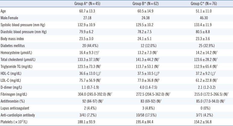
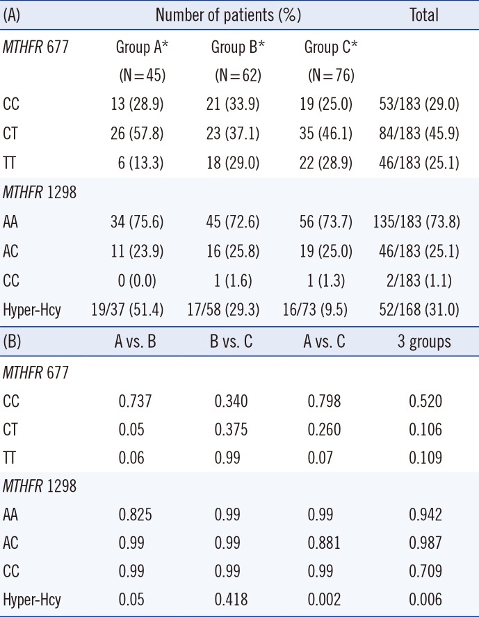
Clinical data and laboratory biomarkers are presented in Table 1. All 183 patients expressed wild type Factor V G1691A and prothrombin G20210A. The genotype distributions of MTHFR C677T and A1298C are presented in Table 2. Hcy levels were significantly higher in MTHFR 677TT genotype than in CT or CC (wild) genotypes (P=0.008, Supplemental Data Fig. S1).

2. Group A vs. group C

The laboratory data were compared between group A and C. The median age of patients in group A (58 yr) was higher than in group C (51 yr) (P=0.04). The hyperhomocysteinemia (Hyper-Hcy), demographic and clinical data, and laboratory biomarkers of both groups are demonstrated in Table 2.
With regard to the impact of thrombophilic polymorphisms, a higher frequency of the homozygous MTHFR 677TT genotype was associated with group A compared with group C. The MTHFR 677TT genotype tended to increase the development of AVD in HTN patients (OR 2.648, CI 0.982-7.143, P=0.05) (Fig. 1). MTHFR 677CT, 1298 AC, and 1298CC were not associated with AVD in HTN patients.
The group A revealed significantly higher Hcy, fibrinogen levels, and platelet count compared with group C (P<0.05, Table 2, Fig. 1). The ORs (95% CI) of Hyper-Hcy (>15 µmol/L), fibrinogen (>380 mg/dL), and platelet count (>176×1012/L) were 3.67 (1.14-2.44), 4.76 (1.63-13.89), and 2.28 (1.03-5.03), respectively (Fig. 1). Of these significant markers, the Hcy level of group A was higher than the reference range of our laboratory. The fibrinogen and platelet count of group A were within reference range (Table 2).
Body mass index (BMI), diabetes mellitus, TC, TG, HDL-C, LDL-C, D-dimer, AT, and the frequencies of LA and aCL did not differ significantly between group A and C (Table 2, Fig. 1).

3. Group B vs. group C

The laboratory markers of HTN patients with VTE were compared with those of HTN-only patients. Patients in the group B were older (median 63 yr) than those in the group C (median 51 yr) (P=0.01, Table 2).
The thrombophilic polymorphisms, Factor V G1691A and prothrombin G20210A could not be analyzed since they were not detected in any patients. However, MTHFR C677T and A1298C polymorphisms were not associated with VTE in HTN patients (Fig. 1).
Furthermore, group B demonstrated higher levels of D-dimers, fibrinogen, LDL-C, and platelet count (Table 2). ORs (95% CI) of high D-dimer levels (>4.0 mg/L), fibrinogen (>380 mg/dL), LDL-C (>100 mg/dL), platelet count (>176×1012/L), and aCL were 13.8 (4.86-39.20), 3.32 (1.18-9.38), 4.57 (1.24-16.92), 4.08 (1.96-8.49), and 4.93 (1.29-18.85), respectively (Fig. 1). Of these significant markers, the D-dimer levels were higher than the reference range in the group B. Fibrinogen, LDL-C, and platelet count were within the reference range. The frequency of aCL was higher in the group B (17.5%) than in the group C (4.2%) (P<0.05, Table 2).
BMI, diabetes mellitus, Hcy, AT, TC, TG, HDL-C, and the frequency of LA were not different between the group B and C (Table 2, Fig. 1).

DISCUSSION

HTN is a well-known risk factor of MI and ischemic stroke. It is also reported that HTN is relevant to the development of VTE [347]. In this study, we investigated various potential risk factors for their reliability in predicting the development of AVD or DVT, particularly in HTN patients whose BP is highly controlled. We examined known genetic predispositions (MTHFR C677T, A1298C, Factor V G1691A, and prothrombin G20210A polymorphisms), Hcy levels, and thrombophilic biomarkers in three HTN groups. As expected, the lipid profile and BMI were generally well managed in the patients enrolled, and BP was not different among the three groups.
We studied the Factor V G1691A and prothrombin G20210A polymorphisms, because they are important and basic mutations in thrombophilia. We could not observe these polymor-phisms, similar to previous reports [2829]. However, we observed that the MTHFR 677TT genotype was associated with increased development of AVD in HTN patients (OR 2.648, P=0.05). Recent meta-analyses demonstrated that the TT genotype is associated with cardiovascular diseases including MI, venous thrombosis, and peripheral arterial thrombosis [1221]. Although previous studies have demonstrated that the MTHFR 677CT genotype and Hyper-Hcy are associated factors of thrombosis when compared with healthy control [14], this genotype did not correlate with VTE in HTN patient in the present study. Similar results were also observed in a previous study [30]. The frequency of the MTHFR genotypes in this study were similar to those of a previous Korean study, which reported that the frequencies of 677CC, CT, and TT were 28.9%, 47.4%, and 23.7%, respectively [31]. The homozygous TT genotype is slightly higher in this study than in western countries (7.7-18.7%) [323334]. Furthermore, MTHFR A1298C was not associated with AVD or VTE in this study.
Hcy is a sulfur amino acid in the blood that is produced when methionine is broken down in the body. Elevated Hcy levels are known to be associated with hardening of the arteries and blood clots in veins [1835]. Of the three groups, the group A presented the highest Hcy levels, which was slightly higher than those in Korean patients with coronary artery disease [36]. Therefore, Hyper-Hcy is a potential risk factor of AVD in patients receiving treatment for HTN. A previous study reported that the Hcy levels in venous and arterial thrombosis groups did not differ significantly. However, these studies considered only arterial thrombosis, and not all AVD were included in that study [37]. It is not yet clear whether Hcy itself causes vascular diseases or whether it is merely a marker of increased risk. It is also not clear whether Hyper-Hcy results from vascular events [38]. The MTHFR 677TT genotype was associated with Hyper-Hcy in the present study. The patient with the TT genotype demonstrated higher Hcy levels (1.1 µmol/L) than the wild type. This corresponded to previous meta-analyses, which also demonstrated that the TT genotype showed higher Hcy levels (3.5 µmol/L) than the wild type [27].
The D-dimer has been known as a useful biomarker for the diagnosis of PE, DVT, and acute aortic dissection [1723]. In the present study, the group B exhibited higher levels of D-dimers than group C. Furthermore, anti-phospholipid antibodies (APLA) including LA and aCL are associated with venous and arterial thrombosis. The frequency of APLA is reported to be 1-5% in the general population, increasing up to 10% in VTE and 17% in MI [24]. In this study, the presence of LA was not significantly higher in the group B, but aCL was more frequently observed in this group (17.5%) compared with the group A (7.2%) or group C (4.2%).
Platelet activity plays an important role in coagulation and thrombus formation. Previous studies reported that platelet count seems to parallel changes in platelet function in MI patients [25]. In this study, the platelet count was significantly higher in the group A and B than in the group C. Although platelet count were within the reference range for all three groups, HTN patients with platelet counts over 176×1012/L demonstrated higher relevance of VTE (OR 4.08) or AVD (OR 2.28). Similar to our findings, platelet count was related to the relevance of MI [39]. In contrast, a study reported that lower platelet count posed a high relevance for PE development in DVT patients [40]. Further research in larger cohorts is required to elucidate the influence of platelet count on the development of AVD or VTE in HTN patients.
In conclusion, this study investigated potential risk factors of AVD and VTE in patients treated for HTN. The MTHFR 677TT genotype tended to increase the risk of AVD events in HTN patients. In addition, the results suggest that Hcy levels could be valuable in estimating the development of AVD. Our findings demonstrated that D-dimers and aCL could also be considered useful to estimate the occurence of VTE. Although our results need to be validated in a larger number of cases, including various ethnic groups, we propose that the above findings will help clinicians to assess the potential risk factors of vascular diseases in treated HTN patients.

Acknowledgments

This study was supported by a grant from the Korean Health Technology R&D Project, Ministry of Health and Welfare, Republic of Korea (HI10C2020).

Notes

Authors' Disclosures of Potential Conflicts of Interest: No potential conflicts of interest relevant to this article were reported.

References

1. 1999 World Health Organization-International Society of Hypertension Guidelines for the Management of Hypertension. Guidelines Subcommittee. J Hypertens. 1999; 17:151–183. PMID: 10067786.
2. Kearney PM, Whelton M, Reynolds K, Muntner P, Whelton PK, He J. Global burden of hypertension: analysis of worldwide data. Lancet. 2005; 365:217–223. PMID: 15652604.
crossref
3. Pedrinelli R, Ballo P, Fiorentini C, Denti S, Galderisi M, Ganau A, et al. Hypertension and acute myocardial infarction: an overview. J Cardiovasc Med (Hagerstown). 2012; 13:194–202. PMID: 22317927.
4. Gaciong Z, Siński M, Lewandowski J. Blood pressure control and primary prevention of stroke: summary of the recent clinical trial data and meta-analyses. Curr Hypertens Rep. 2013; 15:559–574. PMID: 24158454.
crossref
5. Wolf PA, D'Agostino RB, Belanger AJ, Kannel WB. Probability of stroke: a risk profile from the Framingham Study. Stroke. 1991; 22:312–318. PMID: 2003301.
crossref
6. Yano Y, Stamler J, Garside DB, Daviglus ML, Franklin SS, Carnethon MR, et al. Isolated systolic hypertension in young and middle-aged adults and 31-year risk for cardiovascular mortality: the Chicago Heart Association Detection Project in Industry study. J Am Coll Cardiol. 2015; 65:327–335. PMID: 25634830.
7. Ageno W, Becattini C, Brighton T, Selby R, Kamphuisen PW. Cardiovascular risk factors and venous thromboembolism: a meta-analysis. Circulation. 2008; 117:93–102. PMID: 18086925.
8. Mahan CE, Fanikos J. New antithrombotics: the impact on global health care. Thromb Res. 2011; 127:518–524. PMID: 21529897.
crossref
9. Cordoba I, Pegenaute C, González-López TJ, Chillon C, Sarasquete ME, Martin-Herrero F, et al. Risk of placenta-mediated pregnancy complications or pregnancy-related VTE in VTE-asymptomatic families of probands with VTE and heterozygosity for factor V Leiden or G20210 prothrombin mutation. Eur J Haematol. 2012; 89:250–255. PMID: 22642978.
crossref
10. Saemundsson Y, Sveinsdottir SV, Svantesson H, Svensson PJ. Homozygous factor V Leiden and double heterozygosity for factor V Leiden and prothrombin mutation. J Thromb Thrombolysis. 2013; 36:324–331. PMID: 23054468.
crossref
11. Clarke R, Bennett DA, Parish S, Verhoef P, Dötsch-Klerk M, Lathrop M, et al. Homocysteine and coronary heart disease: meta-analysis of MTHFR case-control studies, avoiding publication bias. PLoS Med. 2012; 9:e1001177. PMID: 22363213.
crossref
12. Gao XH, Zhang GY, Wang Y, Zhang HY. Correlations of MTHFR 677C>T polymorphism with cardiovascular disease in patients with end-stage renal disease: a meta-analysis. PLoS One. 2014; 9:e102323. PMID: 25050994.
13. Gariglio L, Riviere S, Morales A, Porcile R, Potenzoni M, Fridman O. Comparison of homocysteinemia and MTHFR 677CT polymorphism with Framingham Coronary Heart Risk Score. Arch Cardiol Mex. 2014; 84:71–78. PMID: 24793554.
crossref
14. Fukasawa M, Matsushita K, Kamiyama M, Mikami Y, Araki I, Yamagata Z, et al. The methylentetrahydrofolate reductase C677T point mutation is a risk factor for vascular access thrombosis in hemodialysis patients. Am J Kidney Dis. 2003; 41:637–642. PMID: 12612987.
crossref
15. Soltanpour MS, Soheili Z, Shakerizadeh A, Pourfathollah AA, Samiei S, Meshkani R, et al. Methylenetetrahydrofolate reductase C677T mutation and risk of retinal vein thrombosis. J Res Med Sci. 2013; 18:487–491. PMID: 24250697.
16. Spiroski I, Kedev S, Antov S, Arsov T, Krstevska M, Dzhekova-Stojkova S, et al. Association of methylenetetrahydrofolate reductase (MTHFR-677 and MTHFR-1298) genetic polymorphisms with occlusive artery disease and deep venous thrombosis in Macedonians. Croat Med J. 2008; 49:39–49. PMID: 18293456.
crossref
17. Kara H, Bayir A, Degirmenci S, Kayis SA, Akinci M, Ak A, et al. D-dimer and D-dimer/fibrinogen ratio in predicting pulmonary embolism in patients evaluated in a hospital emergency department. Acta Clin Belg. 2014; 69:240–245. PMID: 25012747.
crossref
18. D'Angelo A, Selhub J. Homocysteine and thrombotic disease. Blood. 1997; 90:1–11. PMID: 9207431.
19. Lin LJ, Cheng MH, Lee CH, Wung DC, Cheng CL, Kao Yang YH. Compliance with antithrombotic prescribing guidelines for patients with atrial fibrillation--a nationwide descriptive study in Taiwan. Clin Ther. 2008; 30:1726–1736. PMID: 18840379.
crossref
20. Sode BF, Allin KH, Dahl M, Gyntelberg F, Nordestgaard BG. Risk of venous thromboembolism and myocardial infarction associated with factor V Leiden and prothrombin mutations and blood type. CMAJ. 2013; 185:E229–E237. PMID: 23382263.
crossref
21. Bozok Çetintaş V, Gündüz C. Association between polymorphism of MTHFR c.677C>T and risk of cardiovascular disease in Turkish population: a meta-analysis for 2.780 cases and 3.022 controls. Mol Biol Rep. 2014; 41:397–409. PMID: 24264431.
22. Neff MJ. ACEP. ACEP releases clinical policy on evaluation and management of pulmonary embolism. Am Fam Physician. 2003; 68:759–760. PMID: 12952389.
23. Schutgens RE, Ackermark P, Haas FJ, Nieuwenhuis HK, Peltenburg HG, Pijlman AH, et al. Combination of a normal D-dimer concentration and a non-high pretest clinical probability score is a safe strategy to exclude deep venous thrombosis. Circulation. 2003; 107:593–597. PMID: 12566372.
crossref
24. Misita CP, Moll S. Antiphospholipid antibodies. Circulation. 2005; 112:e39–e44. PMID: 16027261.
crossref
25. Fagher B, Sjögren A, Sjögren U. Platelet counts in myocardial infarction, angina pectoris and peripheral artery disease. Acta Med Scand. 1985; 217:21–26. PMID: 3976430.
crossref
26. Pengo V, Tripodi A, Reber G, Rand JH, Ortel TL, Galli M, et al. Update of the guidelines for lupus anticoagulant detection Subcommittee on Lupus Anticoagulant/Antiphospholipid Antibody of the Scientific and Standardisation Committee of the International Society on Thrombosis and Haemostasis. J Thromb Haemost. 2009; 7:1737–1740. PMID: 19624461.
27. Nygård O, Vollset SE, Refsum H, Brattström L, Ueland PM. Total homocysteine and cardiovascular disease. J Intern Med. 1999; 246:425–454. PMID: 10583714.
crossref
28. Chang JD, Hur M, Lee SS, Yoo JH, Lee KM. Genetic background of nontraumatic osteonecrosis of the femoral head in the Korean population. Clin Orthop Relat Res. 2008; 466:1041–1046. PMID: 18350352.
crossref
29. Kim YW, Yoon KY, Park S, Shim YS, Cho HI, Park SS. Absence of factor V Leiden mutation in Koreans. Thromb Res. 1997; 86:181–182. PMID: 9175239.
30. Joachim E, Goldenberg NA, Bernard TJ, Armstrong-Wells J, Stabler S, Manco-Johnson MJ. The methylenetetrahydrofolate reductase polymorphism (MTHFR c.677C>T) and elevated plasma homocysteine levels in a U.S. pediatric population with incident thromboembolism. Thromb Res. 2013; 132:170–174. PMID: 23866722.
31. Lee SY, Kim HY, Park KM, Lee SY, Hong SG, Kim HJ, et al. MTHFR C677T polymorphism as a risk factor for vascular calcification in chronic hemodialysis patients. J Korean Med Sci. 2011; 26:461–465. PMID: 21394321.
32. Aucella F, Margaglione M, Grandone E, Vigilante M, Gatta G, Forcella M, et al. The C677T methylenetetrahydrofolate reductase gene mutation does not influence cardiovascular risk in the dialysis population: results of a multicentre prospective study. Nephrol Dial Transplant. 2005; 20:382–386. PMID: 15618240.
crossref
33. Haviv YS, Shpichinetsky V, Goldschmidt N, Atta IA, Ben-Yehuda A, Friedman G. The common mutations C677T and A1298C in the human methylenetetrahydrofolate reductase gene are associated with hyperhomocysteinemia and cardiovascular disease in hemodialysis patients. Nephron. 2002; 92:120–126. PMID: 12187094.
crossref
34. Wrone EM, Zehnder JL, Hornberger JM, McCann LM, Coplon NS, Fortmann SP. An MTHFR variant, homocysteine, and cardiovascular comorbidity in renal disease. Kidney Int. 2001; 60:1106–1113. PMID: 11532106.
crossref
35. Antoniades C, Antonopoulos AS, Tousoulis D, Marinou K, Stefanadis C. Homocysteine and coronary atherosclerosis: from folate fortification to the recent clinical trials. Eur Heart J. 2009; 30:6–15. PMID: 19029125.
crossref
36. Park KH, Han SJ, Kim HS, Kim MK, Jo SH, Kim SA, et al. Impact of Framingham risk score, flow-mediated dilation, pulse wave velocity, and biomarkers for cardiovascular events in stable angina. J Korean Med Sci. 2014; 29:1391–1397. PMID: 25368493.
crossref
37. Vuckovic BA, Cabarkapa VS, Ilic TA, Salatic IR, Lozanov-Crvenkovic ZS, Mitic GP. Clinical significance of determining plasma homocysteine: case-control study on arterial and venous thrombotic patients. Croat Med J. 2013; 54:480–488. PMID: 24170727.
crossref
38. Brattström L, Wilcken DE. Homocysteine and cardiovascular disease: cause or effect? Am J Clin Nutr. 2000; 72:315–323. PMID: 10919920.
crossref
39. Paul GK, Sen B, Bari MA, Rahman Z, Jamal F, Bari MS, et al. Correlation of platelet count and acute ST-elevation in myocardial infarction. Mymensingh Med J. 2010; 19:469–473. PMID: 20639847.
40. Monreal M, Lafoz E, Casals A, Ruiz J, Arias A. Platelet count and venous thromboembolism. A useful test for suspected pulmonary embolism. Chest. 1991; 100:1493–1496. PMID: 1959389.
Supplementary Material

Supplemental Data Fig. S1

Homocystein levels (means and 95% confidence interval of means) for the MTHFR 677 genotypes. Patients with the homozygous variant (TT) demonstrated significantly higher homocystein levels than those with CT or CC genotypes (P=0.008).
alm-36-215-s001.pdf
Fig. 1

The odds ratios and 95% confidence intervals of MTHFR 677 and 1298 genotypes and laboratory markers between the atheroscleotic vascular disease (AVD) and hypertension (HTN) only groups (top), and between the venous thromboembolism (VTE) and HTN only groups (bottom). The numbers in parentheses on Y-axis show the P values by odds ratio analysis.

*P values were statistically significant.
Abbreviations: 677, MTHFR 677 genotypes; 1298, MTHFR 1298 genotypes; Hyper-Hcy, hyperhomocysteinemia; LA, lupus anticoagulant; aCL, anticardiolipin antibody.
alm-36-215-g001
Table 1

The frequency of MTHFR 677 and 1,298 genotypes and hyperhomocysteinemia in each group (A), and their statistical significance (P values) between two groups or among three groups (B)

alm-36-215-i001
(A) Number of patients (%) Total
MTHFR 677 Group A*
(N = 45)
Group B*
(N = 62)
Group C*
(N = 76)
CC 13 (28.9) 21 (33.9) 19 (25.0) 53/183 (29.0)
CT 26 (57.8) 23 (37.1) 35 (46.1) 84/183 (45.9)
TT 6 (13.3) 18 (29.0) 22 (28.9) 46/183 (25.1)
MTHFR 1298
AA 34 (75.6) 45 (72.6) 56 (73.7) 135/183 (73.8)
AC 11 (23.9) 16 (25.8) 19 (25.0) 46/183 (25.1)
CC 0 (0.0) 1 (1.6) 1 (1.3) 2/183 (1.1)
Hyper-Hcy 19/37 (51.4) 17/58 (29.3) 16/73 (9.5) 52/168 (31.0)
(B) A vs. B B vs. C A vs. C 3 groups
MTHFR 677
CC 0.737 0.340 0.798 0.520
CT 0.05 0.375 0.260 0.106
TT 0.06 0.99 0.07 0.109
MTHFR 1298
AA 0.825 0.99 0.99 0.942
AC 0.99 0.99 0.881 0.987
CC 0.99 0.99 0.99 0.709
Hyper-Hcy 0.05 0.418 0.002 0.006

*Group A, atherosclerosis with hypertension; Group B, venous thromboembolism with hypertension; Group C, hypertension without complication; Mann-Whitney U or Kruskal-Wallis test was used for statistical analyses of MTHFR 677 and 1298 genotypes, and Hyper-Hcy between two groups or among three groups.

Abbreviation: Hyper-Hcy, hyperhomocyteinemia.

Table 2

Clinical factors and laboratory biomarkers associated with thrombotic vascular diseases in each group

alm-36-215-i002
Group A* (N = 45) Group B* (N = 62) Group C* (N = 76)
Age 60.7 ± 13.3 60.5 ± 14.9 51.1 ± 11.0
Male:Female 27:18 24:38 46:30
Systolic blood pressure (mm Hg) 132.9 ± 10.9 129.5 ± 10.2 133.4 ± 11.9
Diastolic blood pressure (mm Hg) 79.9 ± 6.2 78.2 ± 7.5 80.5 ± 8.8
Body mass index 23.5 ± 3.0 24.1 ± 5.1 23.3 ± 3.6
Diabetes mellitus 20 (44.4%) 12 (12.0%) 25 (32.9%)
Homocysteine (µmol/L) 16.4 ± 9.3 (↑) 13.2 ± 7.3 (N) 14.2 ± 14.2 (N)
Total cholesterol (µmol/L) 133.3 ± 37.1(N) 141.3 ± 44.2 (N) 123.6 ± 28.2 (N)
Triglyceride TG (mg/dL) 123.5 ± 73.3 (N) 113.7 ± 53.1 (N) 112.9 ± 65.4 (N)
HDL-C (mg/dL) 36.6 ± 13.0 (↓) 37.5 ± 10.5 (↓)§ 37.2 ± 9.2 (↓)
LDL-C (mg/dL) 75.7 ± 56.9 (N) 77.0 ± 36.8 (N)§ 61.2 ± 22.8 (N)
D-dimer (mg/L) 1.1 (0.7-1.9) 4.0 (1.4-7.5) 2.1 (1.2-3.2)
Fibrinogen (mg/dL) 304.0 (245.0-392.0) (N) 272.5 (204.5-362.0) (N) 215.0 (172.5-266.5) (N)
Antithrombin (%) 92 (84-97) (N) 83 (69-92) (N) 85.0 (77.0-94.0) (N)
Lupus anticoagulant 2 (4.4%) 3 (4.8%) 0 (0%)
Anti-cardiolipin antibody 3/41 (7.2%) 10/58 (17.5%) 3/71 (4.2%)
Platelets ( × 1012/L) 188.1 ± 93.9 195.4 ± 84.4 154.2 ± 56.8

*Group A, atherosclerosis with hypertension; Group B, venous thromboembolism with hypertension; Group C, hypertension without complication; The median (or mean) values of biomarkers are denoted as increased (↑), decreased (↓), or normal (N) on the basis of the reference values. The cut-offs for metric values were determined by reference ranges in our laboratory except for D-dimers and platelets. The cut-off of biomarkers were as follows: Hcy (>15 µmol/L), TC (>200 mg/dL), TG (>200 mg/dL), HDL-C (<60 mg/dL), fibrinogen (>380 mg/dL), and AT (<80%).

Abbreviations: HDL-C, high density lipoprotein cholesterol; LDL-C, low density lipoprotein cholesterol.

TOOLS
Similar articles