Journal List > Ann Lab Med > v.36(1) > 1091357

Park, Ha, Cho, Park, Jang, and Hong: Immature Platelet Fraction in Septic Patients: Clinical Relevance of Immature Platelet Fraction is Limited to the Sensitive and Accurate Discrimination of Septic Patients From Non-Septic Patients, Not to the Discrimination of Sepsis Severity

Abstract

Background

The immature platelet fraction (IPF) reflects the degree of reticulated platelets. We evaluated performances of IPF as a biomarker for the discrimination of septic patients from non-septic patients and sepsis severity.

Methods

Total 312 patients admitted between March and July 2013 were enrolled and samples were obtained at admission. Lactate (LA), procalcitonin (PCT), C-reactive protein (CRP), immature granulocyte fraction (IG), immature reticulocyte fraction (IRF), and IPF were analyzed as sepsis biomarkers and their performances were compared.

Results

The performance of IPF (area under the curve [AUC]=0.868) in the discrimination of septic patients from non-septic patients was comparable to PCT/CRP/LA/IG (AUC=0.923/0.940/0.781/0.812, P=0.233/0.106/0.186/0.353, respectively), and was significantly better than the IRF (AUC=0.658, P=0.007). Sensitivity (89.8%, 95% confidence interval [CI] 84.9-99.8%) and accuracy (83.2%, 95% CI 78.8-90.0%) of IPF were the best among all biomarkers. The performance of IPF in discriminating septic patients from non-septic patients with local infection showed similar results. However, the IPF could not efficiently discriminate sepsis severity (AUC=0.599), similar to other biomarkers (AUC=0.519-0.752).

Conclusions

The IPF possessed high sensitivity/accuracy in discriminating septic patients from non-septic patients, regardless of local infection status. However, the IPF did not efficiently discriminate sepsis severity. The clinical relevance of IPF as a sepsis biomarker is, therefore, limited to sensitive and accurate discrimination of septic patients from non-septic patients, not discrimination of sepsis severity.

INTRODUCTION

Despite improvements in treatment modality and development of various biomarkers used for early detection, sepsis remains an important cause of mortality, especially in intensive care unit (ICU) patients [1]. Current biomarkers used for the detection of inflammation or infection, such as lactate (LA), procalcitonin (PCT), and C-reactive protein (CRP), can reflect the pathologic conditions associated with sepsis development and are useful in sepsis diagnosis or assessment of sepsis severity; however, performance of these biomarkers varies [234].
Inflammation induces coagulation abnormalities and microcirculation disturbances by microthrombi resulted from platelet activation can cause end-organ damage in sepsis pathophysiology [56]. Therefore, it is possible that the parameter reflecting platelet production and activation, specifically the immature platelet fraction (IPF) that indicates the percentage of reticulated platelets, associates with sepsis development and reflects sepsis severity. Since the IPF can measure thrombopoietic activity in bone marrow (BM), increased IPF can be observed in patients with thrombocytopenia owing to peripheral destruction, such as immune thrombocytopenic purpura (ITP), and decreased IPF can be observed in patients with thrombocytopenia owing to BM failure. Several studies confirmed the clinical usefulness of the IPF as a differential diagnostic biomarker in the evaluation of thrombocytopenia [7891011]. Two recent studies suggested that the IPF may help identify patients very early in the course of sepsis and can be used as a screening parameter for bacterial infection, which may allow for initiation of treatment prior to clinical onset [1213]. Another recent study evaluated the performance of the IPF in sepsis diagnosis and discrimination of sepsis severity in 41 ICU patients and suggested that the IPF levels could be used as a sepsis biomarker, especially in the discrimination of septic patients from healthy individuals and assessment of severity [14]. It is also important that a sepsis biomarker be able to discriminate septic patients from non-septic patients with local infections, such as a urinary tract infection (UTI) or an upper respiratory tract infection (URI), and therefore, the performance of the IPF for this purpose also needs to be assessed.
In the present study, we evaluated the performance of the IPF as a sepsis biomarker focusing on three points: (1) the discrimination of septic patients from non-septic patients; (2) the discrimination of septic patients from non-septic patients with local infection; and (3) the assessment of sepsis severity in comparison with nonhematological sepsis biomarkers (LA, PCT, and CRP) and hematological sepsis biomarkers, such as immature granulocyte fraction (IG) and immature reticulocyte fraction (IRF).

METHODS

1. Selection and classification of patients

A total of 312 adult patients admitted to the general ward (N=186) or ICU (N=126) of Asan Medical Center from March to July 2013 were randomly enrolled, and samples were obtained at admission (day 0). They were classified into five subgroups (non-septic [N=47], non-septic with local infection, such as UTI or URI [N=50], uncomplicated sepsis [N=64], severe sepsis [N=61], and septic shock [N=90]) depending on the clinical findings at the time of sampling. Uncomplicated sepsis was defined as the presence of both infection and systemic inflammatory response syndrome (SIRS) without any evidence of organ dysfunction. Severe sepsis was defined as sepsis complicated by organ dysfunction, such as arterial hypoxemia (PaO2/FiO2 <300), oligouria (urine output <0.5 mL/kg/hr or 45 mmol/L for at least two hours), elevated serum glucose (>100 mg/dL), elevated serum creatinine (>1.4 mg/dL or increases >0.5 mg/dL from baseline), thrombocytopenia (<1×10¹¹/L), and hyperbilrubinemia (plasma total bilirubin >4 mg/dL). Septic shock was defined as severe sepsis plus a state of acute circulatory failure characterized by persistent arterial hypotension (systolic blood pressure <90 mm Hg, mean arterial pressure <60 mm Hg, or a reduction of systolic blood pressure >40 mm Hg from baseline) despite adequate volume resuscitation [1516].
Additionally, all sepsis (defined as uncomplicated sepsis+severe sepsis+septic shock; N=215) and complicated sepsis (defined as severe sepsis+septic shock; N=151) subgroups were also generated. All sepsis subgroups included both patients having signs of sepsis, such as fever, and fulfilling the diagnostic criteria of sepsis, but showing negative results in blood cultures (N=106), and blood culture-positive bacteremia patients (N=109).

2. Clinical and laboratory findings from patient samples

For each sample, LA, PCT, CRP, IPF, IG, IRF, white blood cell (WBC) counts, hemoglobin, platelets, WBC differential counts, prothrombin time (PT), activated partial thromboplastin time (aPTT), D-dimer, creatinine, glucose, and total bilirubin were measured. The LA level was measured by using a GEM Premier 3000 instrument (Instrumentation Laboratory, Lexington, MA, USA). The PCT level was measured by immunoluminometric assay (VIDAS B.R.A.H.M.S. PCT; bioMerieux, Saint Laurent, Canada), and the CRP level was measured by latex immunoturbidimetric assay (Tina-quant CRP HS Test System; Roche, Basel, Switzerland). All hematological and coagulation parameters were obtained from an automated blood cell analyzer (Sysmex XE-2100; Sysmex, Kobe, Japan) and coagulation analyzer (Sysmex CA-7000; Syxmex). The creatinine, glucose, and total bilirubin levels were measured by using a clinical chemistry analyzer (TBA-200FR; Toshiba Medical Systems, Tokyo, Japan). This study was approved by the institutional review board of each author's institution.

3. Comparative analysis of clinical and laboratory findings between patient subgroups categorized by the presence of sepsis or sepsis severity

The distribution of gender, age, bacteremia type, and laboratory findings described above were compared between (1) non-septic patients and all septic patients, (2) non-septic patients with local infection and all septic patients, and (3) uncomplicated septic patients and complicated septic patients. Additionally, since the IPF can reflect the status of coagulation activation in sepsis, especially in disseminated intravascular coagulation (DIC) patients who may show increased and decreased platelet production in early and late stage, respectively, correlations between the IPF and PT, aPTT, and D-dimer were analyzed in septic patients.

4. Performance evaluation of six biomarkers in the discrimination of septic patients from non-septic patients and the discrimination of sepsis severity

The performances of six biomarkers (LA, PCT, CRP, IPF, IG, and IRF) in the discrimination of (1) septic patients from non-septic patients, (2) septic patients from non-septic patients with local infection, and (3) sepsis severity were evaluated by ROC curve analysis and compared. Additionally, the sensitivity, specificity, negative predictive value (NPV), positive predictive value (PPV), and accuracy of each biomarker following application of the best cutoff values determined from ROC curve analysis were calculated and compared.

5. Statistical analysis

The chi-square and Mann-Whitney U tests were used to compare dichotomous and continuous variables, respectively, between the two patient subgroups. In the comparison of continuous variables, the median values of each result were used since all these variables did not show normal distribution. ROC curve analysis was performed to evaluate the performance of each biomarker. Spearman's correlation analysis was applied in the evaluation of associations between the IPF and PT, aPTT, and D-dimer. All tests were two-tailed, and P≤0.05 was considered significant. All analyses except for the ROC curve and correlation analyses were performed by using SPSS v13.0.1 for Windows (SPSS Inc., Chicago, IL, USA). MedCalc v9.2.0.2 (MedCalc Software, Ostend, Belgium) was used for ROC curve and correlation analyses.

RESULTS

1. Comparison of clinical and laboratory findings between septic patients and non-septic patients or non-septic patients with local infection (Table 1)

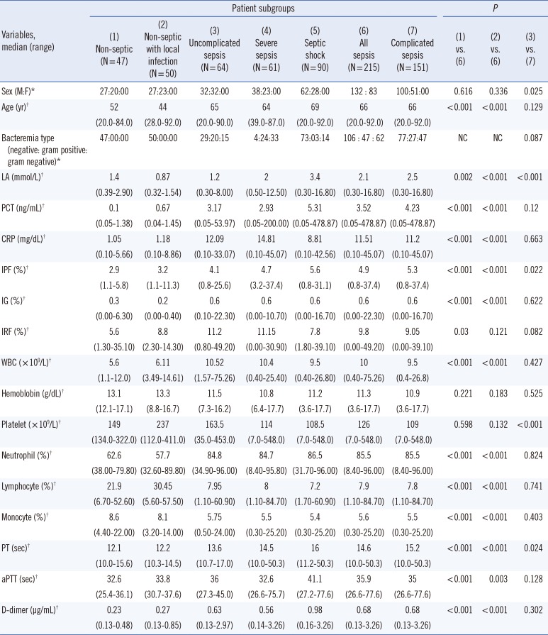
When compared to non-septic patients, septic patients were significantly older (P<0.001). Septic patients also showed significantly higher LA (P=0.002), PCT (P<0.001), CRP (P<0.001), IPF (P<0.001), IG (P<0.001), IRF (P=0.030), WBC counts (P<0.001), neutrophils% (P<0.001), and D-dimer (P<0.001), and significantly lower lymphocytes% (P<0.001) and monocytes% (P<0.001) than non-septic patients. Additionally, septic patients showed significantly prolonged PT (P<0.001) and aPTT (P<0.001) than non-septic patients; however, both hemoglobin and platelet counts were not significantly different between the two patient subgroups. Comparisons between septic patients and non-septic patients with local infection also showed same results, except for the IRF.

2. Comparison of clinical and laboratory findings in septic patients with respect to sepsis severity (Table 1)

Patients with complicated sepsis showed significant male predominance (P=0.025) than those with uncomplicated sepsis. Additionally, patients with complicated sepsis showed significantly higher LA (P<0.001) and IPF levels (P=0.022), lower platelet counts (P<0.001), and prolonged PT (P=0.024) than those with uncomplicated sepsis. However, patient age, PCT, CRP, IG, IRF, WBC counts, hemoglobin, WBC differential counts, aPTT, and D-dimer were not significantly different between the two patient subgroups. The type of bacteremia was not significantly different between the two patient subgroups.

3. Performance evaluation of six biomarkers in the discrimination of septic patients from non-septic patients (Table 2)

Among the six biomarkers, both PCT and CRP performed best, with both their specificities and PPVs being very high. However, their accuracies were not as high as their specificities and PPVs. The IPF showed comparable performance to PCT, CRP, LA, and IG, and showed significantly better performance than the IRF (P=0.007). Notably, the IPF demonstrated the highest sensitivity (89.8%, 95% confidence interval [CI] 84.9-99.8%) and accuracy (83.2%, 95% CI 78.8-90.0%) among all six biomarkers when 3.1% was applied as a cutoff value.
The performance of IG was significantly worse than that of both PCT (P=0.041) and CRP (P=0.013). Although the performance of IG was comparable to that of the IPF (P=0.353), the sensitivity of the IG was lower than that of the IPF. The performance of the IRF was the worst among all six biomarkers.

4. Performance evaluation of six biomarkers in the discrimination of septic patients from non-septic patients with local infection (Table 3)

Among the six biomarkers, both the CRP and IG performed best, with both their specificities and PPVs being satisfactory. The PCT showed significantly worse performance than both CRP (P<0.001) and IG (P<0.001); however, both the specificity and PPV of the PCT were 100.0%, which was the best among the six biomarkers.
Despite the IPF showing significantly worse performance than both CRP (P=0.013) and IG (P=0.011), the IPF showed comparable performance to both LA and PCT, and showed significantly better performance than the IRF (P<0.001). Notably, the IPF demonstrated the highest sensitivity (84.2%, 95% CI 78.7-88.5%) and accuracy (86.0%, 95% CI 78.1-87.3%) among the six biomarkers when 3.4% was applied as a cutoff value. The performance of the IRF was also the worst among all six biomarkers.

5. Performance evaluation of six biomarkers in the discrimination of complicated sepsis from uncomplicated sepsis (Table 4)

Among the six biomarkers, the LA showed significantly better performance than the PCT (P=0.001), CRP (P<0.001), IPF (P=0.003), IG (P<0.001), and IRF (P<0.001). However, the performance of LA was not satisfactory compared with that of other biomarkers, which showed satisfactory performance in the discrimination of septic patients from non-septic patients or non-septic patients with local infection. The IPF also performed poorly in the discrimination of septic patients, similar to the unsatisfactory performances of the other biomarkers.

6. Correlation analysis between the IPF and PT, aPTT, and D-dimer in septic patients

This study included only eight patients with overt DIC according to the International Society for Thrombosis and Haemostasis (ISTH) scoring system [17]. There were no significant correlations between increased IPF and the prolongation of PT (γ=-0.124, P=0.069), aPTT (γ=-0.022, P=0.746), or increased D-dimer (γ=0.026, P=0.708).

DISCUSSION

The majority of previous reports suggested that both PCT and CRP exhibited acceptable performance in the detection of sepsis and discrimination of sepsis severity [18192021] and that the LA is at least a moderately accurate predictor of mortality in complicated septic patients [222324]. However, conflicting data also exist regarding whether the PCT, CRP, or IG can be an independent predictor of sepsis severity and mortality [25262728293031323334]. Since sepsis leads to platelet destruction in the early phase, we hypothesized that the IPF increases before thrombocytopenia occurs and can be a sensitive biomarker in the discrimination of septic patients from non-septic patients. Focusing on this point, we evaluated the performance of the IPF as a biomarker for the discrimination of septic patients from non-septic patients and for sepsis severity.
This study showed that the IPF, along with other biomarkers, was significantly higher in septic patients than in non-septic patients, regardless of whether non-septic patients have local infection (Table 1). Additionally, we found that both PCT/CRP and CRP/IG performed best in the discrimination of septic patients from non-septic patients or non-septic patients with local infection, respectively. However, we found that their sensitivity/accuracy was not as high as expected, which limits their clinical relevance as biomarkers used in the early discrimination of septic patients (Tables 2 and 3).
Notably, we identified that the IPF possesses comparable performance to PCT, CRP, LA, IG, and shows significantly better performance than the IRF in discriminating septic patients from non-septic patients. Also, we found that the IPF possesses the best sensitivity/accuracy among the six biomarkers (Table 2). These results support conclusions from a recent study [14]. We also demonstrated that the IPF performs better than the LA, PCT, and IRF and possesses the best sensitivity/accuracy among the six biomarkers in the discrimination of septic patients from non-septic patients with local infection (Table 3). These results are partially discordant with a recent study [14]; however, the discrepancy may be explained in part by the different patient categorization of non-septic patients (local infection vs. SIRS). Our results suggest that the IPF can be used as a reliable biomarker in the sensitive and accurate discrimination of septic patients and emphasize the importance of increased thrombopoiesis in the initiation of sepsis development [12]. In contrast to the IPF, our study identified that the IRF is not useful as a sepsis biomarker (Tables 2 and 3). These results are consistent with a previous result [14] and suggest that the development of anemia in sepsis is neither hyper-regenerative nor associated with reticulocyte response.
Our study also identified that the IPF is not as useful as other biomarkers in the discrimination of complicated septic patients from uncomplicated septic patients (Table 4). These results are discordant with several studies [18192021]. The discrepancies may be explained in part by the different study populations between our study, which included general ward and ICU patients, and the majority of other studies, which included ICU patients only, or by the different underlying diseases of the included patients. Our results suggest that the IPF does not characterize sepsis severity, given that the IPF reflects a balance between platelet activation and BM inhibition and that these two phenomena are capable of coexisting in the late stage of complicated sepsis.
Our study failed to demonstrate significant correlations between the IPF and PT, aPTT, and D-dimer. These results are consistent with a previous result [14]; however, they are partially discordant with other studies, which showed significant correlations between the IPF and DIC scores in a cohort of critically ill patients [35]. DIC patients may show different severity levels in consumption of coagulation, which may partly explain the discrepancy between results.
Our study has three limitations. First, we did not analyze the consecutive data over a certain period after admission and we could not evaluate the time difference between elevation of the IPF and sepsis development. Further study focused on this issue needs to be performed. Second, both severe sepsis and septic shock patients were categorized into a single "complicated sepsis" subgroup and the differences of each biomarker between the two patient subgroups were not analyzed. Since these two patient subgroups included patients with a broad spectrum of severity, different mortality rates and possibly different BM inhibition rates might be expected. Third, our present study used Sysmex XE-2100 analyzer in the measurement of IPF and recently, this instrument was replaced by XN series. The possible difference between IPF measured by XE-2100 and XN series should be considered especially in the laboratories operating new XN series. In addition, there might be difference between the fraction and absolute number of immature platelets and this point should be also considered when evaluating the results from our present study.
In conclusion, this study showed that the IPF possesses high sensitivity and accuracy in the discrimination of septic patients from non-septic patients, regardless of whether they have local infection. However, the IPF does not efficiently discriminate sepsis severity. The clinical relevance of the IPF as a sepsis biomarker is, therefore, limited to the sensitive and accurate discrimination of septic patients from non-septic patients and not to the discrimination of sepsis severity.

Acknowledgments

This study was supported by a grant (2009-387) from the Asan Institute for Life Sciences, Seoul, Korea.

Notes

Authors' Disclosures of Potential Conflicts of Interest: No potential conflicts of interest relevant to this article were reported.

References

1. Patel GP, Gurka DP, Balk RA. New treatment strategies for severe sepsis and septic shock. Curr Opin Crit Care. 2003; 9:390–396. PMID: 14508152.
crossref
2. Harbarth S, Holeckova K, Froidevaux C, Pittet D, Ricou B, Grau GE, et al. Geneva Sepsis Network. Diagnostic value of procalcitonin, interleukin-6, and interleukin-8 in critically ill patients admitted with suspected sepsis. Am J Respir Crit Care Med. 2001; 164:396–402. PMID: 11500339.
crossref
3. Müller B, Becker KL, Schächinger H, Rickenbacher PR, Huber PR, Zimmerli W, et al. Calcitonin precursors are reliable markers of sepsis in a medical intensive care unit. Crit Care Med. 2000; 28:977–983. PMID: 10809269.
crossref
4. Reinhart K, Bauer M, Riedemann NC, Hartog CS. New approaches to sepsis: molecular diagnostics and biomarkers. Clin Microbiol Rev. 2012; 25:609–634. PMID: 23034322.
crossref
5. Dhainaut JF, Shorr AF, Macias WL, Kollef MJ, Levi M, Reinhart K, et al. Dynamic evolution of coagulopathy in the first day of severe sepsis: relationship with mortality and organ failure. Crit Care Med. 2005; 33:341–348. PMID: 15699837.
crossref
6. Levi M, van der Poll T, Büller HR. Bidirectional relation between inflammation and coagulation. Circulation. 2004; 109:2698–2704. PMID: 15184294.
crossref
7. Briggs C, Kunka S, Hart D, Oguni S, Machin SJ. Assessment of an immature platelet fraction (IPF%) in peripheral thrombocytopenia. Br J Haematol. 2004; 126:93–99. PMID: 15198738.
crossref
8. Kickler TS, Oguni S, Borowitz MJ. A clinical evaluation of high fluorescent platelet fraction percentage in thrombocytopenia. Am J Clin Pathol. 2006; 125:282–287. PMID: 16393688.
crossref
9. Jung H, Jeon HK, Kim HJ, Kim SH. Immature platelet fraction: establishment of a reference interval and diagnostic measure for thrombocytopenia. Korean J Lab Med. 2010; 30:451–459. PMID: 20890075.
crossref
10. Cremer M, Paetzold J, Schmalisch G, Hammer H, Loui A, Dame C, et al. Immature platelet fraction as novel laboratory parameter predicting the course of neonatal thrombocytopenia. Br J Haematol. 2009; 144:619–621. PMID: 19036085.
crossref
11. Zucker ML, Hagedorn CH, Murphy CA, Stanley S, Reid KJ, Skikne BS. Mechanism of thrombocytopenia in chronic hepatitis C as evaluated by the immature platelet fraction. Int J Lab Hematol. 2012; 34:525–532. PMID: 22708981.
crossref
12. De Blasi RA, Cardelli P, Costante A, Sandri M, Mercieri M, Arcioni R. Immature platelet fraction in predicting sepsis in critically ill patients. Intensive Care Med. 2013; 39:636–643. PMID: 23093245.
crossref
13. Di Mario A, Garzia M, Leone F, Arcangeli A, Pagano L, Zini G. Immature platelet fraction (IPF) in hospitalized patients with neutrophilia and suspected bacterial infection. J Infect. 2009; 59:201–206. PMID: 19647322.
crossref
14. Enz Hubert RM, Rodrigues MV, Andreguetto BD, Santos TM, de Fátima Pereira Gilberti M, de Castro V, et al. Association of the immature platelet fraction with sepsis diagnosis and severity. Sci Rep. 2015; 5:8019. PMID: 25620275.
crossref
15. Vincent JL. Clinical sepsis and septic shock--definition, diagnosis and management principles. Langenbecks Arch Surg. 2008; 393:817–824. PMID: 18584205.
crossref
16. Levy MM, Fink MP, Marshall JC, Abraham E, Angus D, Cook D, et al. SCCM/ESICM/ACCP/ATS/SIS. 2001 SCCM/ESICM/ACCP/ATS/SIS International Sepsis Definitions Conference. Crit Care Med. 2003; 31:1250–1256. PMID: 12682500.
crossref
17. Levi M, Toh CH, Thachil J, Watson HG. Guidelines for the diagnosis and management of disseminated intravascular coagulation. British Committee for Standards in Haematology. Br J Haematol. 2009; 145:24–33. PMID: 19222477.
18. Zakariah AN, Cozzi SM, Van Nuffelen M, Clausi CM, Pradier O, Vincent JL. Combination of biphasic transmittance waveform with blood procalcitonin levels for diagnosis of sepsis in acutely ill patients. Crit Care Med. 2008; 36:1507–1512. PMID: 18434897.
crossref
19. Kofoed K, Andersen O, Kronborg G, Tvede M, Petersen J, Eugen-Olsen J, et al. Use of plasma C-reactive protein, procalcitonin, neutrophils, macrophage migration inhibitory factor, soluble urokinase-type plasminogen activator receptor, and soluble triggering receptor expressed on myeloid cells-1 in combination to diagnose infections: a prospective study. Crit Care. 2007; 11:R38. PMID: 17362525.
crossref
20. Gibot S, Béné MC, Noel R, Massin F, Guy J, Cravoisy A, et al. Combination biomarkers to diagnose sepsis in the critically ill patient. Am J Respir Crit Care Med. 2012; 186:65–71. PMID: 22538802.
crossref
21. Tsalik EL, Jaggers LB, Glickman SW, Langley RJ, van Velkinburgh JC, Park LP, et al. Discriminative value of inflammatory biomarkers for suspected sepsis. J Emerg Med. 2012; 43:97–106. PMID: 22056545.
crossref
22. Mikkelsen ME, Miltiades AN, Gaieski DF, Goyal M, Fuchs BD, Shah CV, et al. Serum lactate is associated with mortality in severe sepsis independent of organ failure and shock. Crit Care Med. 2009; 37:1670–1677. PMID: 19325467.
crossref
23. Shapiro NI, Howell MD, Talmor D, Nathanson LA, Lisbon A, Wolfe RE, et al. Serum lactate as a predictor of mortality in emergency department patients with infection. Ann Emerg Med. 2005; 45:524–528. PMID: 15855951.
crossref
24. Howell MD, Donnino M, Clardy P, Talmor D, Shapiro NI. Occult hypoperfusion and mortality in patients with suspected infection. Intensive Care Med. 2007; 33:1892–1899. PMID: 17618418.
crossref
25. Tschaikowsky K, Hedwig-Geissing M, Braun GG, Radespiel-Troeger M. Predictive value of procalcitonin, interleukin-6, and C-reactive protein for survival in postoperative patients with severe sepsis. J Crit Care. 2011; 26:54–64. PMID: 20646905.
crossref
26. Tschaikowsky K, Hedwig-Geissing M, Schmidt J, Braun GG. Lipopolysaccharide-binding protein for monitoring of postoperative sepsis: complemental to C-reactive protein or redundant? PLoS One. 2011; 6:e23615. PMID: 21901123.
crossref
27. Al-Subaie N, Reynolds T, Myers A, Sunderland R, Rhodes A, Grounds RM, et al. C-reactive protein as a predictor of outcome after discharge from the intensive care: a prospective observational study. Br J Anaesth. 2010; 105:318–325. PMID: 20630889.
crossref
28. Ho KM, Lee KY, Dobb GJ, Webb SA. C-reactive protein concentration as a predictor of in-hospital mortality after ICU discharge: a prospective cohort study. Intensive Care Med. 2008; 34:481–487. PMID: 17992507.
29. Litton E, Ho KM, Chamberlain J, Dobb GJ, Webb SA. C-reactive protein concentration as a predictor of in-hospital mortality after ICU discharge: a nested case-control study. Crit Care Resusc. 2007; 9:19–25. PMID: 17352662.
30. Ranzani OT, Prada LF, Zampieri FG, Battaini LC, Pinaffi JV, Setogute YC, et al. Failure to reduce C-reactive protein levels more than 25% in the last 24 hours before intensive care unit discharge predicts higher in-hospital mortality: a cohort study. J Crit Care. 2012; 27:525.e9–525.e15. PMID: 22227090.
crossref
31. Silvestre J, Coelho L, Póvoa P. Should C-reactive protein concentration at ICU discharge be used as a prognostic marker? BMC Anesthesiol. 2010; 27:17. PMID: 20875120.
crossref
32. Hermans MA, Leffers P, Jansen LM, Keulemans YC, Stassen PM. The value of the Mortality in Emergency Department Sepsis (MEDS) score, C reactive protein and lactate in predicting 28-day mortality of sepsis in a Dutch emergency department. Emerg Med J. 2012; 29:295–300. PMID: 21511973.
crossref
33. Ruzicka K, Veitl M, Thalhammer-Scherrer R, Schwarzinger I. The new hematology analyzer Sysmex XE-2100: performance evaluation of a novel white blood cell differential technology. Arch Pathol Lab Med. 2001; 125:391–396. PMID: 11231489.
34. Briggs C, Harrison P, Grant D, Staves J, MacHin SJ. New quantitative parameters on a recently introduced automated blood cell counter--the XE 2100. Clin Lab Haematol. 2000; 22:345–350. PMID: 11318800.
35. Hong KH, Kim HK, Kim JE, Jung JS, Han KS, Cho HI. Prognostic value of immature platelet fraction and plasma thrombopoietin in disseminated intravascular coagulation. Blood Coagul Fibrinolysis. 2009; 20:409–414. PMID: 19528793.
crossref
Table 1

Comparison of clinical and laboratory findings obtained at admission in 312 patients regarding sepsis severity

alm-36-1-i001
Variables, median (range) Patient subgroups P
(1) Non-septic
(N=47)
(2) Non-septic with local infection
(N=50)
(3) Uncomplicated sepsis
(N=64)
(4) Severe sepsis
(N=61)
(5) Septic shock
(N=90)
(6) All sepsis
(N=215)
(7) Complicated sepsis
(N=151)
(1) vs. (6) (2) vs. (6) (3) vs. (7)
Sex (M:F)* 27:20:00 27:23:00 32:32:00 38:23:00 62:28:00 132:83 100:51:00 0.616 0.336 0.025
Age (yr) 52
(20.0-84.0)
44
(28.0-92.0)
65
(20.0-90.0)
64
(39.0-87.0)
69
(20.0-92.0)
66
(20.0-92.0)
66
(20.0-92.0)
<0.001 <0.001 0.129
Bacteremia type
(negative: gram positive: gram negative)*
47:00:00 50:00:00 29:20:15 4:24:33 73:03:14 106:47:62 77:27:47 NC NC 0.087
LA (mmol/L) 1.4
(0.39-2.90)
0.87
(0.32-1.54)
1.2
(0.30-8.00)
2
(0.50-12.50)
3.4
(0.30-16.80)
2.1
(0.30-16.80)
2.5
(0.30-16.80)
0.002 < 0.001 < 0.001
PCT (ng/mL) 0.1
(0.05-1.38)
0.67
(0.04-1.45)
3.17
(0.05-53.97)
2.93
(0.05-200.00)
5.31
(0.05-478.87)
3.52
(0.05-478.87)
4.23
(0.05-478.87)
< 0.001 < 0.001 0.12
CRP (mg/dL) 1.05
(0.10-5.66)
1.18
(0.10-8.86)
12.09
(0.10-33.07)
14.81
(0.10-45.07)
8.81
(0.10-42.56)
11.51
(0.10-45.07)
11.2
(0.10-45.07)
< 0.001 < 0.001 0.663
IPF (%) 2.9
(1.1-5.8)
3.2
(1.1-11.3)
4.1
(0.8-25.6)
4.7
(3.2-37.4)
5.6
(0.8-31.1)
4.9
(0.8-37.4)
5.3
(0.8-37.4)
< 0.001 < 0.001 0.022
IG (%) 0.3
(0.00-6.30)
0.2
(0.00-0.40)
0.6
(0.10-22.30)
0.6
(0.00-10.70)
0.6
(0.00-16.70)
0.6
(0.00-22.30)
0.6
(0.00-16.70)
< 0.001 < 0.001 0.622
IRF (%) 5.6
(1.30-35.10)
8.8
(2.30-14.30)
11.2
(0.80-49.20)
11.15
(0.00-30.90)
7.8
(1.80-39.10)
9.8
(0.00-49.20)
9.05
(0.00-39.10)
0.03 0.121 0.082
WBC ( × 109/L) 5.6
(1.1-12.0)
6.11
(3.49-14.61)
10.52
(1.57-75.26)
10.4
(0.40-25.40)
9.5
(0.40-26.80)
10
(0.40-75.26)
9.5
(0.4-26.8)
< 0.001 < 0.001 0.427
Hemoblobin (g/dL) 13.1
(12.1-17.1)
13.3
(8.8-16.7)
11.5
(7.3-16.2)
10.8
(6.4-17.7)
11.2
(3.6-17.7)
11.3
(3.6-17.7)
10.9
(3.6-17.7)
0.221 0.183 0.525
Platelet ( × 109/L) 149
(134.0-322.0)
237
(112.0-411.0)
163.5
(35.0-453.0)
114
(7.0-548.0)
108.5
(7.0-548.0)
126
(7.0-548.0)
109
(7.0-548.0)
0.598 0.132 < 0.001
Neutrophil (%) 62.6
(38.00-79.80)
57.7
(32.60-89.80)
84.8
(34.90-96.00)
84.7
(8.40-95.80)
86.5
(31.70-96.00)
85.5
(8.40-96.00)
85.5
(8.40-96.00)
< 0.001 < 0.001 0.824
Lymphocyte (%) 21.9
(6.70-52.60)
30.45
(5.60-57.50)
7.95
(1.10-60.90)
8
(1.10-84.70)
7.2
(1.70-60.90)
7.9
(1.10-84.70)
7.8 < 0.001 < 0.001 0.741
Monocyte (%) 8.6
(4.40-22.00)
8.1
(3.20-14.00)
5.75
(0.50-24.00)
5.5
(0.30-25.20)
5.4
(0.30-25.20)
5.6
(0.30-25.20)
5.5
(0.30-25.20)
< 0.001 < 0.001 0.403
PT (sec) 12.1
(10.0-15.6)
12.2
(10.3-14.5)
13.6
(10.7-17.0)
14.5
(10.0-50.3)
16
(11.2-50.3)
14.6
(10.0-50.3)
15.2
(10.0-50.3)
< 0.001 < 0.001 0.024
aPTT (sec) 32.6
(25.4-36.1)
33.8
(30.7-37.6)
36
(27.3-45.0)
32.6
(26.6-75.7)
41.1
(27.2-77.6)
35.9
(26.6-77.6)
35
(26.6-77.6)
< 0.001 0.003 0.128
D-dimer (µg/mL) 0.23
(0.13-0.48)
0.27
(0.13-0.85)
0.63
(0.13-2.97)
0.56
(0.14-3.26)
0.98
(0.16-3.26)
0.68
(0.13-3.26)
0.68
(0.13-3.26)
< 0.001 < 0.001 0.302

*P values were calculated by using a chi-square test; P values were calculated by using a Mann-Whitney U test.

Abbreviations: M, male; F, female; LA, lactate; PCT, procalcitonin; CRP, C-reactive protein; IPF, immature platelet fraction; IG, immature granulocyte fraction; IRF, immature reticulocyte fraction; WBC, white blood cell; PT, prothrombin time; aPTT, activated partial thromboplastin time; NA, not applicable; NC, not calculated.

Table 2

Receiver operating characteristics curve analysis results for each biomarker in the discrimination of septic patients from non-septic patients*

alm-36-1-i002
Tests AUC (95% CI) Best cutoff Sensitivity (95% CI) Specificity (95% CI) NPV PPV Accuracy (95% CI)
LA 0.781 (0.714-0.840) > 2.0 mmol/L 50.5% (35.4-54.3%) 92.3% (75.7-97.9%) 80.8% 98.1% 55.2% (45.7-62.6%)
PCT 0.923 (0.873-0.957) > 1.38 ng/mL 71.1% (56.8-74.3%) 100.0% (87.1-100.0%) 68.7% 100.0% 74.4% (64.5-79.4%)
CRP 0.940 (0.894-0.970) > 5.25 mg/dL 77.5% (74.3-88.5%) 97.9% (88.9-99.6%) 50.5% 99.4% 81.3% (81.0-91.5%)
IPF 0.868 (0.810-0.914) > 3.1% 89.8% (84.9-99.8%) 53.2% (39.2-66.7%) 46.8% 89.8% 83.2% (78.8-90.0%)
IG 0.812 (0.747-0.867) > 0.4% 67.0% (56.8-74.3%) 74.5% (60.5-84.7%) 67.0% 92.3% 68.3% (60.9-75.3%)
IRF 0.658 (0.584-0.727) > 5.6% 77.6% (71.8-87.9%) 56.0% (37.1-73.3%) 75.9% 93.3% 75.1% (67.1-82.6%)

*The sensitivity, specificity, negative predictive value, positive predictive value, and accuracy of each biomarker were calculated from the application of predefined best cutoff values for each biomarker.

Abbreviations: LA, lactate; PCT, procalcitonin; CRP, C-reactive protein; IPF, immature platelet fraction; IG, immature granulocyte fraction; IRF, immature reticulocyte fraction; AUC, area under the curve; CI, confidence interval; NPV, negative predictive value; PPV, positive predictive value.

Table 3

Receiver operating characteristics curve analysis results for each biomarker in the discrimination of septic patients from non-septic patients with local infection*

alm-36-1-i003
Tests AUC (95% CI) Best cutoff Sensitivity (95% CI) Specificity (95% CI) NPV PPV Accuracy (95% CI)
LA 0.854 (0.805-0.895) > 1.18 mmol/L 74.1% (67.6-79.6%) 90.0% (78.6-95.7%) 45.9% 96.8% 77.3% (72.3-82.7%)
PCT 0.814 (0.760-0.861) > 1.45 ng/mL 68.5% (61.7-74.6%) 100.0% (92.9-100.0%) 44.6% 100.0% 74.9% (70.2-81.0%)
CRP 0.922 (0.882-0.952) > 3.39 mg/dL 84.2% (78.6-88.5%) 90.0% (78.6-95.7%) 57.7% 97.2% 85.3% (80.7-89.4%)
IPF 0.857 (0.809-0.897) > 3.4% 84.2% (78.7-88.5%) 78.0% (64.8-87.2%) 53.4% 94.3% 86.0% (78.1-87.3%)
IG 0.934 (0.897-0.960) > 0.3% 78.1% (72.1-83.1%) 98.0% (89.5-99.6%) 51.0% 99.4% 81.9% (77.3-86.6%)
IRF 0.571 (0.507-0.634) > 12.0% 40.3% (33.7-47.3%) 96.0% (86.5-98.9%) 29.1% 97.5% 51.6% (45.8-58.3%)

*The sensitivity, specificity, negative predictive value, positive predictive value, and accuracy of each biomarker were calculated from the application of predefined best cutoff values for each biomarker.

Abbreviations: LA, lactate; PCT, procalcitonin; CRP, C-reactive protein; IPF, immature platelet fraction; IG, immature granulocyte fraction; IRF, immature reticulocyte fraction; AUC, area under the curve; CI, confidence interval; NPV, negative predictive value; PPV, positive predictive value.

Table 4

Receiver operating characteristics curve analysis results for each biomarker in the discrimination of complicated septic patients from uncomplicated septic patients*

alm-36-1-i004
Tests AUC (95% CI) Best cutoff Sensitivity (95% CI) Specificity (95% CI) NPV PPV Accuracy (95% CI)
LA 0.752 (0.687-0.810) > 1.9 mmol/L 64.6% (56.6-71.9%) 78.9% (66.7-87.5%) 53.6% 88.8% 68.6% (62.0-74.6%)
PCT 0.568 (0.496-0.639) > 16.01 ng/mL 28.6% (21.6-36.8%) 89.1% (79.1-94.6%) 62.5% 84.4% 48.2% (41.3-55.2%)
CRP 0.519 (0.449-0.588) > 17.11 mg/dL 35.2% (27.9-43.2%) 81.3% (70.0-88.9%) 64.4% 81.0% 49.3% (42.6-56.0%)
IPF 0.599 (0.530-0.665) > 4.1% 64.9% (57.0-72.1%) 53.1% (41.1-64.8%) 60.9% 76.6% 61.4% (54.7-67.6%)
IG 0.521 (0.452-0.590) > 1.2% 31.1% (24.3-38.9%) 79.7% (68.3-87.7%) 32.9% 78.3% 45.6% (39.1-52.3%)
IRF 0.579 (0.506-0.649) < 6.0% 28.3% (21.4-36.3%) 87.9% (77.1-94.0%) 66.0% 84.8% 45.9% (39.1-52.9%)

*The sensitivity, specificity, negative predictive value, positive predictive value, and accuracy of each biomarker were calculated from the application of predefined best cutoff value for each biomarker.

Abbreviations: LA, lactate; PCT, procalcitonin; CRP, C-reactive protein; IPF, immature platelet fraction; IG, immature granulocyte fraction; IRF, immature reticulocyte fraction; AUC, area under the curve; CI, confidence interval; NPV, negative predictive value; PPV, positive predictive value.

TOOLS
Similar articles