Mast cell leukemia (MCL), a highly aggressive form of systemic mastocytosis, accounts for less than 1% of all cases [1]. It is diagnosed on the basis of the criteria for systemic mastocytosis proposed by the WHO and the presence of at least 20% atypical mast cells of all bone marrow (BM) cells [2]. Here, we present a case of de novo leukemic variant of MCL with KIT D816V; to our knowledge, this is the first report of de novo leukemic variant of MCL in Korea.
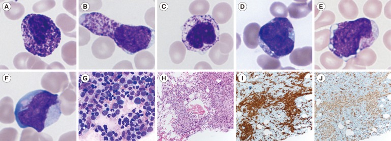
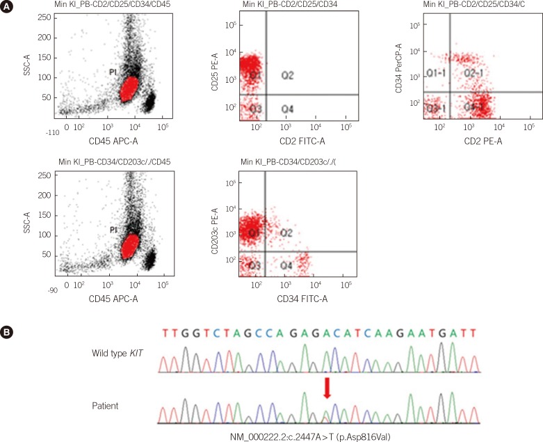
A 47-yr-old man presented with fever, cough, sputum, diarrhea, and abdominal pain of one-month duration. A complete blood count revealed Hb, 11.4 g/dL; leukocyte count, 3.4×10⁹/L; absolute neutrophil count, 1.2×10⁹/L; and platelet count, 44×109/L. Peripheral blood (PB) smear revealed leukoerythroblastosis with 26% abnormal cells, which were found, by immunophenotyping, to have originated from the mast cell lineage. Atypical mast cells displayed various morphological abnormalities (Fig. 1A-F). BM aspirate smears showed ungranulated blasts (12.9%), metachromatic blasts (10.1%), promastocytes (5.9%), and atypical, spindle-shaped mast cells (15.7%) (Fig. 1G). The blasts were negative for myeloperoxidase, periodic acid-Schiff, and α-naphthyl acetate esterase stains. The BM was hypercellular (80%), showing diffuse and interstitial infiltration of spindle-shaped cells (Fig. 1H). Immunohistochemistry indicated that immature cells were positive for CD117 (Fig. 1I) and CD68 (Fig. 1J). PB and BM specimens were positive for CD13, CD33, CD117, CD25 (PB), and CD203c (PB), whereas they were negative for CD2 (PB) (Fig. 2A), as determined by flow cytometry immunophenotyping. FISH analysis with BCR/ABL, PML/RARA, RUNX1/RUNX1T1, CBFB-BAF, D7S486/CEP7 probes showed normal findings. Chromosome analysis of BM showed a normal male karyotype. KIT D816V mutation was identified by PCR and direct sequencing of the gene in exons 8, 10, 11, 12, 13, and 17 (Fig. 2B). Based on these results, the patient was diagnosed as having a de novo leukemic variant of MCL.
The major challenge lay in determining whether the abnormal cells, including metachromatic atypical cells and granulated or ungranulated blasts, originated from the mast cell lineage cells. Both basophil and mast cell lineages stem from a common progenitor, basophil/mast cell progenitor (BMCP), which expresses ectonucleotide pyrophosphatase/phosphodiesterase-3 (ENPP3; CD203c) [3]. By using flow cytometry immunophenotyping, most abnormal cells were found to simultaneously express CD203c and CD25, indicating that they were neoplastic mast cells. Aberrant expression of CD25 and/or CD2 by mast cells in blood or BM is a criterion for the diagnosis of systemic mastocytosis [4]. The CD25+/CD2- immunophenotype shown in this case is a rare occurrence. In a study that reviewed 51 MCL cases, CD25 and CD2 expressions varied, with CD25+/CD2+ (54%), CD25-/CD2- (38%), CD25+/CD2- (13%), or CD25-/CD2+ (4%) phenotypes observed [1]. Another study examining 123 patients with varying subtypes of systemic mastocytosis showed a correlation between the CD25+/CD2- immunophenotype and systemic mastocytosis with poor prognosis, such as aggressive systemic mastocytosis and MCL [5].
Recurrent cytogenetic abnormalities specific for MCL have not yet been reported [1]. Consistent with this, our case also lacked cytogenetic abnormalities. KIT D816V is the most common molecular abnormality, occurring in 13 of 28 (46%) MCL cases [1]; similarly, one of the two cases (50%) previously reported in Korea had the mutation [6, 7]. Other mutations in exons 9, 10, 11, and 13 of KIT have been reported less frequently, including one case in 2004 and six cases since 2011 [1]. The literature indicates that the predominance of KIT D816V mutation in MCL is due to the recent availability of the entire sequence of KIT [1].
Mutations that occur in KIT during the early stages of hematopoietic differentiation lead to multi-lineage abnormalities and an early blockade of maturation [1, 5, 8]. Interestingly, patients with a less aggressive clinical course of MCL exhibit a predominance of mature cells, such as spindle-shaped or round, well-granulated mast cells, while those with a more aggressive clinical course show a preponderance of immature cells such as promastocytes, metachromatic blasts, or ungranulated blasts [9]. In this case, 68% of atypical mast cells were immature, indicating that the KIT mutation might have occurred during the early stages of hematopoiesis. The median survival time (range) is 4 (0.5-24) and 5.5 (2-3) months among de novo MCL and KIT D816V groups, respectively [1]. Our patient was followed up for 12 months after stem cell transplantation, and he is currently stable.
We conclude that the clinical features, histological analysis of blood and BM, and immunophenotype, together with the KIT D816V mutation detected in this case could be helpful for early diagnosis and treatment of MCL patients.
References
1. Georgin-Lavialle S, Lhermitte L, Dubreuil P, Chandesris MO, Hermine O, Damaj G. Mast cell leukemia. Blood. 2013; 121:1285–1295. PMID: 23243287.

2. Swerdlow SH, Campo E, Harris NL, Jaffe ES, Pileri SA, Stein H, editors. WHO classification of tumours of haematopoietic and lymphoid tissues. 4th ed. Lyon: IARC;2008. p. 54–63.
3. Arinobu Y, Iwasaki H, Akashi K. Origin of basophils and mast cells. Allergol Int. 2009; 58:21–28. PMID: 19153533.

4. Lim KH, Tefferi A, Lasho TL, Finke C, Patnaik M, Butterfield JH, et al. Systemic mastocytosis in 342 consecutive adults: survival studies and prognostic factors. Blood. 2009; 113:5727–5736. PMID: 19363219.

5. Teodosio C, García-Montero AC, Jara-Acevedo M, Sánchez-Muñoz L, Alvarez-Twose I, Núñez R, et al. Mast cells from different molecular and prognostic subtypes of systemic mastocytosis display distinct immunophenotypes. J Allergy Clin Immunol. 2010; 125:719–726. PMID: 20061010.

6. Bae MH, Kim HK, Park CJ, Seo EJ, Park SH, Cho YU, et al. A case of systemic mastocytosis associated with acute myeloid leukemia terminating as aleukemic mast cell leukemia after allogeneic hematopoietic stem cell transplantation. Ann Lab Med. 2013; 33:125–129. PMID: 23483057.

7. Park SH, Chi HS, Cho YU, Jang S, Park CJ, Lee JH. A case of de novo aleukemic mast cell leukemia without c-KIT mutations in exons 8 and 17. Int J Lab Hematol. 2014; 36:e44–e46. PMID: 24034180.
8. Teodosio C, García-Montero AC, Jara-Acevedo M, Alvarez-Twose I, Sánchez-Muñoz L, Almeida J, et al. An immature immunophenotype of bone marrow mast cells predicts for multilineage D816V KIT mutation in systemic mastocytosis. Leukemia. 2012; 26:951–958. PMID: 22051531.

9. Valent P, Sotlar K, Sperr WR, Escribano L, Yavuz S, Reiter A, et al. Refined diagnostic criteria and classification of mast cell leukemia (MCL) and myelomastocytic leukemia (MML): a consensus proposal. Ann Oncol. 2014; 25:1691–1700. PMID: 24675021.

Fig. 1
Serial peripheral blood analysis of mast cells, bone marrow aspirate smear, bone marrow trephine biopsy section, and immunohistochemical staining for CD117 and CD68. Peripheral blood cells stained with Wright-Giemsa (×1,000) (A-F): (A) Circulating round mast cells with eccentric oval nucleus, (B) Spindle-shaped mast cells, (C) Hypogranulated mast cells with coalescent granules, (D) Focal granules accumulated in mast cells, (E) Undifferentiated immature cells with metachromatic granules, (F) Ungranulated blasts with prominent nucleoli. Bone marrow aspirate stained with Wright-Giemsa (×400): (G) Ungranulated and metachromatic blasts, atypical bi-lobed promastocytes, and mature mast cells with elongated nuclei and hypogranular cytoplasm on bone marrow aspirate smear. Bone marrow trephine biopsy section stained with hemolysin and eosin (×200): (H) Hypercellular marrow with diffuse and interstitial infiltration of mast cells. Positive immunohistochemistry for CD117 (I) and CD68 (J).


 PDF
PDF ePub
ePub Citation
 Print
Print


 XML Download