

 PDF
PDF ePub
ePub Citation
 Print
Print


INTRODUCTION
Complete and accurate staging of non-Hodgkin lymphoma (NHL) is essential in determining the extent of disease, which may affect both the prognosis and treatment strategies [1, 2, 3, 4, 5]. Although there has been a continual growth in the number of ancillary tools that can be used in the laboratory to evaluate malignant lymphoma over the last decade [6, 7, 8, 9], evaluation of bone marrow involvement of malignant lymphoma is still an important aspect, and bilateral trephine biopsies have been regarded as the standard method [10].
The utility of flow cytometric analysis in the routine staging of NHL has been evaluated by several previous studies [11, 12, 13, 14, 15, 16, 17, 18, 19, 20]; however, sufficient data and standardization of protocols are lacking. Moreover, with the advance of technology, the usefulness of a multi-color strategy for diagnosis has been increasingly recognized, but has not been fully evaluated. For this reason, we evaluated the roles of six-color multiparameter flow cytometric analysis of bone marrow aspirate in the staging of B-cell NHL in the Korean patient population.
METHODS
1. Study population
We collected 248 bone marrow specimens from 232 patients (137 men and 95 women) who were diagnosed as having B-cell malignancy between December 2012 and July 2013 at our center: 198 at diagnosis and 50 during the course of the disease. Regarding diagnoses, diffuse large B-cell lymphoma was most common (44.0%), followed by mucosa-associated lymphoid tissue (MALT) lymphoma (28.2%) and follicular lymphoma (10.9%) (Table 1).
2. Bone marrow aspirate and biopsy
Wright-Giemsa-stained slides of bilateral bone marrow aspirate smears were examined for atypical lymphoid/lymphoma cells independent of immunophenotypic studies. Of 248 bone marrow aspirate specimens, five had no cellular component and were excluded from the study. Bilateral bone marrow trephine biopsies were obtained and tissue samples were fixed in 10% neutral-buffered formalin, decalcified, and paraffin-embedded. Hematoxylin and eosin (H&E) staining and CD3 and CD20 immunostaining were performed to determine lymphoma involvement.
3. Flow cytometry
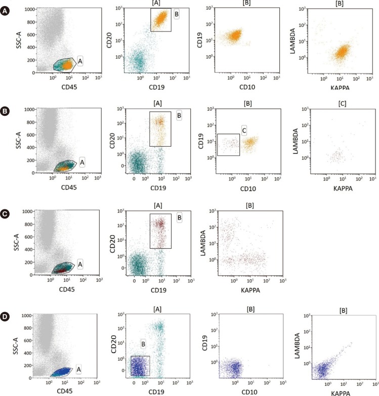
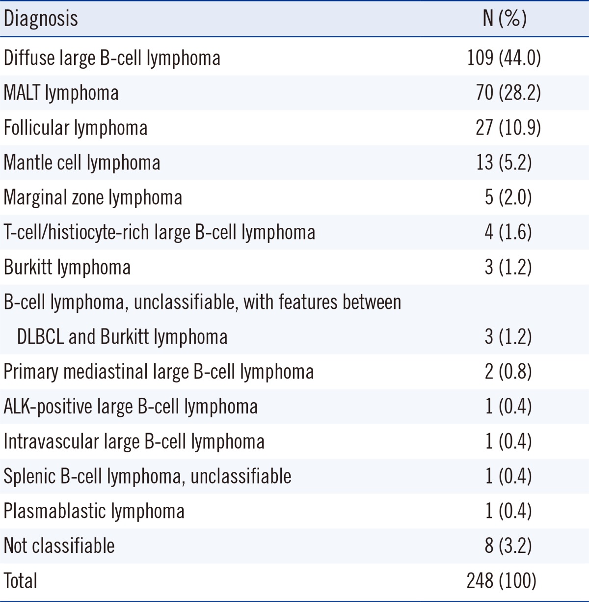
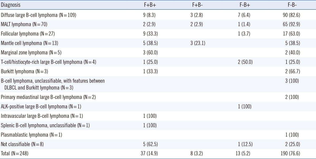
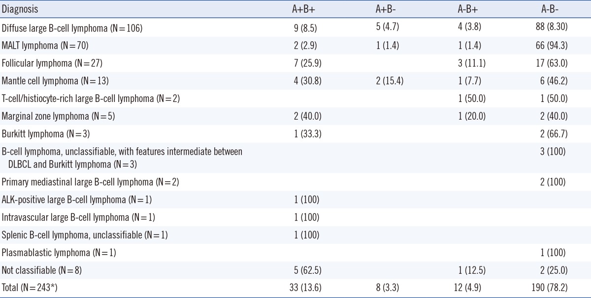
Flow cytometric immunophenotyping of an EDTA anticoagulated bone marrow aspirate specimen was performed in each case. A standard bone marrow assay with erythrocyte cell lysis was used for all bone marrow aspirate specimens. Aspirate specimens from one side or a mixture of both right and left sides were used depending on the quality and amount of specimens. The number of cells analyzed was between 50,000 and 250,000, and the instrumental sensitivity was 0.1%.
Samples were analyzed with a two-step protocol; screening was done first with six-color multiparameter flow cytometry to detect the abnormal lymphoid cell population followed by second-line, detailed immunophenotyping for specific characterization of lymphoma cells. For the first step, an analysis with six markers for B-cell lymphoma was performed with monoclonal antibodies directed against CD19, CD20, CD10, and κ and λ immunoglobulins (Igs). In the case of mantle cell lymphoma, a monoclonal antibody against CD5 was used instead of CD10. These antibodies were combined as κ/λ-fluorescein isothiocyanate/lambda-phycoerythrin (PE), CD19-peridinin chlorophyll (PerCP), CD10-allophycocyanin (APC), CD20-PE-cyanine7 (Cy7), and CD45-APC-Cy7. In the case of mantle cell lymphoma, CD5-PerCP and CD19-APC were used. Antibodies were supplied by Becton Dickinson immunocytometry systems (Becton Dickinson, San Jose, CA, USA) except for CD5, which was supplied by Beckman Coulter (Beckman Coulter, Miami, FL, USA). Because lymphomas associated with these cases were classified on the basis of lymph node or tissue biopsies, the first bone marrow immunophenotypic analyses were solely centered on involvement of malignant lymphoma, i.e., the presence of a monoclonal B-cell population. Cutoffs of κ-to-λ ratios have been pre-established as 0.25:1 and 4:1 in our laboratory with normal bone marrow specimens.
The second-line flow cytometric analysis was performed with positive cases in the initial clonality test. This step is aimed at evaluating the detailed immunophenotype of lymphoma cells. A selected panel of antibodies was used and combined as the following: FMC (developed at Flinders Medical Centre) 7/CD23/CD10/CD45, CD5/CD20/CD22/CD45, CD38/CD3/CD56/CD45, and terminal deoxynucleotidyltransferase (TdT). Both forward/side light scatter and CD45/side light scatter were used in each case as primary gating methodologies. Further gating was performed as necessary on either lymphoid subpopulations based on cell size or backgating in the event of CD19+ B-cell staining.
RESULTS
Approximately 24.6% of bone marrow evaluations showed evidence of NHL; bone marrow trephine biopsies showed histologic involvement of lymphoma cells in 50 cases (20.2%) and flow cytometric analysis of bone marrow aspirate showed the presence of lymphoma cells in 45 cases (18.1%).
1. Flow cytometric immunophenotype
Forty-five cases (18.1%) showed bone marrow involvement by NHL by flow cytometry, with monoclonal B-cell populations ranging from 0.09% to 60.00% (median 5.12%) of total cell populations. Among the positive cases, 19 (42.2%) showed minimal bone marrow involvement (malignant cells less than 5%; Fig. 1). The 203 cases with a negative flow cytometric study typically showed a mixture of polyclonal B cells and a small population of CD19+/Ig- cells representing normal CD19+ precursor B cells. In these cases, the κ-to-λ ratios of polyclonal B cells ranged from 0.98 to 1.85, and the median was 1.35, which was concordant with previously established normal ranges.
2. Discrepancies between flow cytometry and histopathology
The concordance rate between flow cytometry and trephine biopsy histopathology was 91.5%; both were negative in 190 cases (76.6%) and both were positive in 37 cases (14.9%). Discrepant results were found in 21 cases (8.5%) (Table 2). There were eight cases with histomorphologically negative and immunophenotypically positive results, and the diagnosis in these cases included diffuse large B-cell lymphoma (n=3), mantle cell lymphoma (n=3), and MALT lymphoma (n=2). Of those eight cases, six showed minimal bone marrow involvement (0.09-2.2%). Thirteen cases were histopathologically positive and immunophenotypically negative, and the diagnoses in these cases included diffuse large cell lymphoma (n=7), T-cell/histiocyte-rich large B-cell lymphoma (n=2), anaplastic lymphoma kinase (ALK)-positive large B-cell lymphoma (n=1), follicular lymphoma (n=1), MALT lymphoma (n=1), and unclassifiable lymphoma (n=1).
3. Bone marrow aspirate and biopsy
Morpholgic examination and analysis of immunostaining of both bone marrow biopsy and aspirate showed 91.8% concordance rate; both were negative in 190 cases (78.2%) and both were positive in 33 cases (13.6%) (Table 3).
DISCUSSION
Complete and accurate staging of malignant lymphoma is essential in providing adequate management and predicting outcomes. As a part of lymphoma staging, the evaluation of bone marrow involvement is an important process, and morphological assessment of bone marrow biopsy specimens remains the standard method. Recently, flow cytometric analysis of bone marrow aspirate has been performed as a routine procedure. Although flow cytometry provides more objective results with higher sensitivity, previous studies did not support a significant benefit beyond morphological examination alone [18, 19, 20]. However, the low detection rate by flow cytometry in those studies may be partly related to the use of less sensitive and less specific single- or dual-color analyses, which were the standard at that time, compared with the four- or six-color multiparameter analysis technology that is currently used. With the advanced technology currently available, six-color or eight-color multiparameter analysis can improve the sensitivity and specificity [21, 22]. At this point, evaluation of the utility of flow cytometric analysis on bone marrow using multiparameter analysis could be helpful. We performed both morphological examination of bone marrow biopsy specimens and flow cytometric analysis of bone marrow aspirates concurrently, and compared the results.
The immunophenotyping in this study was performed with six-color multiparameter flow cytometry, which has advantages compared to examination with only CD19, κ, and λ light chains. Including CD10 enabled increased detection sensitivity by excluding normal hematogones, and the inclusion of both CD19 and CD20 was additionally helpful for a more specific selection of B-cell populations according to differentiation status. Moreover, the specificity may be improved by using CD10 in follicular lymphoma and by using CD5 in mantle cell lymphoma. This will result in an improved sensitivity by lowering the possibility of masking small portions of malignant cells by the presence of many polyclonal cells.
The flow cytometric analysis is based on the clonality test. Detection of immunoglobulin light chain-restriction expression represents the most reliable evidence for the presence of malignant cells. In this study, κ-to-λ ratio ranged from 0.98 to 1.85 and the median was 1.35, confirming the normal range previously determined. The second-line, detailed immunophenotyping to detect abnormal immunophenotypes of malignant B-cell populations may increase the sensitivity when the role of screening immunophenotyping is limited, as in cases of malignant B cells with no light chain expression. Detailed immunophenotyping can also provide complementary information, which may be helpful in the accurate subclassification of NHL [23, 24]. This is very useful in cases without easily accessible lymph nodes or in cases with no definite lymphadenopathy.
There were 21 discrepant cases between the results of the bone marrow biopsy and flow cytometric analysis. Among those, eight cases were reported, in which lymphoma cells were not detected by morphological examination. The discrepancy could be explained in part by a low sensitivity of morphological examination in detecting minimal bone marrow involvement. Indeed, six of eight cases showed minimal bone marrow involvement of malignant lymphoma. In 13 cases, however, the involvement of the lymphoma was detected only in the biopsy specimen. This discrepancy may be attributed to differences in the specimens analyzed. For example, when the disease does not diffusely involve the bone marrow and the lymphoid infiltration foci are small, the bone marrow may produce aspirate samples free of disease while the biopsy may be positive for lymphoma cells. Evidence for this is the improved sensitivity of bilateral bone marrow biopsy over unilateral biopsy. The same principle can be applied in the cases, in which lymphoma involvement was not detectable on bone marrow aspirate despite positive results on bone marrow biopsies. The presence of these cases confirms previously published results, which concluded that there is no substitute for a bone marrow biopsy [13, 14].
Additionally, collection of bone marrow aspirate is a simple and minimally invasive procedure that is considered an ancillary tool in the staging of NHL. The utility of collecting bone marrow aspirate has been advocated by some groups [25, 26], while others have not supported the utility of this procedure [27]. To evaluate the usefulness of this specimen, we simultaneously compared bone marrow aspirate and biopsy samples. When aspirate was compared with biopsy specimens by morphological examination and immunostaining, the concordance rate was 91.8% with 20 discrepant cases. On morphological examination of aspirate specimens, it was difficult to confirm the involvement of NHL when a minimal number of lymphoma cells were present or the specimen was diluted with peripheral blood. The utility of flow cytometric examination could be maximized when the cytomorphological findings are equivocal.
We conclude that multi-color flow cytometry and examination of bone marrow aspirate are useful methods for assessing bone marrow in NHL staging. Although they are not a substitute for bone marrow biopsy, they do have a complementary role in detecting a small portion of lymphoma cells in a small subset of patients. Particularly, multi-color flow cytometric analysis can increase the sensitivity of this method, enabling more specific gating of abnormal cell populations. It may also be useful in cases where the morphological evaluation of the bone marrow biopsy is inconclusive or unavailable. Since the clinical significance of such a minimal bone marrow involvement is not fully established, further clinical studies are needed to elucidate the effect of minimal bone marrow involvement on the clinical course and prognosis of patients.
 XML Download