

 PDF
PDF ePub
ePub Citation
 Print
Print


Nevus sebaceus is a benign hamartoma of the epidermis, hair follicles, and sebaceous and apocrine glands. It typically appears as a yellow-orange to pink, finely papillomatous alopecic plaque that is often oval or linear. It usually presents at birth, preferentially affects the scalp and face, and is not uncommon, occurring in approximately 1 in 1,000 live births [1].
A recent study by Groesser et al. [1] reported that 95% of patients with nevus sebaceus lesions had somatic mutations in the HRAS gene and 5% had mutations in the KRAS gene, and the HRAS c.37G>C mutation was present in 91% of all lesions as the predominant mutation; approximately 3% of lesions had KRAS c.35G>A (p.Gly12Asp) as the next common mutation. These mutations were not detected in either non-lesional skin tissue or in whole blood, confirming nevus sebaceus as a 'mosaic RASopathy'. We report the case of a female infant with nevus sebaceus, in whom a somatic KRAS mutation was identified by Sanger sequencing in a cutaneous lesion.
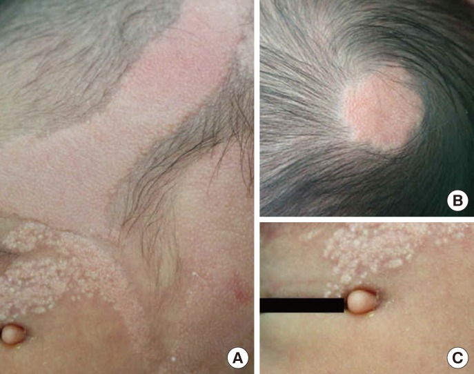
A 3-day-old female infant was transferred to our institution owing to skin lesions on the scalp, especially over the left parietal and occipital areas. The patient had a slippery, skin-colored, hairless, orange-peel-like linear plaque on her left parietal scalp and face (Fig. 1A) and a round plaque on her occipital scalp (Fig. 1B). Facial examination revealed a limbal dermoid with a skin-colored pedunculated papule on the left eyelid (Fig. 1C). Magnetic resonance imaging of the head showed an arachnoid cyst in the anterior portion of the left temporal lobe, and computed tomographic scanning of the orbit showed a calcified nodular lesion in the medial portion of the left orbit. Echocardiography showed that the patient had an atrial septal defect. A biopsy specimen taken from the plaque showed hyperkeratosis, acanthosis, numerous sebaceous glands, and malformed hair follicles (Fig. 2), consistent with nevus sebaceus.
After obtaining informed consent from the patient's parents, tissue samples (~5 mm) were collected from a nevus sebaceus lesion of scalp and from normal skin by elective punch biopsy and were subjected to fibroblast culture for expansion. Genomic DNA was extracted from cultured skin fibroblasts as well as from peripheral blood leukocytes using the Wizard Genomic DNA Purification Kit (Promega, Madison, WI, USA), following the manufacturer's instructions. Sanger sequencing was performed on DNA extracted from nevus sebaceus tissue, normal skin tissue, and peripheral blood. We initially analyzed the specimens for HRAS c.37G>C (p.Gly13Arg), the most common mutation detected in nevus sebaceus, but were unable to detect this mutation in any of the specimens. Subsequently, we analyzed them for the second most common mutation in nevus sebaceus, KRAS c.35G>A (p.Gly12Asp), and detected this mutation in nevus sebaceus tissue from the patient, but not in her normal skin tissue or peripheral blood, confirming somatic mosaicism (Fig. 3).
RAS proteins regulate cell growth, proliferation, and survival. Mutations in HRAS and KRAS affect mitogen-activated protein kinase (MAPK) and phosphoinositide 3-kinase (PI3K) signaling pathways, resulting in alterations in cellular growth, proliferation, and differentiation. RAS mutations occur in 30% of human cancers [2] and the most common HRAS c.37G>C and KRAS c.35G >A mutations found in nevus sebaceus are also the most frequent somatic RAS mutations detected in various malignant tumors, according to the catalogue of somatic mutations in cancer (COSMIC) database [1, 3].
Activating RAS mutations have been detected in genomic DNA from nevus sebaceus lesional keratinocytes, but not in DNA derived from adjacent non-lesional skin or peripheral blood, suggesting that cutaneous mosaicism for postzygotic activating mutations in the HRAS and KRAS genes causes nevus sebaceus development. Although germline mutations in these genes have been shown to underlie Costello (OMIM18040) and Noonan syndromes (OMIM163950) [6, 7], these mutations are different from the most common HRAS and KRAS mutations found in nevus sebaceus. It is likely that the common HRAS and KRAS somatic mutations in nevus sebaceus are nonlethal in a mosaic state but lethal when present in the germline and that they are selected as acquired somatic mutations in human cancers [1].
In approximately 7% of nevus sebaceus patients, the nevus sebaceus lesions may be more extensive with extracutaneous manifestations, and the condition has been defined as nevus sebaceus syndrome [3]. The most frequent extracutaneous manifestation of nevus sebaceus syndrome is central nervous system involvement, and ophthalmologic abnormalities are also common [8]. Our patient had extracutaneous manifestations in the ocular and central nervous system, suggesting the diagnosis of nevus sebaceus syndrome. According to the studies by Groesser et al. [1] and Sun et al. [9], extensive mosaicism for activating HRAS and KRAS mutations involving the skin, skeletal, ocular and central nervous systems is considered to be the underlying genetic cause of nevus sebaceus syndrome.
In conclusion, although nevus sebaceus and nevus sebaceus syndrome are relatively common skin disorders, there has been no prior report on a genetically confirmed case in Korea. In this study, we present the first case of a female infant who had nevus sebaceus accompanied by extracutaneous manifestations, suggesting the diagnosis of nevus sebaceus syndrome, and in whom a somatic KRAS mutation (c.35G>A; p.Gly12Asp) was identified in the cutaneous lesion. Close monitoring for the potential development of secondary tumors is advised for this patient.


 XML Download