

 PDF
PDF ePub
ePub Citation
 Print
Print


INTRODUCTION
Hepatitis B virus (HBV) infection remains a global health problem, with more than 350 million chronically infected people; every year approximately 1 million people die from HBV-related diseases [1]. Notably, bacterial infections are a common cause of morbidity in patients with chronic severe hepatitis B (CSHB). Widespread bacterial infections can decompensate hepatic status and lead to death in patients with CSHB [2, 3]. Various infections lead to death in 30-50% patients with liver cirrhosis [2, 4]. Furthermore, bacterial infections are known to trigger complications of CSHB, including spontaneous bacterial peritonitis (SBP), variceal bleeding, hepatic encephalopathy, renal failure, and impairment in clotting factors [5].
A new inflammatory marker that could predict infections and assist physicians in making treatment decisions would be extremely useful. Inflammatory markers, such as C-reactive protein (CRP) and procalcitonin (PCT) [6] and white blood cells (WBCs) could be easily used for diagnosis and follow-up of several morbidities [6, 7]. CRP is synthesized mainly in the liver. Serum CRP concentrations were reported to be associated with metabolic syndrome and diabetes [8]. CRP has also been suggested as a predictive marker of cardiovascular events in patients with metabolic syndrome [8, 9] and may be correlated with the degree of tissue damage and the activity of the basal malignant disease [10-13]. A 116-amino acid prohormone of calcitonin, PCT is normally synthesized in the C cells of the thyroid gland. Other sources of PCT include liver and inflammatory cells. Thyroid-excised subjects continue to have PCT response during acute inflammation; therefore, the main site of PCT synthesis and its function are not yet completely clear [14-19]. The serum PCT concentrations increase in patients with bacterial infections or sepsis [20]. Similarly, altered levels of serum PCT have been well documented in chronic liver diseases and cirrhosis. However, serum PCT is not elevated in viral or autoimmune diseases of the liver [21].
Although some authors have used PCT as an inflammatory marker in the diagnosis of SBP [22], serum PCT has not been evaluated in the diagnosis of SBP in chronic hepatitis B patients. Therefore, we aimed to study the diagnostic role of serum PCT in CSHB patients with SBP and compared its diagnostic efficacy with those of CRP and WBCs.
METHODS
1. Study population
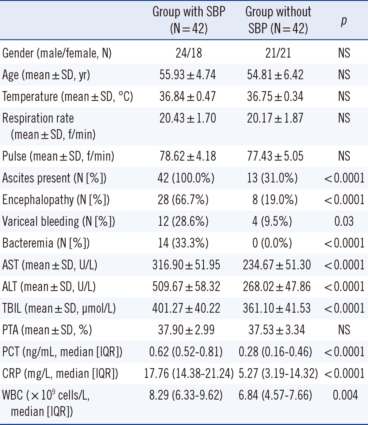
Between June 2010 and June 2012, 84 well-characterized, consecutive patients with CSHB (male/female: 45/39, age: 48.31±2.74 yr) from the Infection Department of Renmin Hospital (University of Wuhan) were considered. Of these, 42 CSHB patients with SBP without any infection in other organs or sites were enrolled as the study group. The control group was defined as 42 CSHB patients without SBP, and these patients did not have any overt infection. The clinical and laboratory characteristics of the patients are presented in Table 1.
2. Study design
Chronic hepatitis B viral carriers was diagnosed based on (a) positive tests for hepatitis B surface antigen (HBsAg) for at least 6 months before hospitalization or (b) positive tests for HBsAg, anti-hepatitis B core antibody (HBcAb) at a high titer and negative tests or a low titer of IgM anti-hepatitis B core antibody (IgM-HBc) in patients with follow-up periods of >6 months before admission. Blood samples and clinical data, including the presence of ascites, encephalopathy, bacteremia, recent episode of variceal bleeding, temperature, respiration rate, pulse, and blood pressure, were taken at enrollment. The diagnosis of SBP was established by assessment of clinical symptoms, appearance of fever, and laboratory reports (polymorphonuclear cell count in ascites >0.25×10⁹/L, a positive culture for any organism, or absence of an intraabdominal infection). Bacteremia was considered present, if the blood culture was positive for at least one organism when a documented primary infection was absent; blood cultures were performed for all patients. Furthermore, a contaminant was defined as a nonpathogenic microorganism [23], and cases of contamination were excluded from the analysis of bacteremia.
Clinical data were obtained retrospectively by thorough review of the patients' medical charts and included age, presence of ascites, encephalopathy, recent episode of variceal bleeding, bacteremia, AST, ALT, total bilirubin, prothrombin activity (PTA), etc.
3. Measurement of PCT, CRP, and WBC count
At the time of admission, venous blood was drawn from all the patients and centrifuged at 1,000×g for 10 min at 4℃ after a full blood cell count. All samples were tested for PCT, CRT, and WBCs within 30 min after acceptance by the clinical laboratory. PCT quantification was performed using automated immunoanalysis with the Liaison analyzer (Diasorin, Saluggia, Italy). For CRP, an immunoturbidimetric assay with ADVIA Chemistry CRP_2R (Siemens Medical Solutions Diagnostics, Tarrytown, NY, USA) was used. The WBC count was determined using DxH 800 (Beckman Coulter, Pasadena, CA, USA). The limits of detection of these techniques were 0.04 ng/mL, 4 mg/L, and 0.5×10⁹ cells/L for PCT, CRT, and WBC count, respectively.
4. Statistical analysis
Continuous variables were reported as mean (SD) or as median (interquartile range [IQR]), according to their homogeneity. Categorical variables were compared with the χ2-test, and continuous variables were compared with Student's t-test for parametric data or a Mann-Whitney test for medians of nonparametric data. To test the accuracies and cut-off values for different inflammatory markers, ROC curves were generated. The Spearman's correlation coefficient (rs) between PCT and other inflammatory markers was calculated for each group. The optimal cutoff value was determined by calculating the point on the ROC curve with the maximum Youden index (sensitivity-[1-specificity]) and the point with the shortest distance from the point (0,1) [(1-sensitivity)2+(1-specificity)2] for each inflammatory marker. Subsequently, the sensitivity, specificity, likelihood ratio of a positive (LR+) result, likelihood ratio of a negative (LR-) result, and diagnostic odds ratio were calculated for the different inflammatory markers. The optimal cutoff values of the different inflammatory markers were investigated for the diagnosis of CSHB patients with SBP. A 2-sided probability value of <0.05 was considered statistically significant. Data were analyzed using GraphPadPrism 5 (San Diego, CA, USA) and SPSS 17.0 (SPSS Inc, Chicago, IL, USA).
RESULTS
1. Comparison of patient characteristics in the 2 groups at baseline
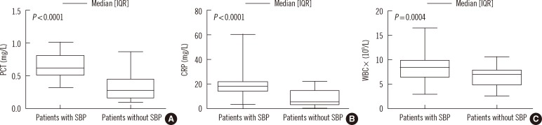
A total of 42 consecutive CSHB patients had clinically proven SBP, whereas the other 42 had no SBP at the time of polymorphonuclear cells counting (Table 1). There were no significant differences in gender, age, temperature, respiration, pulse, blood pressure, or PTA between the 2 groups. However, patients with SBP had specific complications (such as ascites, encephalopathy, variceal bleeding, and bacteremia) more frequently than patients without SBP. ALT, AST, and total bilirubin, were significantly higher in the SBP group than in the non-SBP group.
3. Correlations between CRP, WBC count, and PCT
A significant positive correlation was observed between PCT and CRP concentrations (P<0.0001, rs=0.69), and a less strong, but still significant, correlation was observed between PCT concentration and WBC count (P=0.001, rs=0.36).
4. Diagnostic accuracy of PCT, CRP, and WBC count
As determined by ROC curve analysis (Fig. 2), the accuracies of PCT concentration (area under the curve [AUC], 0.89; 95% (confidence interval) [CI], 0.81-0.96) and CRP concentration (AUC, 0.86; 95%CI, 0.78-0.94) for identifying CSHB patients with SBP were significantly higher (P<0.01) than those for WBC count (AUC, 0.68; 95% CI, 0.57-0.78). The AUC-ROC for the PCT concentration was not significantly different from that for the CRP concentration.
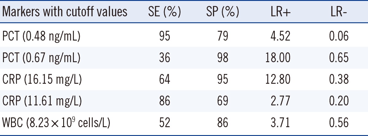
The optimal cutoff value for PCTwas 0.48 ng/mL. Meanwhile, the sensitivity and specificity of PCT for CSHB patients with SBP were 95% and 79%, respectively, at the cutoff of 0.48 ng/mL. The optimal diagnostic cutoff value of CRP was 16.15 mg/L. The sensitivity and specificity of CRP in CSHB patients with SBP were 64% and 95%, respectively, at the cutoff of 16.15 mg/L. The optimal cutoff value for WBCs was 8.23×10⁹ cells/L.The sensitivity and specificity of WBCs for CSHB patients with SBP were 52% and 86%, respectively, at the cutoff of 8.23×10⁹ cells/L. Table 2 shows a comparison of the parameters of serum PCT, CRP, and WBC count for the diagnostic accuracy of CSHB patients with SBP at different cutoff values. The PCT had a maximum LR+ for the diagnosis of CSHB patients with SBP at the cutoff of 0.48 ng/mL. The LR+ of PCT for CSHB patients with SBP was higher than that of CRP at a cutoff of 16.15 mg/L and WBC count at the cutoff of 8.23×10⁹ cells/L.
DISCUSSION
CSHB is a serious chronic liver disease that eventually results in acute decompensation of liver function. Usually, one or more reversible precipitating events such as superimposed acute hepatic necrosis, SBP, worsening renal function, or gastrointestinal bleeding lead to the acute decompensation of liver function. Chronic severe hepatitis often involves acute-on-chronic liver failure and chronic liver failure [24]. In China, most fulminant hepatitis patients have chronic liver failure caused by hepatitis virus B. Patients with CSHB are immunocompromised and thus are highly susceptible to the dissemination of infections such as SBP that worsen hepatic function and results in severe disease complications [25, 26]. Therefore, early diagnosis and active treatment of infections are vital. Obviously, it is crucial to determine a new clinical laboratory test for predicting infections and assist the physician in making treatment decisions. In the present study, we assessed the clinical utility of different inflammatory markers (PCT, CRP, and WBCs) for identifying bacterial infections in CSHB.
In our study, gender, age, temperature, respiration rate, pulse, blood pressure, and PTA were similar between the 2 groups. The disease-specific complications (ascites, encephalopathy, variceal bleeding, and bacteremia), including ALT, AST, and total bilirubin were strikingly different between groups (P<0.05).
The concentrations of PCT and CRP and the WBC count were significantly higher in the CSHB patients with SBP than in those without SBP. Furthermore, there were significant correlations between PCT and other inflammatory markers such as the WBC count and CRP. However, PCT and CRP concentrations were found to be better than WBC count for predicting CSHB with SBP, and the accuracy of the PCT concentration was not significantly different from that for the CRP concentration.
The diagnostic and prognostic value of serum PCT level in liver diseases has been evaluated in several studies, with relatively consistent results [27-32]. In patients with decompensated liver cirrhosis, a high PCT concentration showed a high sensitivity and specificity for bacterial infections [33]. In these studies, the serum PCT was significantly elevated in CSHB patients with SBP compared with those without SBP, but the median values varied: 0.74 ng/mL [28], 2.8 ng/mL [29], 3.2 ng/mL [7], 9.8 ng/mL [11], and 10.1 ng/mL [12]. We also found significantly elevated PCT concentrations in our CSHB patients with SBP, but the median value was low: 0.62 ng/mL. Furthermore, in this study, PCT concentration at the optimal cutoff of 0.48 ng/mL had the best sensitivity (95%), specificity (79%), and AUC-ROC (0.89) for SBP in CSHB patients. In the present cohort, the PCT concentration increased significantly in CSHB patients with SBP compared to those without SBP. These discrepancies can be due to the presence of bacterial infections; on the other hand, patients with acute viral hepatitis in with a CSHB background without proven bacterial infections show a high serum PCT with disease progression [28]. Therefore, we should measure serum PCT when the severity of disease is similar between groups.
CRP and WBC have been used as inflammatory markers of bacterial infections, even in liver diseases [34]. However, many questions on the optimal application of CRP and WBC in this patient population have not been addressed. It is vital to assess the determination power of CRP and WBC. In previous clinical studies involving 20 to 127 patients, CRP and WBC proved to be effective markers of bacterial infections in patients with liver diseases, but they had diverse diagnostic accuracies and cutoff values [27-29, 35-37]. A possible explanation for this variance may be the differences in the type of liver disease in between patients. In the present study, CRP at the optimal cutoff of 16.15 mg/L had the best sensitivity (64%), specificity (95%), and AUC-ROC (0.86). The optimal cutoff value of WBC count was 8.23×10⁹ cells/L, with a lower sensitivity (52%), specificity (86%), and AUC-ROC (0.68). This suggests that the WBC count is inferior to the serum PCT for the diagnosis of CSHB patients with SBP.
In conclusion, serum PCT and CRP concentrations were more accurate than WBC count for the diagnosis of CSHB-associated SBP. However, this study only included a small cohort (n=42) of patients with SBP. Larger samples and more homogeneous groups of CSHB patients with SBP should be used for further studies in order to confirm our results.

 XML Download