

 PDF
PDF ePub
ePub Citation
 Print
Print


INTRODUCTION
Hereditary hemolytic anemia comprises a group of disorders in which red blood cells (RBCs) are destroyed faster than they are produced in the bone marrow; various hereditary factors can cause this condition, including production of defective Hb and erythrocyte membrane. Unstable Hb, which includes defective hemoglobin molecules, should be considered as an important causative factor in patients with hereditary nonspherocytic hemolytic anemia [1]. Unstable Hb may be detected by the presence of an additional abnormal band in Hb electrophoresis or by demonstration of increased instability in Hb stability tests. However, the hemoglobin may be missed unless it is sufficiently stable to be detected by these methods [2-4]. Therefore, in patients showing hereditary nonspherocytic hemolytic anemia, further genetic workup for identification of unstable Hb is required even if the patients show normal results in Hb electrophoresis or stability tests [5].
Recently, we identified Hb Koriyama, a rare Hb variant that was undetectable in Hb electrophoresis and stability tests, in a patient with severe hemolytic anemia. Hb Koriyama is a highly unstable Hb variant and is formed by the tandem insertion of a 5-residue segment in the FG region of the Hb β-chain. The molecular structure of this variant was first modeled at the peptide sequence level and further presumptive changes were introduced in the nucleotide sequence [6] Here, we present the first case report confirming a change in the nucleotide sequence of Hb Koriyama.
CASE REPORT
A 6-yr-old girl was referred for a genetic workup of unexplained severe hereditary hemolytic anemia. She was born at 37 weeks' gestational age at a local hospital, and her birth weight was 2, 750 g; no morphological abnormality was observed at birth. She developed an icteric appearance with a total bilirubin level of 13.0 mg/dL on the second day after birth; the condition was successfully treated using phototherapy. At 3 months of age, she presented with sudden-onset hematuria and pallor. Laboratory test results showed severe hemolytic anemia with hyperbilirubinemia. The initial laboratory test results were as follows: Hb, 4.4 g/dL; hematocrit, 12.2%; reticulocyte count, 12.2%; total bilirubin, 5.8 mg/dL; direct bilirubin, 1.8 mg/dL; aspartate transaminase, 65 IU/L; and alanine transaminase, 15 IU/L. She received regular blood transfusions to raise her Hb level.
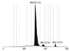
All tests for evaluating the peripheral blood-cell parameters were performed after the patient was referred to our hospital: Hb, 7.2 g/dL; mean corpuscular volume (MCV), 89.7 fL; mean corpuscular Hb (MCH), 29.6 pg; hematocrit, 26.1%; reticulocyte count, 29.0%; red cell distribution width (RDW), 35.3%; and nucleated RBC count, 143/100 white blood cells (WBCs). Peripheral blood smear showed normocytic and normochromic RBCs with severe anisocytosis, mild polychromasia, nucleated RBCs, and moderate poikilocytosis with elliptocytes, acanthocytes, and burr cells. To rule out hereditary spherocytosis, we performed an osmotic fragility test and erythrocyte membrane protein analysis; we obtained normal results for both these tests. For the latter, the levels of erythrocyte membrane proteins such as spectrin, ankyrin, band 3, and proteins 4.1 and 4.2 were analyzed using sodium dodecyl sulfate polyacrylamide gel electrophoresis. Hb electrophoresis was performed using a capillary electrophoresis system (Capillarys; Sebia, Lisses, France); the results showed low HbA (92.7%) levels, normal HbA2 (3.2%) levels, and elevated HbF (4.1%) levels (Fig. 1). Instability and isopropanol precipitation tests showed normal results.
The β- and α-globin genes (HBB and HBA) were analyzed to rule out the possibility of hemoglobinopathy. For this analysis, first, the entire HBB sequences were analyzed. Genomic DNA was extracted from blood using Puregene DNA isolation kit (Gentra Systems, Inc., Minneapolis, MN, USA). We performed gene-specific PCR and direct sequencing using a DNA analyzer (3730xl; Applied Biosystems, Foster City, CA, USA). Second, deletions or duplications in the β- and α-globin gene clusters were screened using a multiplex ligation-dependent probe amplification (MLPA) kit (P102-B1/P140-B2; MRC-Holland, Amsterdam, the Netherlands).
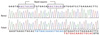
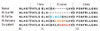
Gene sequence analysis revealed a heterozygous mutation, HBB c.280_294dup, p.(Cys94_His98)dup. We assume that this mutation led to the duplication of 5 amino acids from codons 94 to 98 (Fig. 2). The aberrant amino acid sequence created by this mutation was identical to that of Hb Koriyama, although Kawata et al. [6] had originally described the sequence as an insertion of 5 amino acids (Leu-His-Cys-Asp-Lys) between codons 96 and 97 (Fig. 3). Any large deletion or duplication was not detected in both globin genes on MLPA test.
DISCUSSION
Our case is the second report of Hb Koriyama in the literature; the first one was published by Kawata et al. In the study by Kawata et al. [6], the patient had severe chronic hemolytic anemia, splenomegaly, and required blood transfusion. Interestingly, the isopropanol test for their patient yielded positive results and thus, unstable Hb could be suspected easily; however, the isopropanol test was negative in our patient. Therefore, the Hb stability test cannot be relied upon to produce repeatable results for certain unstable Hb variants, and a negative result in this test does not necessarily exclude further genetic workup.
More than 185 unstable variants of both α- and β-globin chains, which produce varying degrees of hemolytic anemia (http://globin.cse.psu.edu/), have been identified [2-4]. Many unstable variants are associated with mild hemolytic features with minimal or even no anemia in the steady state. Furthermore, extremely unstable Hb is not detectable by the commonly used methods of Hb separation, as in the case of our patient [7]. Therefore, unstable Hb should be considered as a possible cause when unexplained hemolytic anemia i s diagnosed in patients.
By performing an amino acid analysis, Kawata et al. [6] showed that a 5-amino acid residue segment, which included cysteine at position 93, was repeated in tandem in the abnormal β chain. However, nucleotide sequence changes leading to the production of this abnormal Hb were not mentioned in their original study. Our results show that duplication of the 15-nucleotide segment from positions 280 to 294 can result in a 5-residue duplication seen in Hb Koriyama. Two 8-bp identical AGCTGCAC sequences that are present on both sides of the 280-294-bp segment may be a potential core of a replication misalignment event. It can result in either a 15-bp tandem duplication, such as that observed in our patient and Hb Koriyama, or a 15-bp deletion, such as in Hb Gun Hill [8].
In conclusion, our study is the first case of Hb Koriyama where the sequence change has been identified at the nucleotide level. It has to be emphasized that unstable Hb may not be detectable by conventional Hb electrophoresis or stability tests. Thus, genetic workup should be performed
 XML Download