

 PDF
PDF ePub
ePub Citation
 Print
Print


INTRODUCTION
In very elderly patients with acute leukemia, poor clinical condition or severe cytopenia may not allow bone marrow (BM) examination for diagnostic purposes. In such cases, alternative diagnostic approaches should be considered, such as the use of peripheral blood (PB) specimens for flow cytometric analysis to determine the lineage of leukemic cells and/or conventional cytogenetic studies to discover crucial genetic aberrations. AML with myelodysplasia-related changes (AML-MRC) is one of the new/expanded disease entities in the 2008 WHO classification, accounting for 24-35% of all cases of AML and usually occurring in the elderly [1-3]. Several gene rearrangements or chromosomal translocations can aid in the diagnosis of AML-MRC when ≥20% PB or BM blasts are present, even in the absence of immunophenotyping results [1]. Here, we describe a rare case of AML-MRC with t(3;21)(q26;q22) and subsequent RUNX1-MECOM (previously AML1-MDS1-EVI1) fusion transcript in a very elderly patient who was diagnosed on the basis of PB sample since BM examination could not be performed because of her advanced age and poor clinical status.
CASE REPORT
An 87-yr-old woman without any significant medical history was admitted to our hospital for pneumonia treatment in April 2011. The initial complete blood count showed Hb level of 5.9 g/dL (reference range, 12-16 g/dL), platelet (PLT) counts of 27×109/L (reference range, 150-350×109/L), and white blood cell (WBC) counts of 85.33×10⁹/L (reference range, 4-10×109/L) with 55% blasts. Blasts had high nuclear cytoplasmic ratio (N/C ratio), basophilic cytoplasm, and a fine chromatin pattern. Dysplastic features were observed in the PB smear, such as marked anisopoikilocytosis with teardrop cells in the red blood cells (RBCs), hypogranulation, and the Pelger-Huët anomaly in segmented neutrophils (Fig. 1). BM examination could not be performed because of the patient's advanced age and poor general condition; therefore, all of the following studies were conducted using PB specimens.
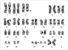
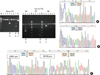
Flow cytometric analysis revealed the blasts to be positive for CD13, CD33, CD34, HLA-DR, and CD45, which indicated AML. Chromosome study showed 46,XX,t(3;21)(q26;q22) in 16 out of 20 analyzed metaphase cells (Fig. 2). FISH was performed according to the manufacturers' instructions with commercially available EVI1 three-color break-apart FISH probe (MetaSystems, Altlussheim, Germany) and AML1/ETO dual color dual fusion FISH probe (Abbott Molecular/Vysis; Des Plaines, IL, USA). The EVI1 break apart probe showed one split-out signal with one fusion signal in 91.5% of analyzed cells (194/212), consistent with EVI1 gene rearrangement (Fig. 3A). The AML1/ETO FISH probe showed nuc ish(ETOx2)(AML1x3)[187/216] (86.6%) (Fig. 3B).
Multiplex reverse transcriptase-PCR (RT-PCR) with the Hemavision kit (DNA Technology, Aarhus, Denmark) showed 3 different sized bands, each, in lanes 4, 7, and 8 (Fig. 4). Cloning and sequencing analyses were conducted using post-PCR products. Comparison with the Ensembl database v.64 (http://www.ensembl.org) revealed that these 3 fusion transcripts were as follows: the fusion transcript in lanes 4 (291 bp) and 8 (856 bp) showed breakpoints between exon 3 of RUNX1 (ENST00000344691) and exon 2 of MECOM (ENST00000494292) and the second one in lane 7 (127 bp) revealed a breakpoint between exon 3 of RUNX1 and exon 2 of RPL22 (previously EAP) (ENST00000494292).
The patient was initially treated with hydroxyurea for a week. Thereafter, her blast count decreased gradually, and the patient was discharged to a hospice facility.
DISCUSSION
In some cases of AML-MRC, identification of unique chromosomal aberrations is essential for accurate diagnosis, especially when morphologic findings or medical histories are not conclusive or proper specimens are absent. Chromosomal aberrations that allow the diagnosis of AML-MRC include unbalanced abnormalities -7/del(7q), -5/del(5q), i(17q)/t(17p), -13/del(13q), del(11q), del(12p)/t(12p), del(9q), idic(X)(q13); and balanced abnormalities t(11;16)(q23;p13.3), t(2;11)(p21;q23), t(5;12)(q33; p12), t(5;7)(q33;q11.2), t(5;17)(q33;p13), t(5;10)(q33;q21), t(1;3) (p36.3;q21.1), t(3;5)(q25;q34), and t(3;21)(q26.2;q22.1) when ≥20% PB or BM blasts are simultaneously present [1]. Our research group recently reported, along with extensive literature review, 2 unique AML-MRC cases exhibiting balanced abnormalities involving the long arm of chromosome 3, which were t(1;3) and t(3;5), respectively [4, 5]. We also reported a case of AML with t(3;21) producing a RUNX1-MECOM (MECOM is also known as MDS1 and EVI1 complex locus) fusion transcript in acute promyelocytic leukemia which relapsed as secondary AML [6].
The t(3;21) is a rare recurring translocation found in therapy-related myeloid neoplasm, CML with accelerated or blastic phase, and, rarely, in de novo AML [6]. In the present case, the patient had no significant medical history or a history of prior chemotherapy with agents such as an alkylating agent or DNA topoisomerase II, enabling a specific diagnosis of AML-MRC. In the Mitelman database (submitted on December 26, 2011), a total of 144 cases with t(3;21) were found to include 60 CML cases, 16 MDS cases, and 62 AML cases, which were mostly therapy-related. The oldest reported AML-MRC patient is an 89-yr-old woman with a complex karyotype, including t(3;21) (q26.2;q22) [7]. In our study, the RUNX1-MECOM fusion transcript was confirmed by breakpoint analyses using subsequent RT-PCR, cloning, and sequencing methods. It is well known that in most t(3;21) cases, RUNX1 could be fused with 3 genes located within the 3q26 region, RPL22 (previously EAP), MDS1, and EVI1, as a result of alternative intergenic splicings [8-10]. Studies have indicated that the RUNX1-MECOM fusion transcript contributes directly to leukemogenesis or leukemic transformation [8, 11, 12].
In this study, AML-MRC in a very elderly patient was successfully diagnosed by detecting chromosomal translocation t(3;21) and gene rearrangement (the RUNX1-MECOM fusion gene) along with immunophenotyping results of the PB specimen, although the clinical condition of the patient did not allow BM examination. Because cytogenetic abnormalities associated with AML-MRC on the basis of the 2008 WHO classification can be applied as very useful diagnostic markers, alternative active diagnostic approaches, such as the use of PB samples for chromosome, FISH, and RT-PCR analyses can lead to a proper conclusion. This is true even in cases in which BM examinations or flow cytometric analyses are not available and also when morphologic evidence of dyspoiesis is not apparent in very elderly patients.


 XML Download