

 PDF
PDF ePub
ePub Citation
 Print
Print


The discovery of the 46 human chromosomes by Tijo and Levan in 1956 is one of the most remarkable discoveries of the century, thus defining the starting point of conventional cytogenetics [1]. After the Philadelphia chromosome was found by Nowell and Hungerford in 1960, Rowley identified several major chromosomal translocations in human leukemias in the 1970s, including t(8;21), t(15;17) and t(9;22) [2]. By the middle of 1980s, the Philadelphia chromosome, defined at the chromosome level, was finally identified as originating from BCR-ABL1 rearrangement. In the following molecular biology era that was guided by passionate researchers and their contributions, many genetic aberrations known at the chromosomal level were revealed to be associated with gene fusions such as PML-RARA, RUNX1-RUNX1T1, CBFB-MYH11, and MLL-AF4. Therefore, it can be said that from the inception of the field of cancer cytogenetics and for nearly half a century, chromosomal translocations or gene rearrangements have been the main research focus. However, efforts to search for second hit genetic aberrations were made throughout the recent decades with the aid of remarkable technical advancements such as high throughput sequencing technologies, which led to increased interest in gene mutations. Furthermore, recent discoveries related to gene expression profiles, the identification of disease-specific gene signatures, epigenetic mechanisms, and the role of microRNA in acute leukemias are filling in this emerging genetic picture [3].
The current molecular diagnostic approach for leukemias in Korea is mainly focused on identifying chromosomal translocations or gene rearrangements by 3 different technologies: 1) conventional cytogenetics, 2) FISH analysis using particular probe sets, and 3) specific reverse transcriptase-PCR (RT-PCR) assays. All of these methods are useful for detecting known gene rearrangements such as the MLL gene translocations, although each method has its own merits and disadvantages. Conventional cytogenetic analysis has the advantage being able to evaluate the number and integrity of chromosomes. Moreover, it has the potential to indicate certain chromosome abnormalities. However, it is quite difficult to detect cryptic gene rearrangements of submicroscopic deletions or inversions with conventional cytogenetic analysis. Both split-signal FISH technologies and RT-PCR assays have the advantage of being fast and target gene-specific. However, commonly employed multiplex RT-PCR kits such as "HemaVision" (DNA Technology, Aarhus, Denmark) are also limited in the number of rearrangements they can detect [4, 5]. For example, if an 11q23 abnormality is suspected by chromosome analysis, an MLL rearrangement can be detected by split-signal FISH without knowledge of the partner genes involved. Therefore, both of these technologies (RT-PCR and split-signal FISH) will not allow the detection of currently unknown MLL rearrangements, as further studies are required to identify the actual partner genes. In this letter, we would like to introduce an alternative, compensatory diagnostic method, termed long distance-PCR (LD-PCR) or long distance inverse-PCR (LDI-PCR), which can be used in combination with the current molecular diagnostic tools.
As previously described, LDI-PCR is a modified LD-PCR method that utilizes genomic DNA [6-8]. Briefly, the DNA sample is treated and analyzed as previously described [6, 7]. First, 1 µg of genomic DNA is digested with restriction enzymes and re-ligated to form circular DNA before performing LDI-PCR using myeloid/lymphoid leukemia (MLL)-specific primers. Then, restriction polymorphic PCR amplimers are isolated from the gel and subjected to DNA sequence analysis to obtain patient-specific fusion sequences. Detailed LDI-PCR methods were previously provided elsewhere [6, 7].
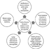
By applying this technique, we have successfully identified the MLL-CASP8AP2 rearrangement in a Korean AML patient, as well as MLL-related 3-way and 4-way translocations using precise genomic breakpoint analysis of LDI-PCR, with additional findings regarding the involvement of the NRXN1 and CCDC6 genes in fusion breakpoints [9-12]. Thus, this technique seems to have several advantages over the currently established diagnostic procedures (Fig. 1). The combined use of these previously described techniques allows for routine diagnostic work to be completed more quickly, while aiding researchers in making novel discoveries.
Importantly, LD-PCR is useful for recognizing unusual cryptic cytogenetic findings or discrepancies in molecular results that are hard to identify through common molecular diagnostic methods. A cryptic PML-RARA rearrangement in acute promyelocytic leukemia (APL) patient and another unique case of PML-ADAMTS17-RARA rearrangement in APL were identified by using LD-PCR [13, 14]. The molecular identification of a PML-RARA genomic fusion breakpoint using genomic DNA from the PML-RARA FISH-negative cell pellet is a representative example of the utility of this method [13]. Of note, gene rearrangements with alternative splicing (such as variant PML-RARA and CBFB-MYH11) are indicated for applying multiplex LD- or LDI-PCR.
Lastly, we have applied LD-PCR to the clonal eosinophilic disorder, which was first introduced in the 2008 WHO classification, for discovery of the genomic fusion breakpoint of ZMYM2-FGFR1 rearrangement [15]. This method was particularly useful as FGFR1-rearrangement has multiple fusion partner genes such as MLL rearrangement and cDNA was not available in this lymphoma case, suggesting that future utilization of LD-PCR as a method of choice for such circumstances is possible. However, PCR-based analyses of genomic DNA are not always feasible, as large intron size can be a limitation for applying LD-PCR. Further studies aimed at the usage of multiplex procedures based on LD- or LDI-PCR techniques will unveil the potential for this analysis in new disease states, such as in solid tumor DNA samples, for example [16, 17]. Furthermore, using the established sequences of genomic fusion sites for patient-specific minimal residual disease monitoring of acute leukemia has also been reported [7, 18].
LD- and LDI-PCR are supplementary methods that overcome the limitations of conventional cytogenetics, FISH, and RT-PCR, and exactly how to introduce this new technology into routine diagnostic procedures should be discussed among laboratory physicians in the field of hematology in Korea. Cutting edge methods derived from advanced molecular technologies in the field of genomics such as whole genome/exome sequencing or microarray are important, but are also expensive. LD- and LDI-PCR are relatively inexpensive procedures that complement the existing routine methods. We hope that these advanced diagnostic methods will eventually be adopted as tailored, target-specific genomic analyses in the near future.
 XML Download