

 PDF
PDF ePub
ePub Citation
 Print
Print


INTRODUCTION
Chromosome 5 abnormalities, which are involved in various rearrangements (including deletions, translocations, insertions, and rings), are a common aberration that is associated with AML or MDS [1-3]. Acquired ring chromosomes have been observed as part of a complex karyotype or, less frequently, as the sole change in human neoplasias [4, 5].
Sometimes, chromosomes that form a corresponding ring cannot be clearly defined by conventional cytogenetics (CC) owing to the complexity of the rearrangements, the suboptimal banding quality, and/or the shortage of material. FISH studies with region-specific probes provide only partial information that is confined to the target regions that are examined. Single nucleotide polymorphism arrays (SNP-A) were initially developed for genotyping DNA sequence polymorphisms and determining genome-wide allelic information. Because the strength of the hybridization signal is proportional to the copy numbers of the genomic region corresponding to a given probe, copy number changes can also be identified by SNP-A [6, 7]. Karyotypic analyses using high-density, whole-genome SNP-A may result in the better resolution of chromosomal defects, the identification of previously cryptic lesions, and more precise definitions of the breakpoints [6, 7].
Here, we describe a case of AML with a ring chromosome 5, resulting in terminal deletions of both 5p and 5q. To the best of our knowledge, this is the first report of a ring chromosome 5, in which the breakpoint and deleted segments were defined by genome-wide SNP-A-based karyotyping.
CASE REPORT
1. Case history
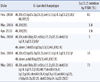
A 55-yr-old male presented with complaints of dizziness and dyspnea. The initial laboratory findings revealed a leukocyte count of 3.4×10⁹/L, a hemoglobin level of 8.1 g/dL, and a platelet count of 37×109/L. On a peripheral blood smear, 63% immature cells were found with 2% segmented neutrophils and 35% lymphocytes. The bone marrow aspirate was hypercellular and replaced by 90% blasts. The remaining cells were lymphocytes. Megakaryocytes were decreased in number. Accordingly, our case did not have sufficient non-blast cell elements in the bone marrow to adequately assess for multilineage dysplasia. The biopsy could not be evaluated due to an inadequate specimen. Flow cytometry showed that the blasts were positive for cytoplasmic myeloperoxidase, CD13, CD33, CD34, HLA-DR, and CD7 and negative for CD3, CD5, CD19, CD20, CD22, CD10, CD56, and CD14. Cytochemistry demonstrated that nonspecific esterase was positive. Finally, the patient was diagnosed with AML with myelodysplasia-related changes based on the 2008 WHO classification, because he had a MDS-related cytogenetic abnormality, the -5/del(5q). He was treated with induction chemotherapy involving idarubicin and arabinofuranosyl cytidine (Ara-C). Complete hematologic remission with a normal karyotype was achieved. After the second consolidation chemotherapy (daunorubicin+Ara-C), the patient received an autologous peripheral stem cell transplantation and remained in remission status for 3 months. However, 9 months after the diagnosis, relapse occurred (Table 1). Even after 3 reinduction therapies, remission could not be achieved and clonal evolution developed. The proportion of blasts increased up to more than 80% by 12 months after the diagnosis. The patient subsequently refused to receive continuous treatment and was lost to follow up.
2. CC, SNP-A, and FISH
For the cytogenetic analysis, unstimulated short-term cultures were set up using bone marrow aspirates, and at least 20 metaphases were analyzed. G-banded karyotype results were described according to the International System for Human Cytogenetic Nomenclature (ISCN) 2009 [8].
We applied a genome-wide SNP 6.0 array (Genome-Wide Human SNP array 6.0; Affymetrix Inc., Santa Clara, CA, USA) using genomic DNA according to the manufacturer's instructions, and the data were analyzed using Genotyping Console 3.1 software (Affymetrix Inc.). Aberrations that were identified by SNP-A were described according to ISCN 2009 [8]. In order to detect the somatic origin of the copy number alterations that were distinguished from constitutional polymorphic copy number variants (CNVs), we adopted our stringent and conservative algorithm. We selected only the lesions that were more than 100 kb and with more than 10 SNP/CNV probe markers of the array involved within those regions. The lesions that were then identified by SNP-A were compared with the Database of Genomic Variants (http://projects.tcag.ca/variation/) and with an internal control series from healthy normal controls with Korean ethnicities (N=200) in order to exclude known CNVs. For the copy neutral loss of heterozygosity (CN-LOH), we excluded homozygous stretches of the DNA region that were less than 25 megabases (Mb) in the interstitial chromosomal regions, except for those encompassing the telomeric regions of the chromosome, according to the algorithm adopted in the previous studies [6].
Interphase FISH studies were done on the bone marrow aspirates using commercially available probes (BCR/ABL1, RUNX1/RUNX1T1, PML/RARA, CBFB/MYH11, MLL, EGR1/D5S23, D5S721, and CEP7/D7S486; Abbott Laboratories, Abbott Park, IL, USA), according to the manufacturer's instructions.
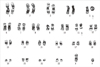
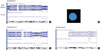
The cytogenetic analysis showed that 90% of the metaphase cells were bearing -5, del(11)(q22), +r in 20 metaphase cells analyzed (Fig. 1). Genomic aberrations identified by SNP-A were arr 5p15.33p15.2 (423,554-11,003,428)×1, 5q33.2q35.3 (153,225,007-180,652,396)×1, 11q14.1q23.2 (84,514,776-113,765,153)×1, and 21q22.13q22.2 (37,018,986-39,492,610)×3. These results indicated a 5p-terminal deletion (11 Mb), a 5q-terminal deletion (27 Mb), an 11q-interstitial deletion (29 Mb), and a 21q gain (3 Mb) (Fig. 2A, C, D). Therefore, the G-banded karyotype was finally revised as 46,XY,r(5)(p15.2q33.2),del(11)(q14.1q23.2),dup(21) (q22.13q22.2)[18]/46,XY[2].
As for the chromosome 5 abnormality, FISH revealed nuc ish (D5S721×1, D5S23×1, EGR1×2)[129/200]. This result corresponded to the signal patterns of the 5p15.2 deletion in 65% of the nuclei examined, whereas 2 EGR1 (early growth response 1) signals were indicative of no deletion of 5q31 on both chromosomes 5 (Fig. 2B). This finding was compatible with the result that the deleted region of 5q that was identified by SNP-A was more telomeric to 5q31, which was spanned by the commercially available EGR1/D5S23, D5S721 FISH probe.
Interphase FISH using the commercial MLL (mixed lineage leukemia) probe demonstrated normal hybridization patterns of the 11q23 regions, indicating that the deleted region of 11q by SNP-A was located more proximal to the MLL gene. FISH using BCR/ABL1, RUNX1/RUNX1T1, PML/RARA, CBFB/MYH11, and CEP7/D7S486 probes demonstrated normal hybridization signal patterns.
During the follow up, additional chromosomal abnormalities, including add(4)(q32) were found, suggesting clonal evolution (Table 1).
DISCUSSION
In this case, cytogenetic analysis showed metaphase cells bearing monosomy 5 and a ring chromosome. By SNP-A, 5p- and 5q-terminal deletions could be identified. However, FISH showed a normal hybridization pattern for the 5q31 region, although the common deleted region of the long arm of chromosome 5 is known to be located on 5q31 [1, 2]. Finally, in the present case, r(5) resulting from the partial deletion of the distal part of the short and long arms of the chromosome 5 and 5q31 chromosomal regions were included in part of the ring chromosome. Similar to our case, another study reported that monosomy 5 that was detected by CC was not true monosomy 5. Instead, some regions of chromosome 5 were retained in part of the ring or derivative chromosomes, as determined by molecular cytogenetics [3].
Ring chromosome formation may occur through breaks in the chromosome arms and fusion of the proximal broken ends, leading to the loss of distal material [1, 5]. In addition, rings may also be formed by telomere dysfunction that triggers fusion of the reactive chromosome ends without a major loss of genetic material [1, 5]. In our case, chromosome 5 appeared to be broken and fused at the 5p15.2 and 5q33.2 subtelomeric bands, leading to the formation of r(5).
A review of the Mitelman database revealed 12 cases carrying r(5) with variable breakpoints and the loss of various parts of chromosome 5 [1]. Most of the ring chromosomes were embedded in highly complex karyotypes. This suggests that the ring chromosomes were secondary changes in the course of disease progression. Rings were reported to be associated with a poor prognosis in most, but not all, cases [4, 5]. It cannot be determined whether the complex karyotype or the presence of a ring chromosome itself influences the prognosis. In our case, relapse occurred 9 months after diagnosis and progressed to refractory AML with clonal evolution, suggesting that r(5) may be associated with poor prognosis. More data needs to be collected to definitively establish the role and impact of a ring chromosome.
One advantage of SNP-A over other cytogenetic techniques is its higher level of resolution (less than 1 Mb), which allows the detection of cryptic lesions that can harbor clinically relevant genes [6, 7]. Because of the limited resolution of CC, lesions less than 2 Mb are mostly undetectable with CC. In some complex karyotypes, especially those containing marker or ring chromosomes like in our case, it is often impossible to determine the exact nature of the chromosomal rearrangements with certainty based on the banding pattern alone. In the present study using SNP-A, we could determine the exact nature of the r(5) rearrangements and define the precise breakpoint of r(5) and del(11q). Furthermore, the cryptic lesion of the 21q gain (3 Mb) could be detected by SNP-A, and it was not identified by CC.
In particular, another advantage of SNP-A over other genomic analysis techniques, such as CC, is its ability to identify the CN-LOH through the simultaneous measurement of DNA copy number and the detection of genotype calls [6, 7, 9]. CN-LOH cannot be recognized by CC, FISH, or array-based comparative genomic hybridization (aCGH) because CN-LOH occurs without concurrent changes in the gene copy number. Somatic CN-LOH results from mitotic homologous recombination events, or it may represent an attempt to correct for the loss of chromosomal material, followed by reduplication of the remaining chromosome. The existence of CN-LOH has aroused considerable interest in the field of cancer genetics because CN-LOH appears to be an important mechanism by which cells are rendered homozygous for preexisting mutations [6, 7, 9]. For example, CN-LOH of 9p is associated with JAK2 homozygous mutations [6, 7, 9]. In our case, CN-LOH lesions were not found.
In conclusion, in the present case, r(5) resulted from partial deletion of the distal part of the short and long arms of chromosome 5. The breakpoint and deleted segments of ring chromosome 5 could be precisely defined by whole-genome SNP-A. Therefore, SNP-A could be a powerful tool for characterizing the cytogenetic material of various rearranged chromosomes, especially when the involved chromosomes or bands cannot be identified by CC or commercially available FISH.
 XML Download