

 PDF
PDF ePub
ePub Citation
 Print
Print


INTRODUCTION
Chromosomal rearrangements involving the MYC gene, located on band 8q24, are well known characteristic of Burkitt lymphoma, and are also found in subsets of mature B-cell neoplasms [1]. The MYC rearrangement results in dysregulation of the MYC proto-oncogene and plays a key role in the pathogenesis and progression of disease, by juxtaposition of the MYC gene to immunoglobulin genes in particular. The major cytogenetic abnormality is the MYC-immunoglobulin heavy chain gene (IGH) rearrangement t(8;14)(q24;q32), which is followed by MYC-rearrangements t(8;22)(q24;q11) and t(2;8)(p12;q24). Although MYC rearrangements are primarily found in mature B-cell lymphoid neoplasms, rare cases of precursor B-cell ALL carrying the MYC rearrangement have also been reported [2-9]. The majority of these cases had leukemic blasts morphologically reminiscent of Burkitt lymphoma, but had a precursor B-cell immunophenotype (positive for terminal deoxynucleotidyl transferase [TdT]). All of these cases had MYC rearrangements that involved immunoglobulin genes.
Herein we report a pediatric case of precursor B-cell ALL with a MYC rearrangement involving a novel non-immunoglobulin partner locus. To our knowledge, this is the first report of a case of a MYC rearrangement with a non-immunoglobulin partner in precursor B-cell ALL.
Go to : 
CASE REPORT
A 4-yr-old boy was diagnosed with B-cell ALL at an outside hospital. The leukemic blasts were positive for CD19, CD20, CD22, and HLA-DR and were negative for CD10 and CD34 (Table 1). The leukemic blasts co-expressed the T-lymphoid marker CD5 and myeloid markers CD13, CD14, and CD33. Immunohistochemistry showed that a clot section was positive for TdT. Both flow cytometry and immunohistochemistry showed that blasts were negative for myeloperoxidase (MPO). A cytogenetic study revealed del(22)(q11.2). The cerebrospinal fluid (CSF) was negative for leukemic blasts. The patient was enrolled in the high-risk Children's Cancer Group (CCG)-1882 protocol and received induction chemotherapy followed by double-delayed intensification. He attained complete remission and was receiving maintenance therapy.
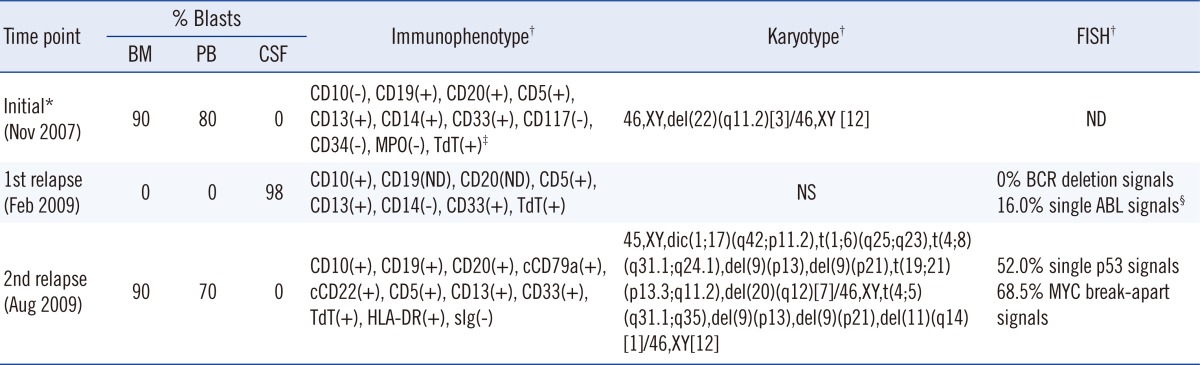
Table 1
Summary of the patient's hematologic, immunophenotypic, and cytogenetic data
*Data from another hospital; †Data from BM aspirate specimens at initial diagnosis and 2nd relapse and from CSF specimen at 1st relapse; ‡Data obtained by immunohistochemistry on a clot section; §The nature of the ABL deletion signal could not be determined because the cytogenetic analysis was not successful.
Abbreviations: BM, bone marrow; PB, peripheral blood; CSF, cerebrospinal fluid; MPO, myeloperoxidase; TdT, terminal deoxynucleotidyl transferase; ND, not done; NS, not successful.
![]()
Fifteen months after the initial diagnosis, the patient was transferred to our institution for evaluation of nausea and vomiting and was diagnosed with isolated central nervous system (CNS) relapse of the disease. In the CSF specimen, the white blood cell count was 1,450/µL with 98% leukemic blasts (Table 1 and Fig. 1A). The leukemic blasts were positive for CD10, CD5, CD13, CD33, and nuclear TdT and were negative for CD14 and CD34. Cytogenetic analyses of leukemic blasts in the CSF failed due to the poor quality of the specimen. FISH analyses for del(22) (q11.2) using the Vysis LSI BCR/ABL Dual Color, Dual Fusion Translocation Probe (Abbott Molecular Inc., Des Plaines, IL, USA) showed no interphase cells with BCR (22q11.2) signal deletion and 16.0% of cells with a single ABL (9q34) signal. There was no evidence of leukemic blasts in the peripheral blood or bone marrow. A cytogenetic study showed no abnormal clones in the bone marrow samples. The patient was treated with the CCG-1882 protocol, along with whole brain irradiation (24 Gy divided into 12 fractions) and whole spine irradiation (6 Gy divided into 3 fractions) during the consolidation period.
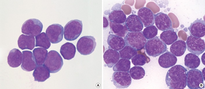
Four months from the initial relapse, the patient experienced a second relapse with ≥90% leukemic blasts in the bone marrow (Fig. 1B). Immunophenotypically, the blasts were positive for CD19, CD10, CD20, cytoplasmic CD79a, cytoplasmic CD22, HLA-DR, and TdT, with co-expression of CD5, CD13, and CD33 (Table 1). Cytogenetic analysis revealed complex structural abnormalities, including t(4;8)(q31.1;q24.1) (Fig. 2A). FISH analysis using the Vysis LSI MYC Dual Color, Break Apart Rearrangement Probe (Abbott Molecular Inc.) revealed MYC rearrangement in 68.5% of interphase cells (Fig. 2B). There was no IGH/MYC fusion signal in a FISH study using the Vysis LSI IGH/MYC/CEP8 Tri-Color Dual Fusion probe (Abbott Molecular Inc.). In addition, a FISH study using the Vysis LSI p53 probe (Abbott Molecular Inc.) showed deletion of the p53 (17p13.1) signal in 52.0% of interphase cells, which was compatible with the presence of dic(1;17)(q42;p11.2) observed in conventional cytogenetics (Table 1). Despite aggressive reinduction chemotherapy, the patient died 3 months after the second relapse of disease.
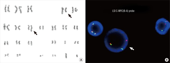 | Fig. 2Cytogenetic analysis showing complex chromosomal abnormalities including t(4;8)( q31.1;q24.1). Black arrows indicate the rearranged chromosomes 4 and 8 (A). FISH analysis using the Vysis LSI MYC Dual Color, Break Apart Rearrangement Probe (Abbott Molecular Inc.), which showed the MYC gene rearrangement. White arrow indicates a interphase cell with 1 fusion and 1 break apart signal (B).
|
Go to :
DISCUSSION
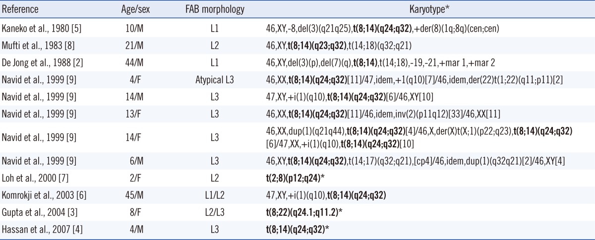
The MYC rearrangement is considered the hallmark of Burkitt lymphoma; however, it also arises in other subsets of mature B-cell neoplasms. In particular, the MYC rearrangement was reported to be a critical event in the progression of follicular lymphoma to higher-grade lymphoma or leukemia [10, 11]. Biologically, the c-MYC protein has a central role in the transcriptional regulation of various processes, including cell growth, cell cycle progression, and apoptosis [12]. Translocation of one MYC allele into the vicinity of an immunoglobulin heavy chain gene on chromosome 14q32, or less commonly, the kappa and lambda light chain genes on chromosomes 2p12 and 22q11, respectively, leads to deregulated expression of c-myc and cell proliferation. According to the experience of the Pediatric Oncology Group, the MYC rearrangement accounts for 0.1% (5/5,280) of cases of pediatric ALL with the precursor B-cell phenotype [9]. Our literature review of precursor B-cell ALL cases showed that all the partners involved in MYC rearrangements were immunoglobulin genes [2-9] (Table 2). The most common partner was t(8;14), followed by 2p12 and 22q11. In contrast, the partner chromosomal locus of the MYC rearrangement in our case was not a conventional immunoglobulin loci, but 4q31. Band 4q31 is a chromosomal locus that has been rarely involved in hematologic malignancies; t(4;5)(q31;q31) and t(4;21)(q31;q22) were reported in MDS/AML and T-cell ALL [13-16]. SH3D19 is the only gene identified as involved in t(4;21)(q31;q22) in AML [14]. Although no genes in 4q31 have been reported to be involved in precursor B-cell ALL, another candidate gene is MAML3, a coactivator of the notch signaling pathway [17].
The MYC rearrangement was detected in leukemic cells in the bone marrow during the second relapse. We speculated that the leukemic cells underwent serial genetic evolution from the first through the second relapse. Unfortunately, we could not ascertain whether the MYC rearrangement was absent at initial diagnosis, because the MYC FISH of the initial bone marrow sample was not successful. However, complex cytogenetic abnormalities, including the translocation involving the MYC locus, which was detected at the second relapse, provided evidence of genetic evolution.
The presence of the MYC rearrangement warrants intensive treatment due to the highly proliferative nature of the neoplastic cells. In the Pediatric Oncology Group experience, 5 patients with precursor B-cell ALL received intensive chemotherapy, being considered the presence of the MYC rearrangement, and 4 achieved long-term survival [9]. The patient in the present report attained a complete response (CR) for less than 6 months after the first relapse, and he never achieved a CR after the second relapse. He was treated based on the protocol for high-risk precursor B-cell ALL, since the MYC gene rearrangement was unexpectedly detected during the second relapse. This suggested that the protocol for high-risk precursor B-cell ALL (CCG-1882) might have been less effective in our patient. In addition to the MYC gene rearrangement, p53 deletion from dic(1;17)(q42;p11.2) was observed at the second relapse of the disease. P53 deletion is a recurrent genetic aberration in Burkitt lymphoma/leukemia [18], and p53 inactivation was reportedly associated with predisposition to oncogenic translocations in B lineage lymphomas and poor prognosis [19]. Immunophenotypically, the leukemic blasts in our patient expressed not only B-lymphoid antigens but also myeloid and T-lymphoid antigens, including CD5. CD5-positive precursor B-cell ALL is extremely rare, and only 4 such cases have been reported in the literature [20-22]. All 4 patients with CD5-positive precursor B-cell ALL were adolescents, and had aggressive disease courses and poor outcomes. From this perspective, the aberrant CD5 expression in our patient in combination with the cytogenetic abnormality might have been a poor prognostic factor.
In summary, we described the first case of precursor B-cell ALL with a MYC rearrangement involving a novel non-immunoglobulin partner locus. The outcome of this case suggested that the presence of the MYC rearrangement in ALL with the precursor B-cell phenotype warrants intensive treatment, regardless of the partner gene. Further identification of patients with this cytogenetic abnormality will allow us to expand our knowledge regarding its prognostic significance and the optimal treatment for this rare subgroup of patients.
Go to :
 XML Download