

 PDF
PDF ePub
ePub Citation
 Print
Print


INTRODUCTION
Malaria is a human infection that has been documented since ancient times. It is a vector-borne disease caused by Plasmodium species and transmitted by Anopheles mosquitoes [1]. Annually, more than 300-500 million cases of malaria are reported worldwide, and 1.5-2.7 million of these patients die from malaria infections every year [2]. In Korea, malaria cases are regularly reported. Since the re-emergence of P. vivax in the early 1990s, most malaria cases occurring in Korea have resulted from P. vivax infections. According to the Korea Centers for Disease Control and Prevention (KCDC), induced malaria comprises 1-4% of all malaria cases in Korea, in the following order of frequency: P. falciparum (41.5%), P. vivax (36.8%), and malaria caused by other species [3].
Many cases of P. malariae infections were diagnosed in Korea from the 1930s to the 1950s, and a case of co-infection with Babesia and P. malariae malaria was reported in 1998. However, these were diagnosed based only on morphologic features [4, 5]. Here, we report a case of imported malaria that was caused by P. malariae. In combination with peripheral blood smear (PBS), conventional and real-time PCR for 18S RNA were also conducted for a conclusive diagnosis.
CASE REPORT
1. Clinical history and laboratory findings
A previously healthy 23-yr-old woman visited the emergency room (ER) because she experienced chills for 3 days, which were exacerbated at night and were accompanied by headaches and rhinorrhea. Three weeks prior to her ER visit, she had travelled on business to the Republic of Ghana for 1 week. At the time of her visit to the ER, her body temperature was 36.9℃, and her other physical examination results were nonspecific. The laboratory tests results were as follows: hemoglobin level, 13.9 g/dL; white blood cell count, 4.4×10⁹/L (neutrophils, 72.0%; lymphocytes, 13.8%; monocytes, 9.6%; eosinophils, 4.4%; and basophils, 0.2%); platelet count, 135×109/L; and C-reactive protein (CRP) level, 1.20 mg/dL (slightly above normal). Wright-Giemsa staining of the PBS was performed to screen for malarial parasites. However, the smear did not show the presence of malarial parasites. Therefore, the patient was discharged, and upper respiratory tract infection was suspected.
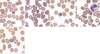
Six days later, she visited the outpatient clinic for follow-up and presented with diarrhea and abdominal discomfort in addition to chills. Her self-recorded body temperature was 40.3℃. Laboratory investigations performed at that time showed that her hemoglobin level was 12.6 g/dL and white blood cell count was 6.3×10⁹/L (metamyelocytes, 1%; segmented neutrophils, 40%; lymphocytes, 42%; monocytes, 9%; eosinophils, 5%; and basophils, 3%); furthermore, her platelet count had decreased to 95×109/L, and her CRP levels had increased to 5.45 mg/dL. The liver function test, which was normal at first visit to the ER, showed a slight increase in AST and ALT levels to 77 IU/L and 84 IU/L, respectively. She was admitted to our medical center for further evaluation, and 3 pairs of blood culture and malaria tests (PBS and 2 different molecular tests) were performed. The blood cultures yielded negative results. The results of Wright-Giemsa staining on the PBS and the molecular tests for malaria are shown in Fig. 1 A-C (at the time of presentation), Fig. 2, and Fig. 3. She was treated with oral hydroxychloroquine (Oxiklorin; immediate dose of 800 mg, followed by 400-mg doses at 12, 24, and 36 hr) and was later discharged. Although malarial parasites were not observed on the PBS 2 days later and the patient was symptom-free until the next follow-up, the PBS and real-time PCR results at the 1-month follow-up showed recurrent parasitemia (Fig. 1 D-E, and Fig. 3). She was successfully treated with oral mefloquine (Lariam; immediate dose of 750 mg, followed by 500 mg at 24 hr), and the results of the PBS and the molecular tests performed 7 days after completing this therapy were negative.
2. Molecular diagnosis
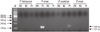
For the 18S rRNA, conventional PCR was performed. DNA was extracted from the blood sample of the patient using the QIAamp DNA Blood Mini Kit (Qiagen, Hilden, Germany). Four pairs of species-specific primers (rFAL1/rFAL3 for P. falciparum, rVIV1/rVIV2 for P. vivax, rMAL1/rMAL2 for P. malariae, and rOVA3/rOVA4 for P. ovale) that had been reported in a previous study were used [6, 7]. Amplification was performed in a 50-µL reaction mixture that contained 5 µL of extracted DNA, 2 µL of 10 pmol/µL primer (each), 4 µL of 2.5 mM dNTP, 0.25 µL of Taq polymerase (Takara, Shiga, Japan), and 5 µL of 10× buffer. All the PCR cycling protocols incorporated an initial denaturation step at 95℃ for 3 min; 35 amplification cycles of 95℃ for 40 sec, 53℃ for 1 min, and 72℃ for 1 min; and a final extension step at 72℃ for 5 min. PCR products were visualized using 2% agarose gel electrophoresis and subsequent ethidium bromide staining. Only P. malariae was detected in the patient's peripheral blood sample (Fig. 2).
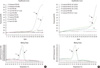
The 18S rRNA real-time PCR assays for the 4 Plasmodium species, which produce 205-bp, 117-bp, 144-bp, and 456-bp amplicons, respectively, were also performed using previously reported primers (the 4 forward species-specific primers were FWfal, FWviv, FWmal, and FWova, respectively) [8]. The components of the real-time PCR reaction mixture were as follows: 2.0 ng of DNA template with 2.0 µL of LC FastStart DNA Master SYBR Green I (Roche, Penzberg, Germany), 0.4 µL of each primer (10 pmol/µL), and 1.6 µL of 25 mM MgCl2 in a final volume of 20.0 µL. The PCR program used was as follows: 94℃ for 15 min and 40 cycles of 94℃ for 5 sec, 58℃ for 5 sec, and 72℃ for 10 sec. The program for analytic melting was as follows: 95℃ for 5 sec and 60℃ for 30 sec, followed by an increase in temperature to 99℃ with a 0.2℃/sec ramp rate. The real-time PCR assay correctly identified the presence of P. malariae (Fig. 3).
DISCUSSION
Malaria is the most common parasitic disease worldwide [1]. P. vivax, P. falciparum, P. ovale, P. malariae, and the recently emerging P. knowlesi are the Plasmodium species known to cause human infections [9]. Among these species, P. vivax is responsible for 80% of all human malaria cases because of its prevalence in a wide range of areas, from tropical to temperate zones. The second most common malaria-causing species is P. falciparum. P. falciparum is limited to only tropical areas; however, it causes severe symptoms and a high mortality rate. Therefore, many countries are involved in a malaria control project sponsored by the WHO to achieve advances in malaria control [10]. The 2 species mentioned above are responsible for more than 95% of human malaria cases, and the WHO eradication projects focus on these species. In contrast, the importance of P. malariae and P. ovale infections is usually overlooked.
The Republic of Ghana is located in West Africa, where the prevalence of malaria is higher than in East Africa [11]. Indeed, in Ghana, the entire nation is at the risk of malaria infection. A very large number of cases (3.7 million cases of suspected malaria and 1.9 million cases of probable or confirmed malaria) were reported in 2009 [12]. The country has been involved in the malaria control program conducted by the WHO since early 2000.
Malaria infection is suspected in patients experiencing the characteristic cyclic fever, having a history of habitation or visit to an endemic area, and presenting with other conditions, such as transfusion history. Details of the medical examination can help identify malaria to the species level. The average incubation time, for example, is influenced by the species of Plasmodium and may provide information about the source of infection. The incubation time for P. falciparum infections is 9-14 days, while P. vivax has an incubation time of 12-17 days and P. malariae has an incubation time of 18-40 days, which is considered relatively long [13]. In this case, although the exact time of exposure was unknown, the supposed incubation time based on travel history was 14-21 days. This information assisted partially in excluding P. falciparum infection.
Among the various diagnostic approaches, the common choice of method for diagnosis is microscopic examination of blood films and confirmation of parasites. Although a physician can check for the presence of parasites and other parameters through a PBS, low parasite burdens are difficult to observe on a PBS [14]. In particular, P. malariae malaria is usually chronic and asymptomatic and exhibits a relatively low parasitic load, usually less than 5,000 parasites/µL of blood [15]. Furthermore, in P. ovale and P. malariae infections, if the infected red blood cells are damaged and their distinguishing morphologic features, such as fimbriated margins or contracted cells, are distorted, the diagnosis is more difficult [16]. Moreover, P. knowlesi, a newly discovered human plasmodia species, is similar in morphology to P. falciparum and P. malariae, according to its blood stage [17]. Therefore, in the diagnosis of P. malariae infections, which are rare in Korea, a molecular test would be helpful for detecting and identifying the specific species involved. In this case, the patient had a low parasite burden, and parasites were not observed in her blood films, although she was symptomatic and had not undergone prophylactic therapy with antimalarial drugs. At the time of emergence of parasites in the peripheral blood, from the low parasite burden, travel history, and incubation time, we could expect the presence of a species uncommon to our domestic area. However, the precise diagnosis at the species level was made with the aid of molecular tests.
Relatively few studies have investigated the treatment of P. malariae malaria compared with the treatment of P. falciparum malaria. Chloroquine is the first-line drug for P. malariae infections as well as for P. vivax and P. ovale. In general, P. malariae is known to be susceptible to chloroquine and to other treatments, including amodiaquine, mefloquine, and artemisinin [14]. It is very unusual for P. malariae to be resistant to chloroquine; however, a study has reported cases of persistent parasitemia 28 days after the initial disease management, suggesting either new infection or late recrudescence of a chloroquine-resistant strain [18]. The parasites in this case, although showing negative conversion in PBS immediately after the initiation of therapy, did not respond to chloroquine. Instead, they responded to mefloquine after a positive PCR result at the 1-month follow-up. Considering that P. malariae malaria is very rare in Korea, we could exclude the possibility of a new infection occurring in our patient, and late recrudescence of a chloroquine-resistant P. malariae strain was strongly suggested.
In summary, an imported P. malariae infection showing late recrudescence of a chloroquine-resistant strain was confirmed by species-specific conventional and real-time PCR assays for 18S rRNA. Due to difficulties in the diagnosis of the rare Plasmodium species in Korea, molecular tests combined with morphologic examination of the PBS would help to correctly diagnose and identify the parasites and would result in better clinical management of P. malariae infections.
 XML Download