This article has been
cited by other articles in ScienceCentral.
Abstract
An indirect inguinal hernia containing the fallopian tube alone is extremely rare in reproductive-aged women without any genital tract anomalies. Despite this rarity, early diagnosis and adequate management is important to prevent strangulation and recurrence. We present a case of an indirect inguinal hernia containing only the fallopian tube in the hernia sac, which was successfully reduced by using a laparoscopic total extraperitoneal approach and repaired with a polypropylene mesh.
Keywords: Hernia, inguinal, Fallopian tubes, Laparoscopy
Introduction
An indirect inguinal hernia is a protrusion of abdominal cavity components through the inguinal ring, particularly lateral to the inferior epigastric vessels. Indirect inguinal hernias occur in ≤5% of women [
1]. Hernias containing genital organs (e.g., the fallopian tube, ovary or uterus) in the hernia sac account for approximately 2.9% of all hernia cases [
2]. Moreover, an indirect inguinal hernia containing only the fallopian tube is extremely rare. Most reported cases of indirect inguinal hernias involving genital organs in infants are accompanied by other genital tract anomalies.
We report an uncommon case of a 22-year-old woman who had an indirect inguinal hernia containing only the fallopian tube without any genital anomalies, in which the hernia was successfully reduced using a laparoscopic total extraperitoneal (TEP) method and repaired with a polypropylene mesh.
Case report
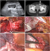
A 22-year-old nulliparous patient with a left groin lump and left lower quadrant pain visited our emergency room. She reported a recurrent palpable mass in the left groin that was present since several months. A lump had appeared 10 days previously and had not disappeared. Two days before her visit, left lower quadrant pain occurred and became aggravated. The patient had no surgical or medical history but was diagnosed with polycystic ovaries 1 year ago. Physical examination revealed a 6×5 cm soft mass in the left groin, but there was no genital anomaly. Ultrasonography showed a normal uterus and both polycystic ovaries. The left fallopian tube was found in the hernia sac as hydrosalpinx (
Fig. 1A). It demonstrated a 6×4 cm cystic structure was observed by computed tomography in the left inguinal area (
Fig. 1B), which was a hernia sac containing the left fallopian tube.
Fig. 1
(A) Ultrasonographic image showing the left hydrosalpinx in the hernia sac. (B) Computed tomography image of the pelvis showing a 6×4 cm cystic structure in the inguinal area (white arrow). (C) Laparoscopic view of herniation of the left fallopian tube in the inguinal canal. (D) Laparoscopic view of the polypropylene mesh used to repair the hernia using the total extraperitoneal hernia repair method. (E) Laparoscopic view of the repaired partial defect of peritoneum in the inguinal internal ring area. (F) Laparoscopic view of the left fallopian tube after reduction. Operative findings showed no ischemic changes and no anatomical abnormalities.

Laparoscopic hernia repair was performed using the TEP method. Edematous left fallopian tube and part of the round ligament were found in hernia sac (
Fig. 1C). The left fallopian tube was incarcerated with the round ligament. Part of the left round ligament was cut, and reduction of the left fallopian tube into the peritoneal cavity was performed. A polypropylene mesh was used to repair the hernia (
Fig. 1D). After treatment of the hernia using the TEP method, we performed laparoscopic intra-abdominal exploration. Both ovaries showed polycystic feature but did not have other abnormal finding. The uterus, right fallopian tube, and right round ligament appeared normal. Laparoscopic suture was performed to repair the peritoneum and anchor the round ligament (
Fig. 1E). Tubal patency test results were normal (
Fig. 1F).
The patient had an uneventful post-operative course and was discharged on postoperative day 2. Follow-up ultrasonography was performed 1 week after the operation. The diameter of left salpinx had decreased and edema was improved.
Discussion
The incidence of inguinal hernia containing genital organs (e.g., the fallopian tube, ovary or uterus) is infrequent. In a retrospective review article of inguinal hernias in 2006, Gurer et al. [
2] reported that among 1,950 cases, ovaries and fallopian tubes accounted for 2.9% of the unusual contents of hernia sacs and only 0.41% of patients showed a hernia sac containing only the fallopian tube [
3]. In adult women, inguinal hernia containing genital organs is generally accompanied by other genital abnormalities [
4]. Only 2 cases of fallopian tube and ovary indirect inguinal hernia in reproductive-aged women without genial anomalies have been reported to the
Obstetrics and Gynecology Science [
56]. This is the first case of indirect inguinal hernia containing only the fallopian tube, without the ovary, in a woman of reproductive age without any genital organ anomaly. Kim et al. [
5] reported a case of indirect inguinal hernia containing left ovary, left fallopian tube and endometriosis in Korea. In that case report, hernia sac size was 4×5 cm. Compare to Kim et al.'s case report [
5], the patient of our case showed larger hernia sac but contained only the left fallopian tube without ovary or any other organ. In addition, the patient was younger and had no history of pregnancy which means the patient had lower risk of abdominal wall or ligamental weakness.
The mechanism of inguinal hernias of genital organs in reproductive-aged women without any genital organ anomalies is unclear. Ozkan et al. [
3] suggested that weakness of the broad ligament and ovarian suspensory ligament may cause this kind of herniation. This can also be aggravated when abdominal pressure is increased. But Ozkan et al.'s hypothesis [
3] cannot explain why recurrent indirect hernias containing the fallopian tube, ovary or uterus occur in reproductive aged woman because weakness of the ligaments increases with age.
Because of the rarity and unclear pathogenesis, it is difficult to predict or diagnose genital organ herniation. However, early diagnosis and timely management is essential to reduce the risk of adnexal damage and preserve fertility.
Most cases of inguinal hernia of the fallopian tube and ovary or hernia of the fallopian tube alone are treated by excision of the hernia sac and its components [
357]. Kim et al. [
5] used a laparoscopic approach to perform left salpingo-oophorectomy because the patient of that case has been had plan of hysterectomy because the patient already had carcinoma
in situ of cervix as underlying disease. Song et al. [
6] reported a high ligation method for a left fallopian tube hernia that saved the left salpinx, but this was an open method. Our case, the patient was young and had no history of delivery, so we performed laparoscopic reduction of the fallopian tube into the peritoneal cavity with TEP hernia repair using polypropylene mesh to preserve future fertility, prevent recurrence and achieve better cosmetic outcomes. Using a laparoscopic approach, patients recover faster, experience less pain during the first day, face fewer complications (e.g., infection or bleeding), have a lower risk of chronic pain and have better cosmetic outcomes than open hernia repair [
8].
In conclusion, we reported a case of inguinal hernia of only the fallopian tube in a woman of reproductive age without any genital anomalies. A laparoscopic approach with using the TEP method was successful, with good cosmetic outcomes and less pain after surgery, and the fallopian tube was preserved. Therefore, before determination of resection of a hernia sac or herniated fallopian tube, we suggest considering a laparoscopic approach to reduce herniated adnexae and prevent recurrence.


