

 PDF
PDF ePub
ePub Citation
 Print
Print


Introduction
A retroperitoneal mass indicates an origin in the retroperitoneum (RP), not from pelvic organs. Masses originating in the RP are either benign, malignant, or metastatic [1]. Approximately 10%–15% of soft tissue sarcomas (STSs) develop in the RP [2345], of which approximately 30%–60% are liposarcomas [56] that arise from adipose cells in deep soft tissue. The prognosis of liposarcomas depends on patient age, location and depth of invasion, size, resectability, histologic grade, and the presence of metastases [3].
Histological liposarcomas are classified into 5 subtypes: well-differentiated, myxoid, round cell, pleomorphic, and de-differentiated (i.e., poorly differentiated) [78910].
Retroperitoneal liposarcoma is rarely metastatic [11]. Therefore, complete en bloc resection with a margin of uninvolved tissue is the mainstay of treatment. The main adjuvant therapy is radiation therapy (RT), but the benefits remain controversial [5]. We present a case of RP liposarcoma that mimicked an ovarian tumor with no other evidence of retroperitoneal origin in the intra-operative field.
Case report
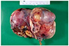
A 74-year-old postmenopausal woman was referred to the gynecology clinic of the Jeonju Presbyterian Medical Center with complaints of a palpable abdominal mass that she first recognized approximately 2 months prior. The patient had a history of gravida 2 and para 2. She had no other medical history but hypertension. First, during routine physical examination, an approximately 16–18 gestational week-sized mass was palpated. It was movable, painless, and was suspected to be a large ovarian mass based upon transvaginal ultrasonography. The echogenicity of the mass was heterogeneous with irregular contours. Pelvic computed tomography (CT) indicated that the mass was 10×8.5×13 cm in size with cystic and solid-enhancing features. Enhancement of the internal component of the mass was complex, but there was no definite pathologic enlargement of lymph nodes and ascites (Fig. 1). Additionally, there was a unilocular right ovarian simple cyst and a uterine myoma. Two suspicious retroperitoneal lipomas were observed in the para-aortic region and infrarenal portion. According to CT findings, the sigmoid colon bordered the huge left ovarian tumor. A colonoscopy was performed and confirmed the absence of tumor invasion into the bowel mucosa. Laboratory findings showed only mild anemia and other laboratory analyses revealed normal values. Tumor markers, including cancer antigen (CA) 125, CA19-9, carcinoembryonic antigen, α-fetoprotein, and β-human chorionic gonadotrophin were also in normal ranges. A laparotomy was performed to assess the pathological findings of the left ovarian mass. During surgery, the large ovarian mass seemed to adhere tightly to the ovarian wall, and its smooth capsule seemed to be connected to the ovarian germinal epithelium. There was no sign of tumor capsule rupture. The tumor looked benign in nature without any ascites or peritoneal carcinomatosis. There was slight adhesion between the tumor base and the peritoneal surface (Fig. 2). Since the patient was elderly with a uterine myoma and a right ovarian cyst, total hysterectomy, and bilateral salpingo-oophorectomy were performed. The 2 suspicious lipomas in the para-aortic and infrarenal area were not dissected because of their benign features in the CT. Final pathology of the left ovary indicated dedifferentiated liposarcoma mixed with well-differentiated, myxoid, spindle cells in the liposarcomatous area. The mitotic rate was 20/10 HPF (French Federation Nationale des Centres, cancer grade 3). No progesterone and estrogen receptors were located in the liposarcoma tissue, as confirmed by immunohistochemical testing. There were also no adipose cells in the ovarian stroma. The liposarcoma was considered to have originated not from ovarian stroma but from fat tissues surrounding the left adnexa. Adjacent retroperitoneal fat tissue was especially strongly suspected. After surgery, positron emission tomography (PET)-CT was performed to assess distant organ metastasis and to determine if remnant tissues were present. The fat-containing masses in the left para-aortic and left infrarenal space did not take up fluorodeoxyglucose (FDG) abnormally. There were also no metastatic lesions on other organs. Furthermore, there was no abnormal FDG uptake in the pelvic cavity. Before deciding to start RT, pathologic findings and radiologic findings were further reviewed. Furthermore, pelvic magnetic resonance imaging (MRI) was performed one month after surgery to assess the patient's status. In doing so, a pelvic fatty mass approximately 8 mm in size in the lower pelvic wall was found. At this point, there were three options for planning the patient's further treatment. First, after histologic differential diagnosis via surgical biopsy of the small mass found on MRI, further treatment could be determined. Second, prophylactic radiotherapy might be recommended for local control if surgery was difficult. Third, the fatty mass could just be monitored. Although the pathologic grade indicated a high risk, if the pathologic resection margin status was negative, postoperative RT would confer no survival benefit. Therefore CT scan monitoring could be another choice. Finally, the patient chose the third option because of her age and the economic burden.
Fig. 1
Intra-pelvic mass mimicking an ovarian tumor with cystic and solid-enhancing internal features.
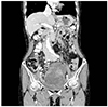
Fig. 2
The liposarcoma was grossly encapsulated without the ruptured sign and tightly adjacent to ovarian tissue.
The radiation oncology department chose to perform a follow-up MRI after 6 months to assess changes in the fatty lesion. If the gross lesion showed growth, RT or secondary en bloc resection would be planned. Until now, the patient had no other complications after surgery and no signs of aggravation of the disease. It was decided to follow up on the progress of the patient's disease every 3 to 6 months for 5 years.
Discussion
Liposarcoma is one of the most common sarcomas arising from soft-tissue [7], with approximately 42% occurring in the RP, 41% occurring in the lower extremities (particularly the thigh), 11% occurring in the upper extremities, and 6% occurring in the head and neck [8].
Liposarcomas develop in a deep, expandable space without any bony boundaries as barriers. Therefore, when they are detected, they are generally larger than 5 cm [1], with approximately 20% of cases measuring larger than 10cm at presentation [131415]. Most patients with RP liposarcomas present with a painless, palpable mass and diverse symptoms due to compression or displacement of adjacent organs [18].
Abdominal and pelvic ultrasonography is the quickest and most useful technique for locating and measuring the size of the RP mass. Contrast-enhanced CT-scanning or MRI of the abdomen and pelvis are useful for evaluating the tumor location, size, origin, and relationship to adjacent tissues and organs [4]. PET-CT, if possible, can be performed to obtain information on metastases and to estimate tumor grade [1].
Prognostic factors include tumor size, anatomical location, surgical resection, and histological subtype, with histological subtype being the most important determining factor [17]. Liposarcoma is categorized into five subtypes according to the WHO classification: well-differentiated, which is non-aggressive; myxoid type, which is also a low-grade tumor; round-cell type with intermediate behavior; pleomorphic; and de-differentiated types with aggressive behavior and a poor prognosis [1].
Surgery is the standard treatment for RP liposarcomas with no metastases [1]; however, because liposarcomas are usually large, located deep in the RP, and cover an anatomically complex area that includes several vital structures, they are sometimes difficult to remove completely [7]. Patients who undergo macroscopic complete resection of the tumor have an improvement in prognosis of up to 54%–70% [2]. Therefore, if it is possible, en bloc removal of adjacent structures, that is excision of the tumor with adjacent involved viscera such as the abdominal wall and psoas, or paravertebral muscles, is required in a margin of uninvolved tissue [1]. Achieving complete resection requires multi-organ resection in approximately 50% of cases [2]. For example, the kidney is removed in approximately 30% of cases [2]. If it is impossible to remove the entire tumor with an abnormal resection margin, a patient may develop locally recurrent disease, which can result in death [12].
Local control is one of the mainstay requirements of treatment to inhibit the recurrence of a tumor. The main adjuvant approach is RT, but the benefits are uncertain. Additionally, RT is an option if there is a high risk of local failure after surgery, but that role here remains controversial [159].
In our case, liposarcoma was mistaken for an ovarian tumor. The tumor and left ovary were not well-differentiated macroscopically. Even during the surgery, the liposarcoma was well demarcated by capsulation, so it was thought that the resection margin was macroscopically clear. The exact origin of the fat tissue was ambiguous, but the RP, a more affected area, was highly suspected. With reference to treatment and diagnostic algorithms for suspected retroperitoneal STS [5], when surgery is completed and if the resection margin is macroscopically or microscopically clear, additional options do exist. This includes following up with the patient, because there is no definite survival advantage to using postoperative RT [5].
In this case, because of variables such as old age, tumor size and its solid character, malignancy should always be excluded although the tumor looked benign. We reconsidered the need to determine the extent of surgery using histologic findings during surgery. This might be provided necessary information when the tumor was not typical, or when the results for the diagnosis were not consistent. The radiologic, clinical, and pathologic findings had important roles and bridged gaps between each other to define the tumor's character and origin.
 XML Download