Journal List > Obstet Gynecol Sci > v.60(6) > 1090862

Jeon and Na: Vacuum extraction vaginal delivery: current trend and safety

Abstract

Operative vaginal birth retains an important role in current obstetric practice. However, there is an increasing trend in the rate of cesarean section in Korea. Surgical delivery is more advantageous than cesarean section, but the rate of operative vaginal delivery is decreasing for various reasons. Furthermore, there is no unified technique for vacuum extraction delivery. In this context, this review was performed to provide details of the necessary conditions, techniques, benefits, and risks of operative vaginal delivery. Future research should focus on overcoming the limitations of operative vaginal delivery.

Introduction

Operative delivery is divided into vaginal and abdominal delivery. Vaginal delivery is further divided into forceps delivery, breech extraction, and vacuum extraction (VE), while abdominal delivery can be divided into cesarean section and postpartum hysterectomy.
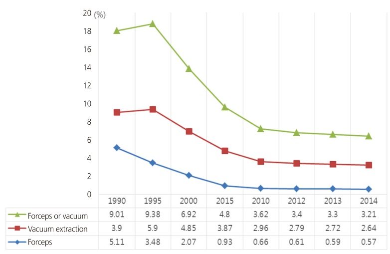
The frequency of cesarean section in Korea increased from 4.9% in 1970 to 39.1% in 2015, but has remained relatively unchanged from the rate of 37.1% in 2005 [1]. This trend is considered to be related to maternal aging, increases in the rate of induced labor, and increases in the incidence of obesity among women. In addition, most breech deliveries are delivered by cesarean section, and the rates of forceps and vacuum deliveries have decreased in the United States (US) (Fig. 1) [2]. About 1 of 4 low-risk women was delivered by cesarean section in US. In response to this growing number of cesarean deliveries and the morbidities associated with cesarean deliveries, the Society for Maternal-Fetal Medicine and the American College of Obstetricians and Gynecologists recently convened a workshop to address preventing the primary cesarean delivery [3].
Fig. 1
Statistics of delivery mode in the United States. Operative vaginal delivery in the United States tends to decrease gradually. Also, vacuum extraction is more dominant than forceps.
ogs-60-499-g001
With the increasing adoption of fetal monitoring systems in most hospitals, fetal distress can be diagnosed earlier than in previous decades, and medical staff makes a decision to perform cesarean section quickly due to concern regarding lawsuits.
Surgical vaginal delivery helps safe and successful vaginal deliveries in women with fetal indications. Although the use of cesarean delivery has grown considerably and is considered safe by many, surgical vaginal delivery has the great advantage of reducing the complications associated with cesarean section, such as death, postpartum hemorrhage (PPH), wound disruption and injury, venous thromboembolism, hemorrhage, infection, recovery time, rising costs, and subsequent repeat cesarean section [45678].
The decrease in surgical vaginal delivery is due to the risk of adverse court judgments against the doctor in the case of a problem [9]. This has led to a lack of experience with these techniques among medical residents, as hospitals and medical schools tend not to focus on training in surgical vaginal delivery. Although there are no statistical data regarding this issue, the rates of surgical vaginal deliveries are decreasing in private hospitals in particular in Korea.
The most important factors in determining surgical vaginal delivery are consideration of which interventions are needed for delivery, the degree of risk associated with a particular technique, and the practitioner's skill level. This review will mainly discuss surgical vaginal delivery [10]. As the frequency of forceps delivery has been decreasing in Korea, we will mostly discuss VE. We review the published findings on maternal and neonatal complications and provide the clinical benefits of VE.

Indications

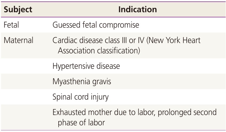
Surgical vaginal delivery can be performed to shorten the second phase of labor during delivery if the fetal heart rate is guessed fetal compromise or if the mother has medical problems, especially heart disease or hypertension. In addition, surgical vaginal delivery can be applied even if the mother is exhausted due to the pain associated with labor (Table 1). In recent years, painless delivery has become more common, and the delayed standard time of the second phase of labor is different between primiparous and multiparous women, depending on whether there is painless delivery. If the delivery has only normal pain, the time is 3 to 4 hours for primiparous women and 2 to 3 hours for multiparous women. Without painless delivery, 2 hours for primiparous women and 1 hour for multiparous women are regarded as delays in the second phase of labor [11].
Table 1

Indications of surgical vaginal delivery

ogs-60-499-i001
Subject Indication
Fetal Guessed fetal compromise
Maternal Cardiac disease class III or IV (New York Heart Association classification)
Hypertensive disease
Myasthenia gravis
Spinal cord injury
Exhausted mother due to labor, prolonged second phase of labor
The use of a second-stage upright or lateral position relative to the supine or lithotomy position was associated with a reduction in operative delivery [12]. Epidural analgesia reduces pain compared to non-epidural analgesia. However, epidural analgesia increases the incidence of operative vaginal delivery [13]. According to a meta-analysis, primiparous women with epidural analgesia are less likely to undergo rotation or surgical intervention when pushing 1 to 2 hours later or until they had a strong impulse to push [14].
Although overweight and obese nulliparous women are more likely to show signs of labor dystocia in the first stage of labor, the possibility of an operative vaginal delivery should be considered in the obese women who has arrested or protracted descent in the second stage. From a maternal standpoint, avoiding abdominal surgery will decrease the risk of complications such as wound infection, venous thromboembolism, and PPH, all complications that are increased in the obese patient. Many studies have shown that obese patients may benefit from operative vaginal deliveries when the second stage of labor is obstructed by soft tissue dystocia, with obese patients having higher rates of operative deliveries than their overweight or normal weight counterparts [15].

Contraindications

Contraindications in the fetus include fetal bone demineralization such as osteogenesis imperfecta, blood clotting disorder, abnormal presentation such as brow presentation or face presentation, not in the engaged state, difficulty to grasp of fetal part and recent fetal scalp blood collection, and suspicion of fetal-pelvic disproportion [11].
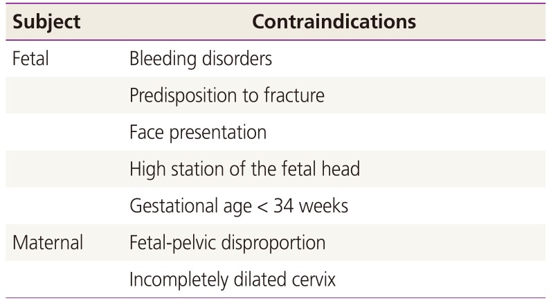
Viral infections caused by maternal blood are not contraindications for surgical vaginal delivery. However, it is best to avoid tough operative delivery where there is a potential for increased fetal skin abrasion or trauma and to avoid fetal scalp clipping or blood sampling during labor [16]. In addition, all obstetrics and gynecology guidelines are limited to 34+0 gestational weeks, so if there is a possibility of intraventricular hemorrhage, cephalohematoma, subgaleal hematoma or neonatal jaundice (Table 2). The use of VE is contraindicated in fetuses <34+0 gestational weeks, and most guidelines state that safety between 34 and 36 gestational weeks is still insufficient [1718].
Table 2

Contraindications of surgical vaginal delivery

ogs-60-499-i002
Subject Contraindications
Fetal Bleeding disorders
Predisposition to fracture
Face presentation
High station of the fetal head
Gestational age < 34 weeks
Maternal Fetal-pelvic disproportion
Incompletely dilated cervix
Obstetrical indications such as placenta previa and primary cesarean delivery, labor arrest in the first stage and nonreassuring fetal tracing before complete dilation are also contraindications of surgical vaginal delivery [6].
Medical staff should be aware of these points to avoid unnecessary criticism.

Necessary combination

A knowledgeable and experienced obstetrician is the most important factor in successful operative vaginal delivery [19]. For VE, the uterine cervix must be fully dilated, the amniotic membrane ruptured, the fetal head engaged with the maternal pelvis, the doctor must know the weight and position of the fetus, there must be no fetal-pelvic disproportion, and the maternal bladder should be empty. In addition, the medical staff should be able to stop and consider cesarean section at any time after informing the mother regarding the risks or benefits and seeking her consent [11].

Technique

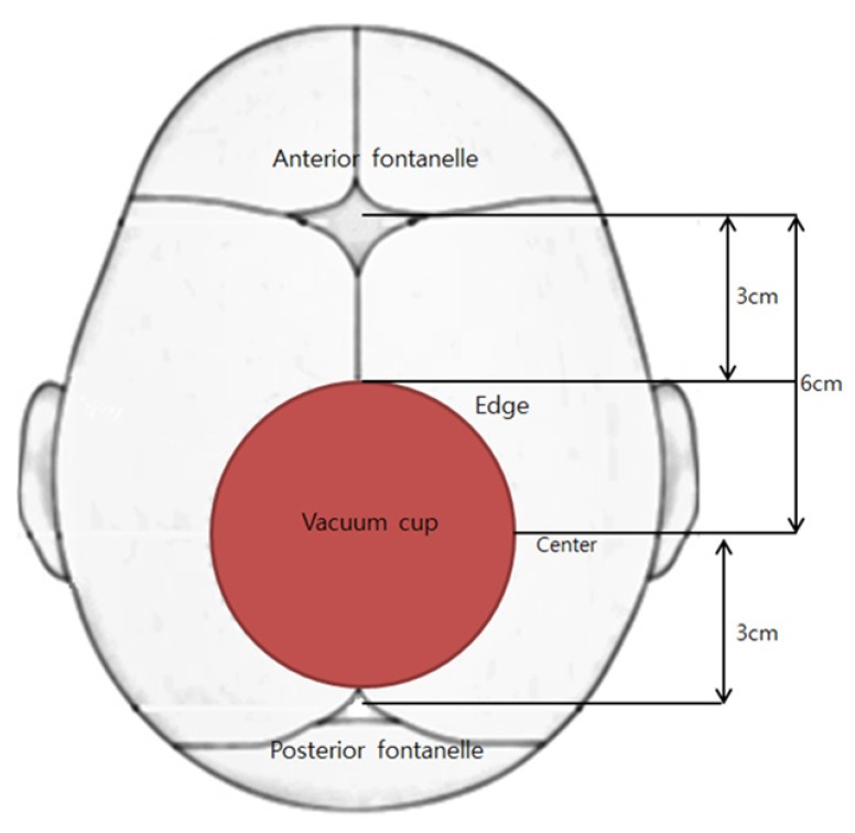
First, the vacuum cup and generator should be confirmed to be working well before the procedure. The pressure scale must not exceed 500 to 600 mmHg [20], and the cup should be placed on the head of the fetus. When using a standard 6-cm cup, the vacuum cup should be centered with the sagittal suture line (Fig. 2), with the vacuum generator inoperative, and the cup edge should be positioned at least 3 cm from the posterior surface, at the flexion point.
Fig. 2
Diagram of appropriate of vacuum cup placement. The vacuum cup must be placed at the flexion point to maximize adhesion. At this time, the center of the vacuum cup should be 6 cm from the anterior fontanelle and 3 cm from the posterior fontanelle on the same line of the sagittal suture line.
ogs-60-499-g002
The cup must be attached flexion point to maximize the traction force, which makes it difficult to remove the adsorbent, and it will deliver to the shortest diameter at the correct fetal head position. In addition, a 360° check should be performed to ensure that the pelvic tissue of the mother is not caught in the vacuum cup. Traction pulls along the curvilinear axis of the pelvis when there is contraction of the uterus.
The maximum number and times of pulling that can be safely performed have not yet been established. The Royal Australian and New Zealand College of Obstetricians and Gynaecologists recommends stopping the attempt if the vacuum cup disengages more than 3 times [11]. The descent should be evaluated after each contraction of the uterus, and the absence of descent in the appropriate technique is evidence of fetal-pelvic disproportion.
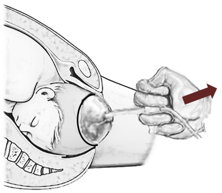
Successful vacuum delivery is dependent on the doctor's skill and appropriate site selection. At the start of traction, the cup should be pulled downward, and when the fetal head begins to appear, the direction of upward traction should be gradually changed (Fig. 3).
Fig. 3
Diagram of vacuum cup traction technique. Vacuum cup applied when the uterine cervix is fully dilated, the amniotic membrane ruptured and the fetal head engaged with the maternal pelvis when the fetal head begins to appear, the direction of traction should be gradually changed to upward.
ogs-60-499-g003
In addition, applying rotational force to rotate the head of the fetus is contraindicated because it can lead to detachment of the cup, cephalohematoma of the fetus, and scalp laceration [2122].

Episiotomy

Most guidelines in other many countries state that episiotomy should not be performed routinely during surgical vaginal delivery. Randomized controlled trials were conducted to reduce perineal injury, but there were no significant differences in the rates of maternal anal sphincter tears or primary PPH between groups with and without episiotomy [2324]. The rates of trauma in the newborn infant were similar between the 2 groups, as were those of maternal fecal or urinary incontinence, perineal infection, and prolonged hospital admission [25]. Routine perineal incision is not recommended, and if necessary, a mediolateral perineal incision can be made to reduce damage to the anal sphincter [2627]. In Korea, the decision of whether to perform episiotomy is left to the discretion of each obstetrician.

Complications

Complications are known to occur more frequently when a metallic cup rather than a soft cup is used [2829]. Fetal and neonatal complications include shoulder dystocia, subdural hemorrhage, facial nerve palsy, subconjunctival hemorrhage, retinal hemorrhage, cranial fracture, intracranial hemorrhage, scalp laceration, and cervical injury [30]. Intracranial hemorrhage was reported to occur in one of 860 newborns delivered by VE delivery. Infants delivered by VE had significantly higher rates of subdural or cerebral hemorrhage than those delivered spontaneously. Operative vaginal delivery was associated with a neonatal encephalopathy rate of 4.2 per 1,000 term neonates, and a rate of neonatal death from intracranial hemorrhage of 3 to 4 per 10,000 operative vaginal deliveries. One study looking at a large California population demonstrated that infants who were delivered by surgical vaginal delivery had a significantly higher rate of subdural or cerebral hemorrhage than those who delivered spontaneously. However, in order for operative deliveries to be a safe alternative to cesarean deliveries, the appropriate comparison is those who underwent assisted vaginal deliveries to those who had cesarean sections during labor. This same study found that those infants who were delivered by cesarean delivery during labor also had an increased odds ratio, whereas those who were delivered by cesarean section before labor did not. In conclusion, that the common risk factor for intracranial hemorrhage is not the operative delivery but rather abnormal labor, is important to acknowledge to promote the increased use of operative vaginal delivery as a safe alternative to cesarean delivery. In addition, compared with VE, cesarean delivery during labor was associated with significantly higher rates of neonatal convulsions, feeding difficulty, and mechanical ventilation [31].
In addition, maternal complications include cervical and vaginal laceration, PPH, urinary tract infections, pelvic floor injuries, and third- to fourth-degree lacerations [3132333435]. The long second stage may also be associated with adverse maternal outcomes. Prolonged second stage, generally considered greater than 3 hours, has demonstrated an increased risk of infection, third and fourth-degree perineal lacerations, and PPH [36].

Conclusion

In Korea, many women have traditionally preferred natural labor or naturalistic labor. However, for a variety of reasons, many women receive cesarean delivery. Because the best decision is made by the obstetrician at the time of delivery, it is important to screen for mothers who are more likely to undergo surgical vaginal delivery.
Operative vaginal delivery can be safely chosen when the second stage of delivery labor is prolonged, when prompt delivery is necessary due to fetal distress, and when the mother has to avoid or minimize labor due to medical illness.
The main points for operative vaginal delivery can be summarized as follows.
  1. Vacuum delivery is defined as the delivery of the fetal head into the pelvis of the mother, and is safe as long as the amniotic membrane has ruptured, the precise location of the fetus is known, the fetus is at least 34+0 gestational weeks, and the bladder of the mother is empty.

  2. Care should be taken as vacuum delivery can cause damage to the fetal head, as it can only pull the head of the fetus, unlike forceps delivery.

  3. If there is difficulty to position the vacuum cup, descent of the head is unsatisfactory or if traction does not progress even after 15 to 20 minutes or 3 attempts, the vacuum delivery attempt should be stopped and cesarean section should be considered.

  4. There are a wide range of possible complications of the mother and the newborn, most of which can also occur during the natural labor process.

  5. Cephalohematoma or retinal hemorrhage of the fetus is more common in vacuum delivery than in forceps delivery or spontaneous delivery, but most cases recover naturally without complications.

  6. The most appropriate choice at the time of delivery should be decided by the obstetrician.

  7. It is important to screen mothers who are more likely to require vacuum delivery.

  8. It is recommended to record what is described in the chart on an outpatient basis, explain at the beginning of the delivery, and consider receiving consent, if possible.

Notes

Conflict of interest: No potential conflict of interest relevant to this article was reported.

References

1. Statistics Korea. Data of statistics Korea [Internet]. Daejeon: Statistics Korea;2017. cited 2017 May 30. Available from: http://kostat.go.kr.
2. Korean Society of Obstetrics and Gynecology. Obstetrics. 4th ed. Seoul: Koonja;2007.
3. Spong CY, Berghella V, Wenstrom KD, Mercer BM, Saade GR. Preventing the first cesarean delivery: summary of a joint Eunice Kennedy Shriver National Institute of Child Health and Human Development, Society for Maternal-Fetal Medicine, and American College of Obstetricians and Gynecologists Workshop. Obstet Gynecol. 2012; 120:1181–1193. PMID: 23090537.
4. Murphy DJ, Liebling RE, Verity L, Swingler R, Patel R. Early maternal and neonatal morbidity associated with operative delivery in second stage of labour: a cohort study. Lancet. 2001; 358:1203–1207. PMID: 11675055.
crossref
5. Contag SA, Clifton RG, Bloom SL, Spong CY, Varner MW, Rouse DJ, et al. Neonatal outcomes and operative vaginal delivery versus cesarean delivery. Am J Perinatol. 2010; 27:493–499. PMID: 20099218.
crossref
6. American College of Obstetricians and Gynecologists. Society for Maternal-Fetal Medicine. Obstetric care consensus no. 1: safe prevention of the primary cesarean delivery. Obstet Gynecol. 2014; 123:693–711. PMID: 24553167.
7. Deneux-Tharaux C, Carmona E, Bouvier-Colle MH, Bréart G. Postpartum maternal mortality and cesarean delivery. Obstet Gynecol. 2006; 108:541–548. PMID: 16946213.
crossref
8. Silver RM, Landon MB, Rouse DJ, Leveno KJ, Spong CY, Thom EA, et al. Maternal morbidity associated with multiple repeat cesarean deliveries. Obstet Gynecol. 2006; 107:1226–1232. PMID: 16738145.
crossref
9. Cheong YC, Abdullahi H, Lashen H, Fairlie FM. Can formal education and training improve the outcome of instrumental delivery? Eur J Obstet Gynecol Reprod Biol. 2004; 113:139–144. PMID: 15063949.
crossref
10. Wen SW, Liu S, Kramer MS, Marcoux S, Ohlsson A, Sauvé R, et al. Comparison of maternal and infant outcomes between VE and forceps deliveries. Am J Epidemiol. 2001; 153:103–107. PMID: 11159152.
11. The Royal Australian and New Zealand College of Obstetricians and Gynecologists. Instrumental vaginal birth. East Melbourne: The Royal Australian and New Zealand College of Obstetricians and Gynecologists;2016.
12. Gupta JK, Hofmeyr GJ, Shehmar M. Position in the second stage of labour for women without epidural anaesthesia. Cochrane Database Syst Rev. 2012; CD002006. PMID: 22592681.
crossref
13. Anim-Somuah M, Smyth R, Howell C. Epidural versus non-epidural or no analgesia in labour. Cochrane Database Syst Rev. 2005; CD000331. PMID: 16235275.
crossref
14. Roberts CL, Torvaldsen S, Cameron CA, Olive E. Delayed versus early pushing in women with epidural analgesia: a systematic review and meta-analysis. BJOG. 2004; 111:1333–1340. PMID: 15663115.
crossref
15. Weiss JL, Malone FD, Emig D, Ball RH, Nyberg DA, Comstock CH, et al. Obesity, obstetric complications and cesarean delivery rate--a population-based screening study. Am J Obstet Gynecol. 2004; 190:1091–1097. PMID: 15118648.
crossref
16. NIH Consensus Statement on Management of Hepatitis C. NIH Consens State Sci Statements. 2002; 19:1–46.
17. American College of Obstetricians and Gynecologists. Delivery by vacuum extraction. ACOG Committee Opinion no. 208. Washington, D.C.: American College of Obstetricians and Gynecologists;1998.
18. American College of Obstetricians and Gynecologists. Operative vaginal delivery. Practice bulletin no. 17. Washington, D.C.: American College of Obstetricians and Gynecologists;2000.
19. O'Mahony F, Hofmeyr GJ, Menon V. Choice of instruments for assisted vaginal delivery. Cochrane Database Syst Rev. 2010; CD005455. PMID: 21069686.
20. Suwannachat B, Lumbiganon P, Laopaiboon M. Rapid versus stepwise negative pressure application for vacuum extraction assisted vaginal delivery. Cochrane Database Syst Rev. 2012; CD006636. PMID: 22895953.
crossref
21. Le Ray C, Deneux-Tharaux C, Khireddine I, Dreyfus M, Vardon D, Goffinet F. Manual rotation to decrease operative delivery in posterior or transverse positions. Obstet Gynecol. 2013; 122:634–640. PMID: 23921875.
crossref
22. Pearl ML, Roberts JM, Laros RK, Hurd WW. Vaginal delivery from the persistent occiput posterior position. Influence on maternal and neonatal morbidity. J Reprod Med. 1993; 38:955–961. PMID: 8120853.
23. Ecker JL, Tan WM, Bansal RK, Bishop JT, Kilpatrick SJ. Is there a benefit to episiotomy at operative vaginal delivery? Observations over ten years in a stable population. Am J Obstet Gynecol. 1997; 176:411–414. PMID: 9065190.
crossref
24. Hirayama F, Koyanagi A, Mori R, Zhang J, Souza JP, Gülmezoglu AM. Prevalence and risk factors for third- and fourth-degree perineal lacerations during vaginal delivery: a multi-country study. BJOG. 2012; 119:340–347. PMID: 22239415.
crossref
25. Murphy DJ, Macleod M, Bahl R, Goyder K, Howarth L, Strachan B. A randomised controlled trial of routine versus restrictive use of episiotomy at operative vaginal delivery: a multicentre pilot study. BJOG. 2008; 115:1695–1702. PMID: 19035944.
crossref
26. de Vogel J, van der Leeuw-van Beek A, Gietelink D, Vujkovic M, de Leeuw JW, van Bavel J, et al. The effect of a mediolateral episiotomy during operative vaginal delivery on the risk of developing obstetrical anal sphincter injuries. Am J Obstet Gynecol. 2012; 206:404.e1–404.e5. PMID: 22425401.
crossref
27. de Leeuw JW, de Wit C, Kuijken JP, Bruinse HW. Mediolateral episiotomy reduces the risk for anal sphincter injury during operative vaginal delivery. BJOG. 2008; 115:104–108. PMID: 17999693.
crossref
28. Baskett TF, Fanning CA, Young DC. A prospective observational study of 1000 vacuum assisted deliveries with the OmniCup device. J Obstet Gynaecol Can. 2008; 30:573–580. PMID: 18644178.
crossref
29. Bofill JA, Rust OA, Schorr SJ, Brown RC, Roberts WE, Morrison JC. A randomized trial of two vacuum extraction techniques. Obstet Gynecol. 1997; 89:758–762. PMID: 9166316.
crossref
30. Vacca A. The trouble with vacuum extraction. Curr Obstet Gynaecol. 1999; 9:41–45.
crossref
31. Towner D, Castro MA, Eby-Wilkens E, Gilbert WM. Effect of mode of delivery in nulliparous women on neonatal intracranial injury. N Engl J Med. 1999; 341:1709–1714. PMID: 10580069.
crossref
32. Gurol-Urganci I, Cromwell DA, Edozien LC, Mahmood TA, Adams EJ, Richmond DH, et al. Third- and fourth-degree perineal tears among primiparous women in England between 2000 and 2012: time trends and risk factors. BJOG. 2013; 120:1516–1525. PMID: 23834484.
crossref
33. Kudish B, Blackwell S, Mcneeley SG, Bujold E, Kruger M, Hendrix SL, et al. Operative vaginal delivery and midline episiotomy: a bad combination for the perineum. Am J Obstet Gynecol. 2006; 195:749–754. PMID: 16949408.
crossref
34. Handa VL, Blomquist JL, McDermott KC, Friedman S, Muñoz A. Pelvic floor disorders after vaginal birth: effect of episiotomy, perineal laceration, and operative birth. Obstet Gynecol. 2012; 119:233–239. PMID: 22227639.
35. Baud D, Meyer S, Vial Y, Hohlfeld P, Achtari C. Pelvic floor dysfunction 6 years post-anal sphincter tear at the time of vaginal delivery. Int Urogynecol J Pelvic Floor Dysfunct. 2011; 22:1127–1134.
crossref
36. Rouse DJ, Weiner SJ, Bloom SL, Varner MW, Spong CY, Ramin SM, et al. Second-stage labor duration in nulliparous women: relationship to maternal and perinatal outcomes. Am J Obstet Gynecol. 2009; 201:357.e1–357.e7. PMID: 19788967.
crossref
TOOLS
Similar articles