

 PDF
PDF ePub
ePub Citation
 Print
Print


Introduction
Ultrasound has become the essential tool of modern obstetric practice [1]. Estimation of gestational age (GA) and evaluation of fetal growth are the beginning steps of virtually all obstetric decision-making processes [23]. The biparietal diameter (BPD), abdominal circumference (AC) and femur length (FL) are measured to calculate the estimated fetal weight [45]. Normal values of fetal BPD, AC and FL differ at each GA. Therefore, it is required for obstetricians to manually look up the reference values of fetal biometry that are previously established by general population [67]. In recent days, the advanced ultrasound equipment shows built-in reference values of GA for each biometric measurement which provide great convenience for examiners. Therefore, the efforts to estimate the reference values of measurements at a glance during ultrasound examination may not be considered to be worthwhile. However, such convenience may not always be present. Thus, it would be very useful to have an extremely simple equation in mind for an ultrasound examiner, so that he or she can quickly figure out fetal growth problems and make decisions such as the need for further evaluation including level II ultrasound, or even the timing of delivery [1]. Several mathematical models have previously been suggested by some researchers [8910], nevertheless the methods for calculation were too complex and difficult to memorize.
The aim of this study was to propose simple mathematical formulae to estimate median values of fetal biometry including BPD, AC and FL at each GA easily without looking up the previously established reference values.
Materials and methods
1. Inference
We had an impression that the positive correlation between GA and median (defined as 50th percentile) biometric values could be expressed in simple mathematical formulae. Looking through the values of GA and biometric values in the references [11] we discovered that some of the values can be expressed in rather simple mathematical equations. For instance, at GA 26 weeks, the median value of BPD in centimeters which is 6.5, was exactly one fourth the number of gestational weeks. As for AC, the median value of AC in centimeters which is 15 at GA 20 weeks, was exactly 5 less than the number of gestational weeks. Lastly for FL, at GA 30 weeks, the median value of FL in centimeters which is 6, was exactly one fifth the number of gestational weeks.
2. Observed data in Korean women
We retrospectively reviewed medical records of 832 pregnant Korean women who underwent serial ultrasound examinations and delivered at Seoul National University Bundang Hospital from January 2008 to December 2008. A cross-sectional study was performed among singleton pregnancies with available records of at least 3 or more ultrasound evaluations of fetal biometry after GA 20 weeks. Those who delivered at term (from GA 37 weeks) were included regardless of delivery mode. Patients with multifetal gestation, intrauterine growth restriction, hypertensive disorders such as preeclampsia, gestational diabetes mellitus, overt diabetes mellitus, major fetal anomalies (including heart, central nervous system, skeletal, gastrointestinal and genitourinary anomaly), systemic disease or medication history which might affect fetal growth and fetal death in utero cases were excluded. After excluding the aforementioned cases, a total of 194 cases and 1,157 sonographic measurements were selected for analysis. Prenatal visits were scheduled at 4-week intervals until 28 weeks, then every 2 weeks until 36 weeks, and weekly thereafter [2]. Ultrasound examinations were performed by certified obstetricians in our institute on regular schedules. GA was truncated to the number of completed weeks based on ultrasound or last menstrual period (e.g., GA 20+3 weeks truncated to GA 20 weeks). The use of clinical data for this study was approved by the institutional review board of Seoul National University Bundang Hospital (B-1502-288-101).
3. Validation of inferential equations
Simple linear regression was done using previously known reference values to validate the inferential equations between GA and each biometric value. Reference values were cited from Ultrasonography in obstetrics and gynecology [11]. After gathering the sonographic data from 194 patients, we calculated the median value of each biometric parameter (BPD, AC, and FL) for every gestational week. These data were named 'observed data'. Biometric values were calculated using inferential equations and were named 'calculated data'. Mean absolute errors were evaluated to compare the difference between the observed and calculated data.
Results
1. Inference
We came up with inferential mathematical formulae between GA (wk) and median fetal biometric values (cm). Median BPD=GA/4. Median AC=GA-5. Median FL=GA/5
2. Validation of inferential equations with known reference values
In attempt to validate these inferential equations, we performed simple linear regression analysis with known reference values (Table 1). The regression equations are as follows: BPD (cm)=0.228×GA (wk)+0.479, AC (cm)=1.013×GA (wk)-4.634, FL (cm)=0.221×GA (wk)-0.935. The R2 values which show the degree of ability to implicate the relationships were over 0.98 in all three biometric parameters, and the P-values were all less than 0.001. When comparing these equations to our inferential equations which are: BPD (cm)=0.25×GA (wk), AC (cm)=1.0×GA (wk)-5.0, FL (cm)=0.20×GA (wk), it is true that the coefficients and constants are not exactly the same. However, given that the purpose of this study is not to find "exact" formulae, but "quick and simple" ones, we decided that such differences were in acceptable range.
3. Application of inferential equations to observed data in Korean women
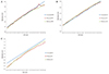
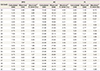
In addition to validating the inferential equations through known reference values, we applied the inferential equations to observed data to see how capable these formulae were in explaining real measurements. Observed data were collected from 194 cases, and the differences between observed and calculated data were analyzed. The calculated, observed and reference values are depicted in graph form in Fig. 1. By looking at the graphs for each biometric value, we can easily compare the range of differences between the data. Although there are parts where the graphs do not coincide, overall, the graphs schematically demonstrate that the values are quite approximate. The median, minimum and maximum values of errors between observed and calculated data are shown in Table 2. The absolute percentage errors are shown in Table 3. The maximum values of errors were 0.6, 1.1 and 0.7 cm for BPD, AC and FL, respectively. And except for FL in earlier gestational weeks (GA 20 to 23 weeks), the percentage errors were all under 10%. Although these values may be accepted as substantial errors if mathematical accuracy was our main pursuit, we concluded that these values of errors were acceptable and small enough to confirm the applicability of our equations. The observational data were recategorized by rounding off the gestational week (e.g., GA 20+4 weeks rounded off to GA 21 weeks), and the values of errors and absolute percentage errors were not significantly different (data not shown).
Discussion
Evaluating adequacy of fetal growth is one of the most essential part of prenatal care. Evaluation of not only estimated fetal weight but also each biometric parameter (BPD, FL, and AC) is also important. There have been several mathematical formulae proposed by different authors which estimate the relationship between GA and fetal biometric values [8910]. Some authors have proposed formulae which estimate GA with measured biometric values. Yaghoobian [8]'s formula estimated GA with measured BPD, and the formula by Honarvar et al. [9] assessed GA with measured FL after the 1st trimester. Others have proposed formulae which estimate fetal biometric values according to each GA. Rosati and Guariglia [10] proposed two linear mathematical models to estimate the length of femur and humerus length using measured BPD and GA in early pregnancy. Of the three formulae, the one suggested by Rosati and Guariglia is similar in concept to our equation and is shown in 4 [10]. To our knowledge, this is the first and only study suggesting a formula estimating median values of BPD, AC and FL with GA as the only independent variable. The formulae in Table 4 may be mathematically precise, but they are complex and thus very hard to memorize. The biggest strength of our study is the simplicity of the three suggested equations. Due to their simplicity, it is easy for examiners to memorize the equations. Furthermore, the examiners can estimate median fetal biometric values without the aid of any electronic device and evaluate fetal growth roughly before or even without looking up any reference.
Another significant finding of this study was discovering the coefficient of the formula for each biometric value. By knowing the gradient of each graph, the derived formulae let us take notice of other aspects of fetal growth rate and its prediction. For instance, through the equation BPD=GA/4, we can assume that fetal BPD increases approximately 1 cm every 4 weeks on average. In the same way, we can expect fetal AC to increase approximately 1 cm every week, and fetal FL to increase approximately 1 cm every 5 weeks on average. Knowledge of such biometric growth trends helps obstetricians in counseling patients by providing simple and easy explanations to those who are curious about their baby's growth trend. Also, in situations when the BPD plane is inevitably measured inadequately due to fetal position, by knowing the growth trend of fetal BPD, the examiner can have in mind the estimated value.
The limitation of this study is the relatively small number (n=194) of patients whose biometric values were used to validate our equations. Also, our observed data were limited to women who delivered at a single tertiary hospital which may not adequately represent the whole Korean population. Lastly, the proposed equations may seem oversimplified and thus not completely fit mathematically, especially if compared to other previously proposed models [10]. It is natural that fetal biometric values increase with advancing GA, but the growth rates differ at each trimester [12]. Therefore, other authors have described that extrapolating a formula determined from one trimester to another would yield inaccurate results [13]. This is the reason why other studies have suggested mathematical models restricted to a certain gestational period or trimester [8910]. We were well aware that accurately estimating biometric values with a single, simple mathematical formula throughout all GAs would be infeasible. However, we emphasize once more that mathematical accuracy was not our main pursuit, and that the significance of this study lies in the simplicity and practicality of the equations. In conclusion, the three mathematical formulae proposed in the present study: BPD=GA/4, AC=GA-5, FL=GA/5 show an extremely easy way to estimate median values of fetal biometry at each gestational week (after GA 20 weeks) with good reliability and clinical applicability.


 XML Download