Abstract
We present a case of retained placenta accreta treated by high-intensity focused ultrasound (HIFU) ablation followed by hysteroscopic resection. The patient was diagnosed as submucosal myoma based on ultrasonography in local clinic. Pathologic examination of several pieces of tumor mass from the hysteroscopic procedure revealed necrotic chorionic villi with calcification. HIFU was performed using an ultrasound-guided HIFU tumor therapeutic system. The ultrasound machine had been used for real-time monitoring of the HIFU procedure. After HIFU treatment, no additional vaginal bleeding or complications were observed. A hysteroscopic resection was performed to remove ablated placental tissue 7 days later. No abnormal vaginal bleeding or discharge was seen after the procedure. The patient was stable postoperatively. We proposed HIFU and applied additional hysteroscopic resection for a safe and effective method for treating retained placenta accreta to prevent complications from the remaining placental tissue and to improve fertility options.
Retained placenta is a condition where the placenta or membranes remain in the uterus during or after the third stage of labor. It is one of the major causes of delayed postpartum hemorrhage. Many retained placenta cases involve placenta accreta, an abnormal adherence of placental villi to the myometrium. The increasing number of cesarean sections has caused the incidence of placenta accreta to increase [1]. Ultrasonography has traditionally been a good method for diagnosing placenta accreta. However, magnetic resonance imaging (MRI) has become more popular for diagnostic accuracy [2]. Additionally, the incidence of conservative treatment such as uterine artery embolization (UAE) or methotrexate has gradually increased, replacing hysterectomy. Furthermore, although patients prefer to undergo conservative treatment rather than hysterectomy, the disadvantages of conservative treatment include the risk of hemorrhage or infection and the risk of needing a secondary hysterectomy [3].
We presented a case of retained placenta accreta. It was diagnosed by ultrasound and MRI and was treated by high-intensity focused ultrasound (HIFU) ablation followed by hysteroscopic resection.
A 33-year-old woman presented with intermittent vaginal bleeding, menorrhagia and dysmenorrhea after a cesarean section at 38 gestational weeks in October 2010. The patient was diagnosed as submucosal myoma based on ultrasonography and was transferred for further evaluation and management on July 28, 2011. The obstetrics history of the patient included 1 cesarean section and 1 artificial abortion. She also received a myomectomy in 2003. She had irregular menstrual cycles, menorrhagia and severe dysmenorrhea. There was nothing remarkable about her general appearance or nutritional state. Her vital signs were stable. On physical examination, the abdomen was soft and the previous operation scar was noted. A fist-sized uterus was palpated and vaginal spotting was noticed on pelvic examination. There was a 3.2×2.0×1.5-cm hyperechogenic shadow in the endometrial cavity on transabdominal and transvaginal ultrasonography, and a thickened posterior uterine wall (Fig. 1A). Both adnexae were intact. Initial laboratory findings were normal, including serum beta-human chorionic gonadotropin, with the exception of hemoglobin, which was 10.9 mg/dL. Based on the history and sonographic findings, an endometrial polyp or an endometrial problem with a submucosal myoma with degeneration was suspected.
Under intravenous propofol sedation, we attempted to remove the uterine mass with hysteroscopic resection (RIWO Resectoscopes, Richard Wolf GmbH, Knittlingen, Germany). However, because of bleeding from the tumor site, the mass could not be removed completely (Fig. 1B). Pathologic examination of several pieces of tumor mass from the hysteroscopic procedure revealed necrotic chorionic villi with calcification.
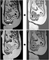
Even though she had stable vital signs after the procedure, her vaginal bleeding continued and additional management was required. In addition, the patient refused methotrexate treatment because she anticipated future pregnancies. Combination therapy with HIFU and curettage was recommended for the first management approach. Radiological images, such as MRI, were necessary to obtain more information prior to HIFU treatment. Subsequently, HIFU was performed using an ultrasound-guided HIFU tumor therapeutic system (Model JC, Chongqing Haifu Tech, Chongqing, China). The ultrasound machine (MyLab, Esaote, Genoa, Italy) had been used for real-time monitoring of the HIFU procedure. A urinary Foley catheter was inserted into the patient's bladder and connected with sterile degassed water through an infusion set to control the bladder volume and to correctly set the uterine position. Vaginal packing was performed to prevent contamination of the circulating degassed water. During the procedure, the patient was placed in the prone position on the system under conscious sedation. Total energy of HIFU ablation was 24552 J with 250 W of power and the ablation time was 10 minutes. After HIFU treatment, no additional vaginal bleeding or complications were observed. A hysteroscopic resection was performed to remove ablated placental tissue and prevent endometrial adhesion after 7 days later because she anticipated future pregnancies. There was no bleeding in hysteroscopy, because that might be due to coagulation necrosis of the lesions. No abnormal vaginal bleeding or discharge was seen after the procedure. The patient was stable postoperatively. The lesion was not seen in MRI finding 3 months later after HIFU combined with 2nd hysteroscopic resection (Fig 2).
Retained placenta accreta is a challenging obstetrical problem; it is rare, but remains a serious condition if not managed correctly. Although hysterectomy has been the recommended treatment for placenta accreta, conservative treatment is proposed as an alternative for patients who desire future fertility options. Traditionally conservative treatment for placenta accreta includes methotrexate and UAE; however, both methods involve the risk of subsequent infection and hemorrhage that could require a hysterectomy because of remaining placental tissue [13]. Thus, some authors have proposed hysteroscopic resection alone or in combination with UAE as a conservative treatment method for retained placenta accreta. They reported that the hysteroscopic method was not only safe and effective, but reduced the rate of synechiae and improved fertility prognosis [456]. However, treatment with hysteroscopic resection only possibly leads to repeat hysteroscopic procedure (50.0%) or delayed hysterectomy (8.3%) [6].
HIFU is a non-invasive treatment technique that is directed harmlessly across skin and damaged target tumor tissue. HIFU applies selective tissue necrosis through heating and cavitation by ultrasound [7]. This method is highly effective, with few side effects and has been reported to have an effect on solid tumors of the liver [8], pancreas [9], breast [10], uterus [11] and other organs.
Paek et al. [12] showed in a pilot study on fetal tissue in a sheep model that HIFU ablation could be successfully applied to placental tissue. Huang et al. [13] and Xiao et al. [14] also showed that HIFU is an effective treatment method for cesarean pregnancy scarring. Additionally, Bohlmann et al. [15] suggested HIFU treatment for fibroids, which can be recommended to women with fibroid-associated subfertility who strictly reject surgical treatment or who have high surgical risk because there is no evidence of any relevant impairment of fertility after HIFU treatment.
Based on the observations described above, we selected HIFU ablation as an alternative treatment method for patients with placental disease who may wish to have fertility options in the future. We decided to apply HIFU treatment for this patient's retained placenta accreta because she refused a hysterectomy or methotrexate treatment and had experienced a failed hysteroscopic resection. We proposed HIFU and applied additional hysteroscopic resection to prevent complications from the remaining placental tissue and to improve fertility options.
There are many established management options for retained placenta accreta with inherent advantages and disadvantages. Additional studies should be performed to assess the application of HIFU, but we propose using this new technology as a new safe and effective method for treating retained placenta accreta.
Figures and Tables
Fig. 1
Pre and post 1st hysteroscopic resection sono finding. (A) Transabdominal sonographic finding before 1st hysteroscopic resection (coronal and sagittal view): 3.2×2.0×1.5-cm hyperechogenic shadow in the endometrial cavity was shown (arrow). (B) Transabdominal sonographic finding after 1st hysteroscopic resection (coronal and sagittal view): remained hyperechogenic shadow was shown (arrow).

Fig. 2
Pre and post high-intensity focused ultrasound (HIFU) combined with 2nd hysteroscopic resection magnetic resonance imaging finding (MRI). (A) T2W1 and (B) contrast enhanced image was MRI finding before HIFU ablation. The retained placenta accreta was shown as low signal intensity on (A) and low enhanced image on (B). (C) T2W1 and (D) contrast enhanced image was MRI finding 3 months later after HIFU combined with 2nd hysteroscopic resection. The lesion was not seen.

References
1. Committee on Obstetric Practice. Committee opinion no. 529: placenta accreta. Obstet Gynecol. 2012; 120:207–211.
2. Tanaka YO, Shigemitsu S, Ichikawa Y, Sohda S, Yoshikawa H, Itai Y. Postpartum MR diagnosis of retained placenta accreta. Eur Radiol. 2004; 14:945–952.
3. Verspyck E, Resch B, Sergent F, Marpeau L. Surgical uterine devascularization for placenta accreta: immediate and long-term follow-up. Acta Obstet Gynecol Scand. 2005; 84:444–447.
4. Greenberg JA, Miner JD, O'Horo SK. Uterine artery embolization and hysteroscopic resection to treat retained placenta accreta: a case report. J Minim Invasive Gynecol. 2006; 13:342–344.
5. Legendre G, Zoulovits FJ, Kinn J, Senthiles L, Fernandez H. Conservative management of placenta accreta: hysteroscopic resection of retained tissues. J Minim Invasive Gynecol. 2014; 21:910–913.
6. Rein DT, Schmidt T, Hess AP, Volkmer A, Schondorf T, Breidenbach M. Hysteroscopic management of residual trophoblastic tissue is superior to ultrasound-guided curettage. J Minim Invasive Gynecol. 2011; 18:774–778.
7. Kennedy JE, Ter Haar GR, Cranston D. High intensity focused ultrasound: surgery of the future? Br J Radiol. 2003; 76:590–599.
8. Shen HP, Gong JP, Zuo GQ. Role of high-intensity focused ultrasound in treatment of hepatocellular carcinoma. Am Surg. 2011; 77:1496–1501.
9. Khokhlova TD, Hwang JH. HIFU for palliative treatment of pancreatic cancer. J Gastrointest Oncol. 2011; 2:175–184.
10. Zhao Z, Wu F. Minimally-invasive thermal ablation of early-stage breast cancer: a systemic review. Eur J Surg Oncol. 2010; 36:1149–1155.
11. Kim YS, Kim JH, Rhim H, Lim HK, Keserci B, Bae DS, et al. Volumetric MR-guided high-intensity focused ultrasound ablation with a one-layer strategy to treat large uterine fibroids: initial clinical outcomes. Radiology. 2012; 263:600–609.
12. Paek BW, Vaezy S, Fujimoto V, Bailey M, Albanese CT, Harrison MR, et al. Tissue ablation using high-intensity focused ultrasound in the fetal sheep model: potential for fetal treatment. Am J Obstet Gynecol. 2003; 189:702–705.
13. Huang L, Du Y, Zhao C. High-intensity focused ultrasound combined with dilatation and curettage for Cesarean scar pregnancy. Ultrasound Obstet Gynecol. 2014; 43:98–101.
14. Xiao J, Zhang S, Wang F, Wang Y, Shi Z, Zhou X, et al. Cesarean scar pregnancy: noninvasive and effective treatment with high-intensity focused ultrasound. Am J Obstet Gynecol. 2014; 211:356.e1–356.e7.
15. Bohlmann MK, Hoellen F, Hunold P, David M. High-intensity focused ultrasound ablation of uterine fibroids: potential impact on fertility and pregnancy outcome. Geburtshilfe Frauenheilkd. 2014; 74:139–145.


 PDF
PDF ePub
ePub Citation
 Print
Print


 XML Download