

 PDF
PDF ePub
ePub Citation
 Print
Print


Introduction
Preterm delivery <37 weeks is a great problem of public health in worldwide with high rates of neonatal morbidity and mortality [1]. The preterm delivery complicated 12.3% of all pregnancies in the USA in 2008 [2] and despite efforts to reduce its incidence, the preterm delivery has been increasing even in developed countries [3]. Despite the etiology of preterm delivery is not known in 20% to 40% [4], the association between short cervix length (CL) in the second trimester and spontaneous preterm delivery is well recognized since 90' [5].
With a publication of randomized controlled trial demonstrating that vaginal progesterone significantly reduced the risk of preterm birth in women found to have a CL assesses by transvaginal ultrasound ≤15 mm between 20 and 25 weeks [6], an universal screening of CL measurement in the second trimester of pregnancy has been proposed [7]. Furthermore, the CL measurement by transvaginal ultrasoun has showed high intra- and inter-observer reproducibility [8].
There are reference ranges for the CL measurement to the American and Swedish populations [910], because it is not known the influence of ethnic factor in the CL measurement [11]. There is an unique study with established reference ranges for the CL measurement using the transvaginal ultrasound in the second and third trimester of pregnancy in a Brazilian population [12].
The objective this study is to establish reference values for the CL measurement by transvaginal ultrasound between 20 and 24+6 weeks of gestation in a large heterogeneous Brazilian population.
Materials and methods
We performed a retrospective cross-sectional study between February 2012 and February 2015 with pregnant women underwent routine second ultrasound examinations at 20 to 24+6 weeks of gestation. This study was approved the Ethic Committee of Uberaba University, the consent form was not necessary because it was a retrospective study. The low-risk pregnant women were selected randomly and were from public and private health services of metropolitan region of Uberaba-MG, Brazil.
The inclusion criteria were the following: singleton pregnancies and gestational age (GA) determined by last menstrual period and confirmed by ultrasound examination performed before 20 weeks (crown-rump length, between 11 and 13+6 weeks; biparietal diameter, between 14 and 20+6 weeks). The exclusion criteria were the following: absence of maternal chronic disease as systemic arterial hypertension, pregestational diabetes, immune disease and renal disease; previous preterm birth; previous cervical surgery; and cervical insufficiency. The pregnant women were assessed only once time and the postnatal outcomes were not available.
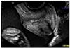
The ultrasound examinations were performed in the Mario Palmério University Hospital and Radiologic Clinic of Uberaba. The pregnant women are asked to empty their bladder and are placed in the dorsal lithotomy position. Ultrasound examinations were performed in a Voluson E6 (General Electric, Healthcare, Zipf, Austria) apparatus equipped with an endovaginal probe (RIC 5-9W) and the exams were carried out by only two examiners (ABP and TMR) both with Fetal Medicine Foundation Certificate accreditation for competence in cervical assessment. The probe was placed in the anterior fornix of the vagina and care was taken to avoid exerting undue pressure on the cervix, which might artificially lengthen the cervix. A sagittal view of the cervix was obtained and the sonolucent endocervical mucosa (cervical gland area) was used as a guide to the true position of the internal os, thereby avoiding confusion with the lower segment of the uterus [13]. The calipers were used to measure the linear distance between the triangular area of echodensity at the external os and the internal os. Each ultrasound exam was performed over a period of about 3 minutes in order to note any dynamic changes in the cervix (Fig. 1).
The data have been transferred to Excel spread sheet (Microsoft Corp., Redmond, WA, USA) and analysed by one of the authors (WPM) using PASW ver. 18.0 (SPSS Inc., Chicago, IL, USA) and GraphPad ver. 5.0 (GraphPad Software, San Diego, CA, USA). The maternal characteristics as height, weight, body mass index (BMI), number of pregnancies, parity and GA at ultrasound exam were expressed by median and ranges. Cigarette smokers and alcohol consumption were assessed as percentage (%). To construct reference ranges for the CL measurement (mm) as a function of GA (weeks), we planned to perform curve fitting using polynomial regression as suggested by Altman and Chitty [14]. However, since the CL does not significantly change with GA, we used all acquired data to determine the 3rd, 5th, 25th, 50th, 75th, 95th and 97.5th percentiles in the evaluated GA. To compare the CL measurement between pregnant women without previous delivery and pregnant women with one or more previous delivery, we used the analysis of variance.
Results
We assessed 996 consecutive pregnant women, and all of them filled the inclusion criteria, being allocated in the statistical analysis. The distribution of pregnant women in each GA period (weeks) was the followed: 20 to 20+6, 84; 21 to 21+6, 254; 22 to 22+6, 426; 23 to 23+6, 181; 24 to 24+6, 51.
The median and ranges of height (cm), weight (kg), body mass index (kg/m2), number of pregnancies, parity, and GA at ultrasound examination (weeks) were 163 (145 to 187), 67 (43 to 122.5), 25.2 (16.3 to 48.3), 1 (1 to 5), 0 (0 to 4), and 22.3 (20 to 24.9), respectively. According to cigarette smokers and alcohol consumption, 1.2%, 3.3%, respectively.
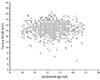
The median±standard deviation and ranges for the CL measurement (mm) was 37.0±10.7 (8 to 51). The CL measurement did not modify significantly with GA (Fig. 2). There was no significant difference in the CL when comparing women without previous deliveries (36.3±5.2 mm, n=620), with one previous delivery (37.1±5.1 mm, n=33), and with two or more previous deliveries (36.8±4.9 mm, n=46; P=0.07).
The observed percentiles for the CL measurement (mm) considering all number case were the following: 3rd, 26 mm; 5th, 28 mm; 25th, 33 mm; 50th, 37 mm; 75th, 40 mm; 95th, 45 mm; 97th, 46 mm.
Discussion
In this study, we established reference values for the CL measurement in the second trimester of pregnancy in a large heterogeneous Brazilian population. A systematic review concluded that a short cervix is predictive of preterm delivery and can identify pregnant women that may benefit from preventive and therapeutic measures [15]. Because of this, the CL measurement using transvaginal ultrasound in the second trimester has been used for the screening of high-risk pregnant women for preterm deliveries to establish preventive attitudes as vaginal progesterone, cervical pessary and cervical cerclage [16].
In this study, we assessed the CL measurement between 20 and 24+6 weeks of gestation, because in this period is performed the second trimester ultrasound examination to assess the fetal morphology. In this period, the CL measurement did not have significantly change with GA, impossible the construction of specific percentiles in each GA. However, we establish reference ranges using all number case, instead a bestfit by polynomial regression. In a previous reference range for the CL measurements between 17 and 41 weeks in a Swedish population, CL measurement did not change substantially between 17 and 32 weeks [10].
In a study witch determined reference ranges for the CL measurement between 16 and 22+6 weeks using transvaginal ultrasound in an American population, the mean was 38.5 mm at 19.9 weeks [9]. This value is higher than mean of our study in the period between 20 and 24+6 weeks of gestation.
In a Brazilian study which established reference ranges for the CL measurement between 20 and 34 weeks of gestation with 1,061 singleton pregnancies, the mean was 35.7 mm between 20 and 24+6 weeks [12]. In our study, the mean was 37.0 mm in this gestational period. In this Brazilian study, the mean of CL measurement also did not significantly change with GA. We decide did not separate our sample in races because of a lot of Brazilian miscegenation population. In agreement of Census 2000, for a total of 168,200,000 inhabitants, the Brazilian population consisted of 53.7% of White; 7.7% of Black and 38.6% of Mixed [17].
The limitation of our study was the absent of perinatal outcomes, however all pregnant women were from low-risk public and private services. Furthermore, all ultrasound examination were performed by only two examiners with accreditation of Fetal Medicine Foundation Certificate which could decrease the inter-observer variability.
In summary, reference values for the CL measurement by transvaginal ultrasound between 20 and 24+6 weeks of gestation in a large heterogeneous Brazilian population were established.
 XML Download