

 PDF
PDF ePub
ePub Citation
 Print
Print


Introduction
Ovarian cancer is a malignant gynecological disease, with >70% of patients diagnosed at an advanced stage [1]. Similar to other cancers, the mechanism of carcinogenesis in ovarian cancer is not yet understood. Despite optimal treatments including debulking surgery and chemotherapy, the 5-year survival rate of these patients are only 45% [2]. Every year, approximately 2,000 women are diagnosed with ovarian cancer in Korea, and about 900 of them die from it [3]. Because of the inability for early diagnosis of ovarian cancer, survival is poor, as compared with other cancers. Thus, a biomarker that could help diagnose ovarian cancer at an early stage is necessitated.
Obesity is a well-known risk factor in several cancers; it is an inflammatory disease that leads to a metabolic syndrome with elevated lipid profiles, glucose, and blood pressure. Inflammatory biomarkers have been implicated in the development of several kinds of cancer [4]. Adipokine is a hormone secreted from adipose tissue and is classified as an obesity-related hormone; adiponectin and leptin are well-known adipokines, and both play an important role in the metabolic system [56].
Adiponectin is an adipokine that can suppress cancer cell growth and invasiveness, and also inhibit angiogenesis by suppressing estrogen receptor α and vascular endothelial growth factor [78]. One of the important mechanisms of limiting tumorigenesis involves the activation of adenosine 5' adenosine monophosphate-activated protein kinase by adiponectin that could restrict tumor growth. An already known theory involves the following: adiponectin and insulin sensitivity, high-density lipoprotein cholesterol was positively correlated, negatively related to triglycerides, along with other health factors such as anti-inflammatory effects and anti-vascular effects [910].
Leptin is a kind of peptide hormone activates in regulation of appetite, formation of bone, functions in reproductive system and angiogenesis [11]. Further, another type of adipokine is directly involved with body fat, and it is the main factor that is associated with obesity-related diseases. Several studies have reported that plasma leptin levels are higher in overweight and obese individuals than normal-weight individuals [1213]. Many studies have shown that decreased adiponectin levels and elevated leptin levels are associated with obesity-linked cancers [14].
A change in adiponectin and leptin levels has been associated with endometrial cancer and breast cancer. To elucidate the relationship between adiponectin, leptin and ovarian carcinogenesis, plasma adiponectin and leptin concentrations in patients with ovarian cancer are necessitated in this pilot study. The aim was to explore differences in plasma adiponectin and leptin concentrations between patients with ovarian cancer and normal women with the same body mass index (BMI) to determine whether adiponectin and leptin are related with ovarian carcinogenesis.
Materials and methods
1. Study population
Between September 2006 and October 2014, a total of 52 patients who were histologically diagnosed with ovarian cancer at the Gynecology and Oncology Center of Ewha Womans University Mokdong Hospital were recruited as subjects. Further, because of the difficulty in collecting absolute healthy female's blood sample, we selected 50 patients clinically diagnosed with benign disease during the same period for the control group. The study was approved by the Ethics Committee of the Ewha Womans University Mokdong Hospital, and informed consent was obtained from all participants. After obtaining approval from the patients and doctors, blood samples were drawn before surgery. Blood samples were drawn from a peripheral vein and placed in heparin containing tubes. The plasma was immediately separated from the blood cells using centrifugation at 3,000 rpm for 30 minutes at 4℃, and the separated plasma was kept at -80℃ until use. The total time between the blood draw and freezing the plasma was no more than 4 hours at all recruitment sites.
Clinical data were reviewed from medical records, including body weight and height, for the calculation of the patients' BMI. Age and cancer history were included. The patients' BMI was categorized as underweight (<18.5), normal (18.5 to 24.9), overweight (25 to 29.9), and obese (>30). Each case of gynecologic cancer was staged based on the FIGO (International Federation of Obstetricians and Gynecologists) staging system [15].
2. Laboratory assays
Plasma adiponectin and leptin levels were measured using a commercially available enzyme linked immunosorbent assay (ELISA) kit (Cusabio Biotech, Newark, DE, USA). The detectable concentration ranges for adiponectin and leptin were 1.56 to 100 ng/mL and 0.156 to 10 ng/mL, respectively. Both concentrations for each sample were calculated according to the standard concentrations, with a corresponding optical density value of 450 nm. Frozen plasma was thawed overnight at 4℃ before measurement.
3. Statistical analysis
Statistical analysis was performed using IBM SPSS ver. 20.0 (IBM Corp., Armonk, NY, USA). Statistical comparison among groups was analyzed using the Student's t-test; correlations were confirmed using the Pearson correlation. A P-value of <0.05 was considered statistically significant.
Results
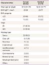
Fifty-two ovarian cancer patients were included in this study. The patients had the following cancer types: serous (n=32, 58.2%); clear cell (n=6, 10.9%); endometrioid (n=2, 3.6%); mucinous (n=1, 1.8%); undifferentiated carcinoma (n=4, 7.3%); carcinosarcoma (n=2, 3.6%); Brenner tumors (n=2, 3.6%); mixed epithelial tumor (n=1, 2%); and others (n=2, 3.6%). And the histology type of benign disease control group as follows: serous cystadenoma (n=7, 14%); mucinous cystadenoma (n=6, 12%); dermoidcyst (n=6, 12%); endometriotic cyst (n=5, 10%), paratubal cyst (n=10, 20%); uterine prolapse (n=6, 12%); hemorrhagic corpus luteum cyst (n=4, 8%); others of unknown histology (n=6, 12%). As seen in Table 1, the median age of the patients with ovarian cancer in this study was 47.9 years (range, 31 to 78 years), while the median age of individuals in the control group (n=50) was 52.3 years (range, 13 to 77 years); no significant difference was found between the two groups (P=0.126). The mean BMI value for patients with ovarian cancer and the control group was 23.34 kg/m2 (range, 17.71 to 33.59 kg/m2) and 23.77 kg/m2 (range, 17.36 to 29.15 kg/m2), respectively; no significant difference was found between the two groups (P=0.659).
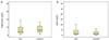
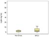
The mean plasma adiponectin concentrations were significantly lower in the ovarian cancer group (8.25±0.97 µg/mL) than those of the control group (11.44±1.13 µg/mL) (P=0.026). Further, the mean plasma leptin concentrations were significantly lower in the ovarian cancer group (7.09±1.46 ng/mL) than the control group (15.4±2.04 ng/mL) (P=0.001) (Fig. 1). However, there was no significant difference in adiponectin and leptin levels between early-stage (I/II) and advanced-stage (III/IV) disease (P=0.078, P=0.675) (Fig. 2). The leptin concentration was higher in non-serous type (7.17±2.75 ng/mL) than serous type (6.94±1.67 ng/mL), but there was no statistical significance (P= 0.941) (Fig. 3).
A significant correlation was identified between leptin and BMI (r =0.343, P=0.001). However, no significant relationship was found between adiponectin and BMI, adiponectin and leptin (P=0.888, P=0.681) (data not shown).
Discussion
On the beginning of this study, we hypothesized that the leptin level of ovarian cancer patients may be higher than normal control group; however in our study we get the totally different result.
The present result provides evidence that the plasma adiponectin and leptin levels are inversely associated with the risk of developing ovarian cancer. Further, this result was different from that of studies conducted on obesity-related cancers such as endometrial cancer and breast cancer.
High serum adiponectin levels were associated with a significantly lower risk of breast cancer in a case-control study [16]. And many studies have indicated that a decreased adiponectin level was associated with cancer development. In our research, we confirmed that a decreased adiponectin level was positively associated with the development of ovarian cancer, and this was consistent with the result of most studies as a low adiponectin level was associated with the risk of developing certain cancers such as breast cancer, endometrial cancer, and renal cell carcinoma [16].
Long-term exposure to leptin enhanced prostate cancer growth according to some studies [17]. Further, another study group concluded that a high leptin expression was significantly associated with lymph node involvement and an advanced tumor stage in esophageal squamous cell carcinoma [18]. Besides, some investigators [19] researched an OVCAR-3 ovarian cancer cell line and found that leptin stimulated the cell cycle and inhibited apoptotic gene and protein expression.
Although higher leptin concentration in cancer patients contrast to healthy control were reported in some publications [20], lower leptin level in ovarian cancer were also reported in more than several reports [2122232425]. In a serum marker study, leptin concentration was significantly decreased in ovarian cancer when analyzed using ELISA and microarray [22].
So we made two explanations, one of them is, the low level of leptin is directly associated with ovarian cancer; the other one is that the low leptin concentration is the result of starvation.
Leptin is a starvation signal; decreasing plasma leptin levels cause neurohumoral and behavioral changes, which contribute to the preservation of energy reserves for vital functions [26]. Therefore, after reducing body fat mass, leptin levels was decreased, following energy expenditure reduction [27]. Starvation is a common feature in most ovarian cancer patients as is reported [28]. And this could help explain the result of our study because more than one-half of the patients in our study were diagnosed with advanced-stage ovarian cancer (nearly 60% was diagnosed with either stage III or IV), meaning the tumor mass was large enough to spread outside the pelvic region. As large pelvic tumors are associated with ascites, most patients with advanced-stage ovarian cancer fail to absorb enough calories to maintain their daily life, and this may induce the low leptin levels observed in patients with ovarian cancer. If the latter explanation was right, then there will have some difference between early stage and advanced stage, because early stage patients have no limitation in energy absorption. But in our result no statistical significant difference was found between them. And this maybe ascribe from small sample size in each group, more sample may have a different result. Conclusively, we cannot support the latter one in this study.
Further, according to a study, endometrioid ovarian cancer was positively associated with obesity, and no relationship was observed between obesity and serous ovarian cancer [27]. In our study, more than one-half of the patients had serous ovarian cancer, and there is no significant difference between serous type and non-serous type. Thus again it supports the first explanation: low leptin level was directly associated with ovarian cancer.
All these studies support our result that low leptin levels, not high leptin levels, are associated with ovarian cancer. Thus, although the ovaries and uterus are anatomically closely positioned, the plasma concentration of leptin in ovarian cancer and endometrial cancer compared with control group was totally different.
From emerging studies on endocrinology, it was proposed that adiponectin and leptin have negative relationship, and also in several cancer-related studies [1329]. However, in other studies, no significant correlation was found between the expression of leptin and adiponectin in esophageal squamous cell carcinoma [30], and it was consistent with our result that there was no significant relationship between adiponectin and leptin.
Further, according to some studies adiponectin and BMI was significantly related, but some other investigators insist that no significant relationship was found between adiponectin and BMI, which agree with our study [30].
The limitations of this study include differing follow-up times for each patient; moreover, it was difficult to obtain blood samples at the same time, as some may have been drawn in the morning, while others may have been drawn in the afternoon. Thus, this factor may have had some influence on plasma adiponectin and leptin levels.
However, this study had many advantages; this study investigated correlations among plasma adiponectin levels, plasma leptin levels, and ovarian cancer. It included circumstantial down-regulated leptin levels in patients with advanced-stage ovarian cancer. Additional study with a larger number of patients is necessitated to further illustrate the relationship between adiponectin, leptin and ovarian cancer.
It is well known that the early detection of cancer is closely associated with prognosis for patients with cancer, even with recurrent cancer. If a tumor is discovered early, more treatment options are available that could help improve patient survival. Clinically, CA 125 and CA 19-9 are biomarkers currently used in clinical practice to monitor the recurrence of ovarian cancer. However, there are some limitations regarding their use, as not every cancer patient experiences a change in CA 125 and CA 19-9 levels that can be discriminated. Thus, adiponectin and leptin could be included as sub-biomarkers to evaluate health status and might help in the follow-up of patients with ovarian cancer.

 XML Download