

 PDF
PDF ePub
ePub Citation
 Print
Print


Introduction
Recurrent miscarriage (RM) is a major reproductive problem because it involves, according to the definition used, about 1% to 2% of women of reproductive age [1] and up to 5% of all couples [2] in which it determines significant personal, psy-chological and relational problems. It also represents a great challenge for the clinicians because too many physiopathologic, diagnostic and therapeutic issues concerning this condition are still unknown, debated and largely unresolved. In fact, even though many causes or risk factors for RM have been identified and RM is believed to be a heterogeneous condition [3] with a multifactorial etiology [4], approximately 50% of RMs are still classified as unexplained recurrent miscarriage (uRM) [456].
A growing body of evidence indicates the fundamental importance of a proper maternal-fetal immune relationship for a successful pregnancy [78] and supports the possibility that several immunologic factors are involved in early pregnancy disorders, such as RM [9101112]. In this context, the role of autoimmunity is believed to be relevant [1314], although the relative importance of many autoimmune factors is still unclear.
Antinuclear autoantibodies (ANA), a group of autoantibodies directed towards nuclear and cytoplasmatic antigens, in several studies have been detected in a significantly higher proportion of women with RM compared with normal healthy control women [151617]. ANA positivity has also been demonstrated to be associated with a markedly impaired outcome of in vitro fertilization, suggesting that ANA could have a detrimental effect on oocyte and embryo development [18]. These findings support the hypothesis that ANA could play a role in early pregnancy complications, including RM, although the mechanisms by which they could cause pregnancy loss are only speculative at present.
There is evidence that RM is associated with high uterine artery impedance [1920] which could be responsible for a suboptimal endometrial and myometrial perfusion, leading to pregnancy loss. Moreover, recent experimental data that suggest a possible correlation between ANA and changes in uterine flow indices in women affected by RM [19]; however it has been shown that ANA positivity is unlikely to impact uterine blood flow in normal women [21]. At present the potential role of ANA in RM, with specific application to uterine blood flow indices, remains unclear because there is scant information, if any, on the possible relationship between ANA status and uterine blood flow and vascularization indices in women with RM. As far as we know, this issue has not been investigated with the use of 3D power Doppler imaging with the VOCAL technique.
Materials and methods
1. Subjects
The present study involved 52 non-pregnant women of reproductive age which were divided into two groups: group 1, formed by 26 women with primary uRM (13 ANA- and 13 ANA+); group 2 (controls), formed by 26 women (13 ANA- and 13 ANA+) with at least two pregnancies at term and no miscarriages.
All the women included in the study attended as outpatients the Complex Operative Unit of Gynecology and Obstetrics at the University Hospital Policlinico Tor Vergata of Rome. Women with uRM were followed at the RM unit, whereas controls were followed at the General Gynecologic Service. The patients were enrolled into the study after giving a written informed consent. The study was approved by institutional review board (no. 90.14) and was carried out according to the principles of the Declaration of Helsinki.
Women with RM underwent a standardized diagnostic workup [2223], which included the following: 1) collection of familial and personal medical, gynecological and obstetrical history with specific application to the previous miscarriages; 2) gynecological examination; 3) transvaginal ultrasound; 4) hysteroscopy and endometrial biopsy; 5) endocrine evaluation panel: assay of luteinizing hormone, follicle-stimulating hormone, prolactin, progesterone in the midluteal phase, thyroid stimulating hormone, free triiodothyronine, free thyroxine, pituitary and ovarian androgens, insulin and glucose curve; 6) karyotype of both partners; 7) immunity panel: anti-phospholipid anti-bodies, lupus anticoagulant, anti-beta-2 glycoprotein I (anti-β2GPI) and anti-annexin V antibodies, anti-thyroid antibodies, ANA, anti-extractable nuclear antigens antibodies, anti-double stranded DNA, anti-smooth muscle antibodies and anti-mitochondrial antibodies; and 8) thrombophilia screening: protein C, protein S, antithrombin III, activated protein C resistance, homocysteine; determination of the following mutations: factor V (G1691A Leiden), factor II prothrombin (G20210 A), plasminogen activator inhibitor (PAI-1 4G/5G), methenyl tetrahydrofolate reductase (MTHFR C677T and A1298C). This workup was aimed to identify proven, probable and doubtful causes of RM. Women were diagnosed with uRM when no known causes for pregnancy loss could be identified.
Group 2 included women with a history of least 2 normal pregnancies at term, without any miscarriage. They were selected in the context of a large cohort of healthy women with at least two pregnancies at term who underwent routine gynaecological checks including pelvic echography. The ANA status (positivity or negativity) of the women of this cohort was established by taking a sample of peripheral blood at the time of the check. Then the women were scheduled for pelvic ultrasonography within one month after having the results of ANA assay. Control women were then selected by matching the major clinical characteristics (age and body mass index) with those of RM women. The first 26 eligible control women (ANA+ and ANA-) who met the above characteristics were then selected for the study, together with the first 26 eligible uRM women found with the ANA status (13 ANA+ and 13 ANA-) of interest for the present study. Then all women underwent the study protocol (further measurement of circulating ANA, P assay, and Doppler investigation).
The exclusion criteria for all groups were the presence of chronic disease and/or uterine abnormalities at ultrasound. None of the women underwent hormonal treatment in the four months before being enrolled into the study.
Then all women included into the study underwent the study protocol, which consisted in three major steps which were performed along one menstrual cycle: 1) measurements of resistance index (RI) and pulsatility index (PI) in the follicular and midluteal phase of the cycle; 2) 3D Doppler investigation; 3) P assay and further measurement of circulating ANA in peripheral venous blood. The steps 2) and 3) ere carried out in the midluteal phase of the cycle at the same time of PI and RI.
2. Ultrasound examinations
Ultrasound examinations were carried out in all the cases by the same operator, which was unaware whether the examined subject was a uRM or a control woman.
3. Transvaginal Doppler ultrasound
All RM patients and control women underwent a transvaginal Doppler ultrasound, in which the PI and the RI of the uterine arteries as well as the uterine flow indexes (vascularisation index [VI], flow index [FI], vascularisation flow index [VFI]), were measured. This investigation has been carried out in both the follicular and midluteal phases of the same menstrual cycle in all women. The phases of the menstrual cycle were dated in all cases according to the following criteria: 1) for the follicular phase: menstrual history of regular 28-days cycles; exact date of the beginning of the menses in the study cycle; ultrasound examination carried out at 9 to 11 day showing the presence of a single follicle of >11 mm diameter; 2) for the midluteal phase: ultrasound examination carried out at 22 to 24 day of the cycle, showing the presence of the corpus luteum within the omolateral ovary; circulating progesterone levels (measured as reported in the assay section) >13 ng/mL. The uterine artery was identified in the sagittal plane with color flow mapping, according to its specific morphology, along the side of the junction between the cervix and uterus. A sampling gate of pulsed wave Doppler was set to cover the whole vessel where it crossed the external iliac artery, with the angle of insonation lesser than 30 degrees.
4. Acqusition of 2D Doppler velocity waveforms
The PI and RI of the left and right arteries were measured by a Voluson E6 GE ultrasound machine (GE Healthcare, Kretztechnik, Zipf, Austria), equipped with a 7.5 MHz 3D transducer with color imaging capabilities. As previously described, the uterine arteries were identified thanks to the color Doppler imaging at its crossing point with the external iliac artery, by applying an angle of insonation less then 30 degrees. Pulsed-wave Doppler was used in order to obtain at least three consecutive flow velocity waveforms for both right and left uterine arteries. PI and RI of both the uterine arteries were automatically calculated by the software installed on the device. All measurements were entered prospectively into a computer database for offline analysis.
5. Acqusition of 3D vascular indices
Ultrasound 3D volume scannings were performed, in the midluteal phase of the menstrual cycle, inmmediately after the measurements of the PI and RI of the uterine arteries, with a 5- to 10-MHz transvaginal probe using a Voluson E6 GE ultrasound machine (GE Healthcare). In all cases the same instrument settings were used (angio mode, cent; smoothing, 4/5; FRQ, low; quality, 16; density, 6; enhancement, 16; balance, G >150; filter, 2; actual power, 2 dB; pulse repetition frequency, 0.9 kHz). Power Doppler ultrasound was applied to the image of the uterine vascularisation: the power Doppler gate was activated and manually adjusted to include the whole uterus. The 3D uterus volume was acquired, with a sweeping angle set at 85°. All volumes were stored and later analyzed with the Virtual Organ Computer-Aided Analysis II program (GE Healthcare, Milwaukee, WI, USA). The contour mode in the Virtual Organ Computer-Aided Analysis II program was set to manual. The rotation steps were set at 15°, 18 and 12 contours of the uterus were drawn manually after a 15° rotation from the previous one as automatically performed by the software. Once all contours had been drawn, the volume of the uterus was automatically calculated. Then the vascularization of the uterus was recorded.
Subsequently each volume was analyzed to calculate the uterine vascularization indexes (VI, FI, and VFI). The VI reflects the density of blood vassels (vascularity) in the analyzed volume. The FI indicates the average intensity of blood flow. VFI is a combination of vascularization and flow information, hence of the overall perfusion. Vascularization of the whole uterus was assessed to make the 2D uterine arteries indexes comparable with the 3D indexes, in relation to uterus perfusion by uterine arteries.
6. Antinuclear autoantibodies and progesterone assays
A peripheral venous blood sample was taken by all study women, in the midluteal phase of the cycle, at the time of the ultrasound examination. It was collected in BD Vacutainer tubes and serum obtained after clotting was stored at -20℃ until the assays.
ANA detection was carried out by an indirect immunofluorescence method, as previously described [16]. This method is based on the use of Hep -2 cells, a human epithelial cell line, as a substrate (INOVA Diagnostics, San Diego, CA, USA). This provides cells in different stages of development, and mitotic figures aid in differential pattern recognition. Specimens were diluted with phosphate-buffered saline with a cutoff dilution of 1:80: this cut-off was chosen to define ANA positivity. Slides were examined with a fluorescence microscope at ×40 magnification. Quality was checked by using negative and positive controls with known antibody titers. Serum progesterone was assayed by a commercially available enzyme-linked immunosorbent assay kit (Enzo Life Sciences Inc., Farmingdale, NY, USA). The sensitivity of the assay is 8.5 pg/mL. The intra- and interassay coefficients of variation are <9%. All the assays were performed at the Policlinico Tor Vergata University Hospital.
7. Statistical analysis
Data are expressed as mean±standard deviation or as median and range as appropriate. Kolmogorov-Smirnov test was used to analyze the distribution of the data. Statistical analysis for normally distributed data has been carried out by using one-way analysis of variance followed by Student-Newman-Keuls test for post hoc multiple comparisons among groups when appr opriate. For data not normally distributed the Kruskal-Wallis test has been used, followed by the Dunn test for multiple comparisons. Significance was set at P<0.05. Statistical analyses were performed using GraphPad Prism ver. 4.0.3.353 (GraphPad Software, San Diego, CA, USA).
Results
1. Clinical characteristics of study women
The main clinical characteristics of study women are reported in detail in Table 1. No significant differences were detected for age and body mass index among women, irrespective of the ANA (ANA+ or ANA-) and RM (RM or control women) status. No significant differences were found in the number of miscarriages as well as in the gestational age at which miscarriages occurred between uRM ANA+ and uRM ANA- women. Serum progesterone levels in the midluteal phase were similar between uRM and control women, irrespective of their ANA status.
2. Uterine arteries flow, vascularization indexes and antinuclear antibodies status
No significant differences could be detected in the PI and RI values of the left and right uterine arteries in any women in all the two groups enrolled into the study. For this reason, the impedance to uterine artery blood flow was reported in terms of the average PI and RI values.
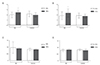
Two-D ultrasound analysis of uterine flux indexes showed that the PI of ANA+ uRM women was significantly higher than that of each the other groups (ANA- uRM women and control ANA+ and ANA- women). This finding has been detected in both the follicular phase and the midluteal phase of the cycle (Fig. 1A, B). Conversely, no differences were found in RI between uRM and control women, irrespective of the ANA status and of the phase of the cycle (Fig. 1C, D). ANA status (ANA+ or ANA-) at the time of sampling during the midulteal phase of the cycle remained unchanged in each woman compared to ANA status at the enrollment into the study.
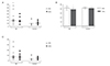
Three-D ultrasound analysis of uterine flux and vascularization indexes revealed that VI in ANA- uRM women was significantly higher than that in ANA+ control women (Fig. 2A). No other differences could be found when VI was compared among any other group (Fig. 2A). FI in uRM ANA+ women was significantly lower than that of each of the other groups (uRM ANA- and control ANA- and ANA+ women) (Fig. 2B). No differences were found in VFI in any of the study groups (Fig. 2C).
Discussion
The role of ANA, as well as of several other factors involved in autoimmunity, in RM is still largely unknown. There is experimental evidence supporting the hypothesis of a possible correlation between ANA and changes in the uterine blood flow in women affected by uRM, even though the available data are conflicting. Habara et al. [19] demonstrated that the PI-measured by 2D pulsed Doppler ultrasonography in the midluteal phase of an untreated non-pregnant cycle-was significantly higher in women with uRM than in control women; moreover, it was higher in ANA+ uRM women than in both ANA- uRM and in ANA+ control women even though no difference was found between PI in control women irrespective of the ANA status. Moreover, in a subsequent study of the same group, carried out on uRM and control women, the uterine artery PI measured in very early pregnancy was higher in uRM women, and particularly in the ANA+ uRM women; however, the finding that even among women ANA- the uterine artery PI was higher in uRM than in control women suggested that increased uterine artery PI could be an ANA-independent risk factor for uRM [21].
The present study investigated again this issue by applying, in addition to the classic 2D ultrasound methodology, also the 3D analysis technique, which has now became available. To our knowledge, this approach in the studies on ANA and uRM is novel. The interest in further studying the potential involvement of ANA in uRM stems from the consideration that recent evidence, including that from our group, found a significantly higher percentage of positivity to ANA in women with uRM compared to control healthy women [1617]. These findings are in agreement also with previous observations [15], including the observations by Nakatsuka al. in the above mentioned study [21] and with those by Ying et al. [18] carried out on women undergoing in vitro fertilization and embryo transfer.
The clinical meaning of 3D uterine flow indices is still incompletely determined and currently is under active investigation. At present, it is generally believed that information obtained by VI reflects the density of blood vassels (vascularity) in the analyzed volume. The FI is an index of the average intensity of blood flow. VFI is a combination of vascularization and flow information, hence it is an index of the overall perfusion of the organ.
The major findings of the present study, when the data obtained by 2D and 3D Doppler indices are considered together, are the following: 1) ANA+ women with uRM have a significantly increased PI throughout all the menstrual cycle and a reduced FI compared with all the other categories of women studied; 2) ANA- women with uRM have a unique increase in VI; 3) no differences could be detected in VFI and RI, irrespective of the uRM and ANA status.
A tentative unitary explanation for these findings could be that ANA in women with uRM are associated with a lesser than normal intensity of uterine blood flow, as evidenced by the reduced FI, that is an index of the intensity of blood flow. To counteract the impairment of uterine blood flow, an in-creased pulsatility of the uterine arteries could occur in these women, as evidenced by the increased PI. This compensatory mechanism could, in turn, explain why no differences could be detected in VFI, which measures both the vascularization and the perfusion. These hypothesized mechanisms could not be operative in uRM women who are ANA-, in which an increase in VI, has been detected. It is possible that in these women, in the absence of ANA, an increased density of vessels (measured by VI) in the uterus has been developed in response to other causative factors for RM different from ANA. Alternatively, since no differences were detected in VI between uRM ANA- women and both uRM ANA+ and controls ANA-, it is also possible that an increased VI could represent a risk factor for RM independent of ANA negativity.
In conclusion, the results of the present study suggest that ANA might have a role in uRM, at least in some women with uRM, possibly by determining, by still unclear mechanisms, an impairment in uterine blood hemodynamic, with specific application to uterine blood flow intensity and uterine artery impedance. Further investigation will clarify whether ANA are actually a causal factor for this and the possible underlying mechanism(s). Another interesting future investigation, suggested by the results of the present study, could be to check the outcome of future pregnancies in women with uRM in relation both to the presence or absence of ANA and to the values of blood flow and uterine artery impedance.

 XML Download